. 2008 Mar 11;105(10):4028-32.
doi: 10.1073/pnas.0800376105. Epub 2008 Mar 5.

The maturing architecture of the brain's default network

Affiliations

The maturing architecture of the brain's default network

Damien A Fair et al. Proc Natl Acad Sci U S A. .

Abstract

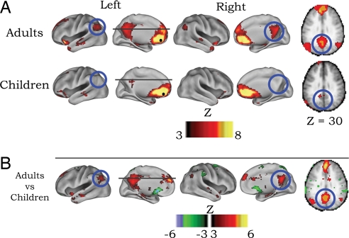
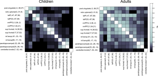
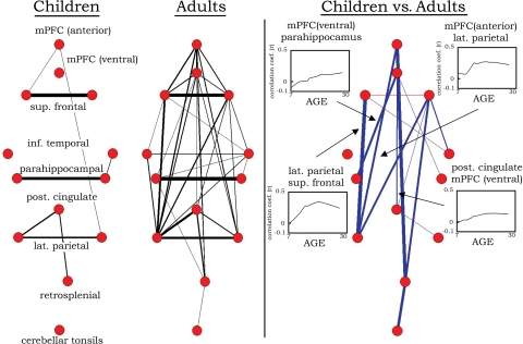
In recent years, the brain's "default network," a set of regions characterized by decreased neural activity during goal-oriented tasks, has generated a significant amount of interest, as well as controversy. Much of the discussion has focused on the relationship of these regions to a "default mode" of brain function. In early studies, investigators suggested that, the brain's default mode supports "self-referential" or "introspective" mental activity. Subsequently, regions of the default network have been more specifically related to the "internal narrative," the "autobiographical self," "stimulus independent thought," "mentalizing," and most recently "self-projection." However, the extant literature on the function of the default network is limited to adults, i.e., after the system has reached maturity. We hypothesized that further insight into the network's functioning could be achieved by characterizing its development. In the current study, we used resting-state functional connectivity MRI (rs-fcMRI) to characterize the development of the brain's default network. We found that the default regions are only sparsely functionally connected at early school age (7-9 years old); over development, these regions integrate into a cohesive, interconnected network.


Conflict of interest statement

The authors declare no conflict of interest.

Figures

Fig. 1.
Fig. 1.
Voxelwise resting-state functional connectivity maps for a seed region (solid black circle) in mPFC (ventral: −3, 39, −2). (A) Qualitatively, the rs-fcMRI map for the mPFC (ventral) seed region reveals the commonly observed adult connectivity pattern of the default network (9, 18, 19). The connectivity map in children, however, significantly deviates from that of the adults. Functional connections with regions in the posterior cingulate and lateral parietal regions (highlighted with blue open circles) are present in the adults but absent in children. (B) These qualitative differences between children and adults are confirmed by the direct comparison (random effects) between adults and children. mPFC (ventral) functional connections with the posterior cingulate and lateral parietal regions are significantly stronger in adults than children.
Fig. 2.
Fig. 2.
Correlation matrices representing default network functional connectivity for children and adults. Darker boxes represent low-correlation coefficients between regions, whereas lighter boxes represent stronger correlations. Qualitatively, adults have stronger correlation coefficients between regions of the default network than children do. The direct comparisons (see Fig. 3) between children and adults confirm this observation.
Fig. 3.
Fig. 3.
Graph visualization of the correlation matrices shown in Fig. 2 represented in a pseudoanatomical organization. Default regions (18) are only very sparsely connected in children aged 7–9 years. In adults aged 21–31 years, the default regions are highly integrated (strongly functionally connected). Only connections with r > 0.15 are shown at Left (line width is proportional to the connection strength). Statistically significant differences in functional connectivity between children and adults are highlighted on the Right. Blue lines represent significantly greater functional connectivity (r) in adults than in children. One connection (between superior frontal regions), although present in both groups, was significantly greater in children than in adults (red line). For the direct comparison, line width is proportional to the significance level (i.e., increased level of statistical significance; all lines meet P < 0.05 multiple comparisons corrected). Selected LOWESS curves for significant connections are also presented showing correlation coefficients (r) between regions as a function of age. For the full complement of LOWESS curves, see SI Fig. 4.

References

    1. Shulman GL, et al. Common blood flow changes across visual tasks: II. Decreases in cerebral cortex. J Cognit Neurosci. 1997;9:648–663. - PubMed
    1. Raichle ME, et al. A default mode of brain function. Proc Natl Acad Sci USA. 2001;98:676–682. - PMC - PubMed
    1. Gusnard DA, Akbudak E, Shulman GL, Raichle ME. Medial prefrontal cortex and self-referential mental activity: Relation to a default mode of brain function. Proc Natl Acad Sci USA. 2001;98:4259–4264. - PMC - PubMed
    1. Raichle ME, Snyder AZ. A default mode of brain function: A brief history of an evolving idea. NeuroImage. 2007;37:1083–1090. - PubMed
    1. Morcom AM, Fletcher PC. Does the brain have a baseline? Why we should be resisting a rest. NeuroImage. 2006;37:1073–1082. - PubMed

Publication types