. 2008 May;294(5):F1146-56.
doi: 10.1152/ajprenal.00592.2007. Epub 2008 Mar 5.

Sensitization of pelvic afferent nerves in the in vitro rat urinary bladder-pelvic nerve preparation by purinergic agonists and cyclophosphamide pretreatment

Affiliations

Sensitization of pelvic afferent nerves in the in vitro rat urinary bladder-pelvic nerve preparation by purinergic agonists and cyclophosphamide pretreatment

Yongbei Yu et al. Am J Physiol Renal Physiol. 2008 May.

Abstract

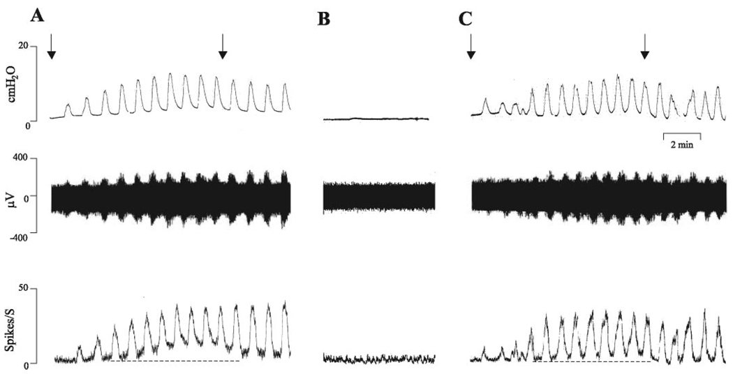
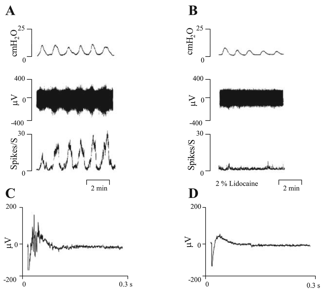
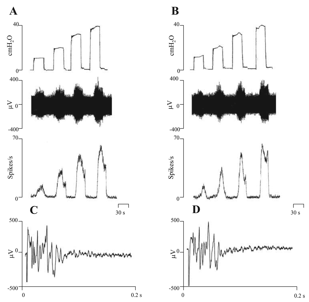
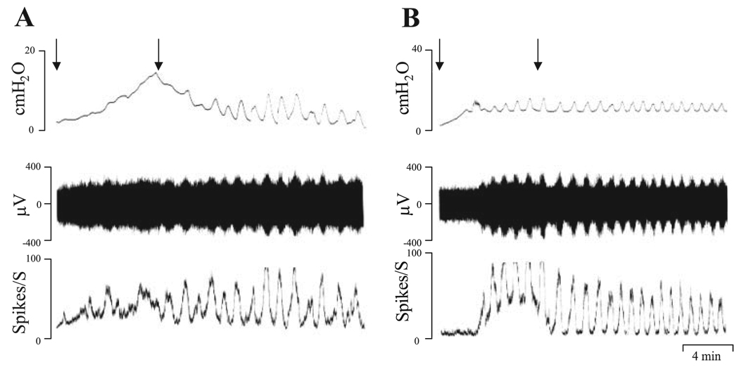
Effects of purinergic agonists (alpha,beta-meATP and ATP) and cyclophosphamide-induced cystitis on bladder afferent nerve (BAN) activity were studied in an in vitro bladder-pelvic nerve preparation. Distension of the bladder induced spontaneous bladder contractions that were accompanied by multiunit afferent firing. Intravesical administration of 40 and 130 microM alpha,beta-meATP increased afferent firing from 27 +/- 3 to 53 +/- 6 and 61 +/- 2 spikes/s, respectively, but did not change the maximum amplitude of spontaneous bladder contractions. Electrical stimulation on the surface of the bladder elicited action potentials (AP) in BAN. alpha,beta-meATP decreased the voltage threshold from 9.0 +/- 1.2 to 3.5 +/- 0.5 V (0.15-ms pulse duration) and increased the area of the APs (82% at 80-V stimulus intensity). These effects were blocked by TNP-ATP (30 microM). ATP (2 mM) applied in the bath produced similar changes in BAN activity. These effects were blocked by bath application of PPADS (30 microM). Neither TNP-ATP nor PPADS affected BAN activity induced by distension of the bladder. Cystitis induced by pretreatment of the rats with cyclophosphamide (100 mg/kg ip) increased afferent firing in response to isotonic bladder distension (10-40 cmH(2)O), decreased the threshold, and increased the area of evoked APs. The increase in afferent firing at 10 cmH(2)O intravesical pressure was reduced 52% by PPADS. These results indicate that purinergic agonists acting on P2X receptors and cystitis induced by cyclophosphamide can increase excitability of the BANs.


Figures

Fig. 1
Fig. 1
Effects of repeated bladder distension by intravesical infusion of Krebs solution at the rate of 0.04 ml/min for 8 min (A and C) on bladder activity and multiunit pelvic afferent nerve firing. Arrows indicate start and stop of infusion. Top traces represent bladder contractile activity measured as intravesical pressure. Middle traces represent afferent nerve firing. Bottom traces represent ratemeter recording of pelvic afferent nerve firing. The dashed lines in the bottom traces indicate basal level of afferent activity and show that phasic and tonic firing increased during intravesical infusion of Krebs solution. B: recording with bladder empty.
Fig. 2
Fig. 2
Effects of 2% lidocaine on bladder contractions and pelvic afferent nerve firing. A: responses after distention of bladder with 0.32 ml Krebs solution. B: responses after distention of bladder with 2% lidocaine in Krebs solution. Top traces represent bladder contractile activity measured as intravesical pressure. Middle traces represent afferent nerve firing. Bottom traces represent ratemeter recording of afferent nerve firing. C: evoked action potentials recorded on the pelvic nerve at the stimulus intensity of 80 V and 0.15-ms duration. D: evoked action potentials after intravesical infusion of 2% lidocaine at the same stimulation parameters as in C. Note that lidocaine blocked pelvic afferent nerve firing and evoked action potentials but did not abolish the bladder contractions. Vertical calibrations in A and B are intravesical pressure in cmH2O, nerve activity in µV, and spikes/s. Horizontal calibration represents 2 min. Vertical calibration in C and D is in µV and horizontal calibration represents 0.3 s.
Fig. 3
Fig. 3
Enhancement of pelvic afferent nerve firing after intravesical administration of α,β-meATP. Top traces represent bladder contractile activity measured as intravesical pressure. Middle traces represent pelvic afferent nerve activity. Bottom traces represent ratemeter recording of afferent firing. A: control recording during bladder distension induced by intravesical infusion of Krebs solution at the rate of 0.04 ml/min for 8 min. B: intravesical infusion of 130 µM α,β-meATP. C: after pretreatment with intravesical infusion of 0.3 ml TNP-ATP (30 µM) for 10 min and then intravesical administration of 130 µM αβ-meATP combined with 30 µM TNP-ATP which suppressed the afferent firing. Vertical calibrations are the same as in Fig. 1. Horizontal calibration represents 4 min. D: intravesical infusion of α,β-meATP did not change bladder pressure (top), but enhanced pelvic afferent nerve firing (40 µM, ***P < 0.001, n = 12; 130 µM, ***P < 0.001, n = 8; bottom). This enhanced afferent nerve firing was blocked by TNP-ATP, a P2X receptor antagonist (30 µM, n = 8).
Fig. 4
Fig. 4
Effects of PPADS (30 µM), a nonselective purinergic receptor antagonist applied in the bath, on afferent nerve activity evoked by isotonic distention of bladder with Krebs solution at 10, 20, 30, and 40 cmH2O for 30 s (A and B) and action potentials in pelvic afferent nerves evoked by electrical stimulation of the bladder (C and D). A and C represent control recordings; B and D represent recording after PPADS (30 µM). Top traces in A and B show intravesical pressures during isotonic distention of the bladder. Middle traces show the afferent nerve activity. Bottom traces represent ratemeter recording of afferent nerve firing. C and D: evoked action potentials recorded on the pelvic nerve evoked by stimulation on the surface of the bladder (80 V, 0.15-ms duration, average of 5 responses) before PPADS (C) and after PPADS (D). Note that PPADS (30 µM) did not alter afferent nerve firing or evoked action potentials.
Fig. 5
Fig. 5
Effects of 2,3-butanedione monoxime (BDM) on bladder contractions and afferent nerve activity induced by ATP (2 mM). Top traces represent bladder contractile activity measured as intravesical pressure with bladder distended by 0.32 ml of Krebs solution. Middle traces represent afferent nerve firing. Bottom traces represent ratemeter recording of pelvic afferent nerve firing. All records were obtained in the same preparation at 45- to 60-min intervals. A: responses to ATP (2 mM) applied in the bath. B: responses to 2 mM ATP after 60 mM BDM. Note that bladder activity was partially blocked and ATP-evoked firing was reduced by BDM. C: responses to 2 mM ATP after 80 mM BDM. Note that ATP-evoked bladder contraction but not ATP-evoked firing was blocked by BDM. D: 60 min after washout of BDM with Krebs solution the bladder spontaneous contractions and afferent nerve activity partially recovered.
Fig. 6
Fig. 6
Effects of α,β-meATP (130 µM) administered by intravesical infusion on evoked compound action potentials. A: threshold for evoked action potentials was reduced by purinergic receptor agonist, α,β-meATP (n = 8), and this effect was blocked by P2X receptor antagonist, TNP-ATP (30 µM, n = 7), in combination with α,β-meATP. B: compound action potentials recorded in pelvic afferent nerves in response to electrical stimulation on the surface of the urinary bladder (80 V, 0.15-ms duration, average of 5 responses). Top trace represents the control response at the stimulus intensity of 80 V, 0.15 ms with the bladder distended with 0.32 ml of Krebs solution. Middle trace was recorded after bladder distention with α,β-meATP (130 µM) at the same stimulus parameters. Bottom trace represents distention of the bladder with 130 µM α,β-meATP and 30 µM TNP-ATP at the same stimulus parameters after 10-min pretreatment with intravesical administration of 30 µM TNP-ATP. Dot above each record indicates the stimulus artifact. C: area of evoked action potentials was increased by α,β-meATP (130 µM) and reversed by TNP-ATP (30 µM) in combination with α,β-meATP. *P < 0.05 in A and C.
Fig. 7
Fig. 7
Effects of ATP on electrically evoked action potentials. A: threshold of evoked action potentials was reduced by ATP (2 mM, bath concentration, n = 7) and this effect was reversed by PPADS (30 µM, bath concentration, n = 6). B: compound action potentials recorded in pelvic afferent nerves in response to electrical stimulation on the surface of the urinary bladder (80 V, 0.15-ms stimulation duration, average of 5 responses). Top trace represents the control response with the bladder distended with Krebs solution. Middle trace was obtained after application of 2 mM ATP (bath concentration). Bottom trace shows responses under the same conditions in the presence of 2 mM ATP (bath concentration) after pretreatment with 30 µM PPADS in the bath. Dot under each record indicates the stimulus artifact. C: area of evoked action potentials was increased by ATP (2 mM) and reversed by PPADS (30 µM). *P < 0.05 in A and **P < 0.01 in C.
Fig. 8
Fig. 8
Pelvic nerve afferent firing induced by intravesical infusion of Krebs solution at the rate of 0.04 ml/min in cyclophosphamide-pretreated preparations (100 mg/kg ip, 17 h before experiments). Top traces represent bladder contractile activity measured as intravesical pressure. Middle traces represent pelvic nerve afferent firing. Bottom traces represent ratemeter recording of afferent firing. A and B recordings in different experiments showing different types of bladder contractions in cyclophosphamide-pretreated preparations. A: hyperactive noncompliant bladder that generates a large intravesical pressure during the initial stage of bladder filling. Tonic pressure declined after filling was stopped and spontaneous contractions emerged. B: bladder activity that was similar to activity in vehicle-treated bladder. However, peak afferent firing in both preparations was higher than in vehicle-treated preparations. Arrows represent start and stop of infusion of the Krebs solution.
Fig. 9
Fig. 9
Effects of PPADS (30 µM, n = 5), a nonselective purinergic receptor antagonist applied in the bath, on afferent nerve activity in cyclophosphamide (100 mg/kg ip, 17 h before the experiments)-pretreated preparations. Top traces in A and B show isotonic distention of the bladder with Krebs solution at 10, 20, 30, and 40 cmH2O for 30 s. Middle traces show the afferent nerve activity. Bottom traces show ratemeter recording of afferent nerve firing. A: before PPADS. B: after PPADS (30 µM). C and D: action potentials evoked by a stimulus intensity of 80 V and 0.15-ms pulse duration (average of 5 responses) before PPADS (C) and after PPADS (30 µM; D) in a cyclophosphamide-pretreated preparation. Note that PPADS reduced afferent nerve firing. E: summary of 5 experiments conducted using the protocol illustrated in A and B showing that PPADS (30 µM) applied in the bath reduced afferent nerve firing induced by isotonic distention of the bladder at 10, 20, 30, and 40 cmH2O for 30 s (n = 5). However, the effect of PPADS was only statistically significant during the 10 cmH2O pressure stimulus (n = 5, P < 0.05). F: area of action potentials evoked by a stimulus intensity of 80 V and 0.15-ms pulse duration as shown in C and D was reduced by PPADS (30 µM, n = 5, P < 0.05). *P < 0.05 in E and F.

Similar articles

Cited by

References

    1. Ahluwalia A, Maggi CA, Santicioli A, Lecci A, Giuliani S. Characterization of the capsaicin-sensitive component of cyclophosphamide-induced inflammation in the rat urinary bladder. Br J Pharmacol. 1994;111:1017–1022. - PMC - PubMed
    1. Birder LA, Barrick SR, Roppolo JR, Kanai AJ, de Groat WC, Kiss S, Buffington CA. Feline interstitial cystitis results in mechanical hypersensitivity and altered ATP release from bladder urothelium. Am J Physiol Renal Physiol. 2003;285:F423–F429. - PubMed
    1. Birder LA, Ruan HZ, Chopra B, Xiang Z, Barrick S, Buffington CA, Roppolo JR, Ford AP, de Groat WC, Burnstock G. Alterations in P2X and P2Y purinergic receptor expression in urinary bladder from normal cats and cats with interstitial cystitis. Am J Physiol Renal Physiol. 2004;287:F1084–F1091. - PubMed
    1. Borlak J, Zwadlo C. The myosin ATPase inhibitor 2,3-Butanedione monoxime dictates transcriptional activation of ion channels and Ca2+-handling proteins. Mol Pharmacol. 2004;66:708–717. - PubMed
    1. Borvendeg-Khrasani M, Rubini P, Fischer W, Allgaier C, Wirkner K, Himmel HM, Gillen C, Illes P. Subsensitivity of P2X but not vanilloid 1 receptors in dorsal root ganglia of rats caused by cyclophosphamide cystitis. Eur J Pharmacol. 2003;474:71–75. - PubMed

Publication types

MeSH terms

Substances

LinkOut - more resources