Comparative Study
. 2008 Mar 5;28(10):2485-94.
doi: 10.1523/JNEUROSCI.5369-07.2008.

Transient receptor potential A1 is a sensory receptor for multiple products of oxidative stress

Affiliations
Comparative Study

Transient receptor potential A1 is a sensory receptor for multiple products of oxidative stress

David A Andersson et al. J Neurosci. .

Abstract

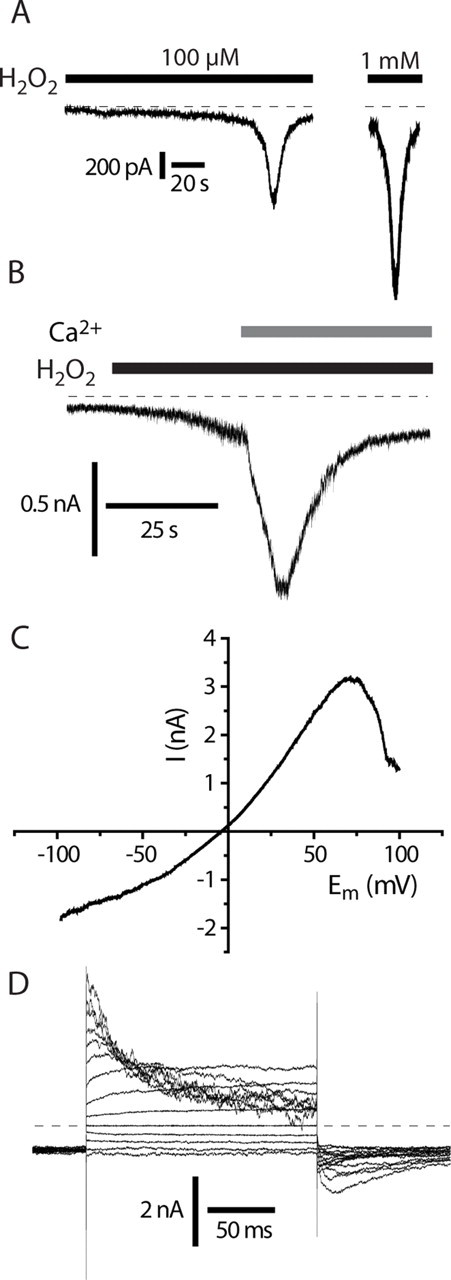
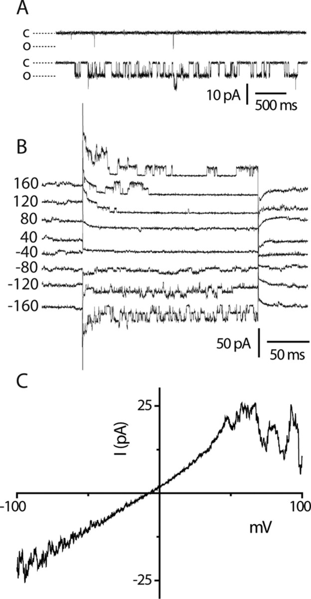
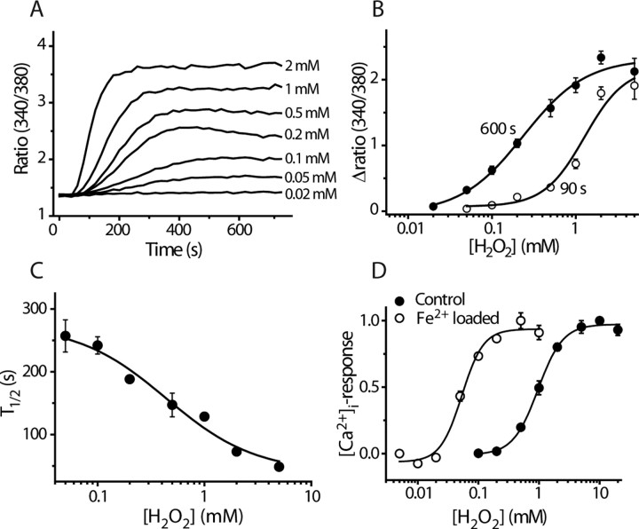
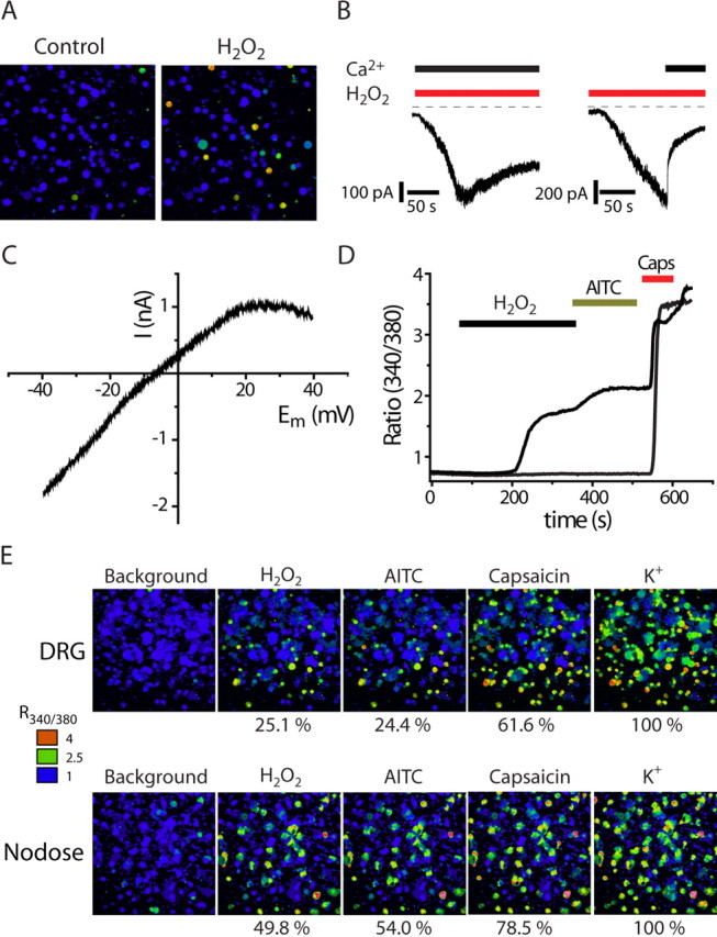
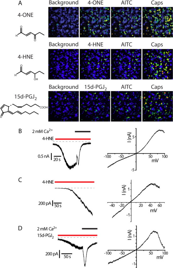
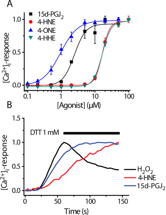
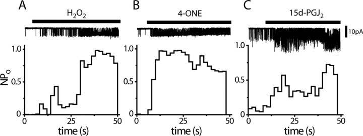
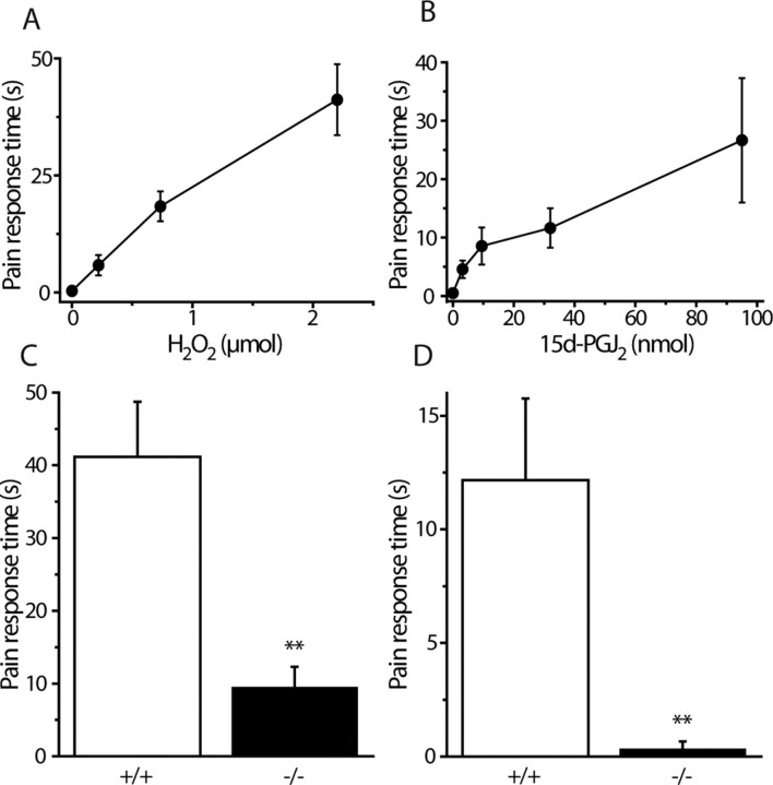
Transient receptor potential A1 (TRPA1) is expressed in a subset of nociceptive sensory neurons where it acts as a sensor for environmental irritants, including acrolein, and some pungent plant ingredients such as allyl isothiocyanate and cinnamaldehyde. These exogenous compounds activate TRPA1 by covalent modification of cysteine residues. We have used electrophysiological methods and measurements of intracellular calcium concentration ([Ca(2+)](i)) to show that TRPA1 is activated by several classes of endogenous thiol-reactive molecules. TRPA1 was activated by hydrogen peroxide (H(2)O(2); EC(50), 230 microM), by endogenously occurring alkenyl aldehydes (EC(50): 4-hydroxynonenal 19.9 microM, 4-oxo-nonenal 1.9 microM, 4-hydroxyhexenal 38.9 microM) and by the cyclopentenone prostaglandin, 15-deoxy-delta(12,14)-prostaglandin J(2) (15d-PGJ(2), EC(50): 5.6 microM). The effect of H(2)O(2) was reversed by treatment with dithiothreitol indicating that H(2)O(2) acts by promoting the formation of disulfide bonds whereas the actions of the alkenyl aldehydes and 15d-PGJ(2) were not reversed, suggesting that these agents form Michael adducts. H(2)O(2) and the naturally occurring alkenyl aldehydes and 15d-PGJ(2) acted on a subset of isolated rat and mouse sensory neurons [approximately 25% of rat dorsal root ganglion (DRG) and approximately 50% of nodose ganglion neurons] to evoke a depolarizing inward current and an increase in [Ca(2+)](i) in TRPA1 expressing neurons. The abilities of H(2)O(2), alkenyl aldehydes and 15d-PGJ(2) to raise [Ca(2+)](i) in mouse DRG neurons were greatly reduced in neurons from trpa1(-/-) mice. Furthermore, intraplantar injection of either H(2)O(2) or 15d-PGJ2 evoked a nocifensive/pain response in wild-type mice, but not in trpa1(-/-) mice. These data demonstrate that multiple agents produced during episodes of oxidative stress can activate TRPA1 expressed in sensory neurons.


Figures

Figure 1.
Figure 1.
H2O2 activates TRPA1 expressed in CHO cells. A, H2O2 activates TRPA1 with a concentration-dependent latency in Ca2+-containing solutions. Currents recorded in calcium- containing solution showing characteristic “threshold” with a sudden increase in membrane current. B, Ca2+ potentiates H2O2-induced TRPA1 currents. Current response to 10 mm H2O2 in a TRPA1 CHO cell, −60 mV. Note the slow increase in current in calcium-free solution followed by a rapid current increase when Ca2+ (2 mm) is added. C, Current–voltage relationship of H2O2-evoked current with 2 s voltage ramp in a TRPA1 CHO cell in calcium-free solution revealed a reduced current at positive potentials. D, Kinetics of H2O2-evoked TRPA1 current in calcium free solution. Note the time and voltage-dependent inactivation at more positive potentials that accounts for reduced conductance seen with voltage ramp protocols (C) (Figs. 2C, 4C). Holding potential −60 mV with 20 mV interval steps to from −80 to +180 mV.
Figure 2.
Figure 2.
Single-channel activity evoked by H2O2. Cell attached TRPA1 single-channel currents. A, Few brief openings seen in the absence of H2O2 (top trace), but robust channel activity elicited by 1 mm H2O2 (lower trace) at −100 mV. B, Voltage-dependent single-channel current activity showing inactivation at positive membrane potentials. Holding potential, −60 mV; traces are offset for clarity. C, Voltage ramp illustrating single-channel inactivation at positive membrane potentials (the trace shown is the average of 5 sweeps).
Figure 3.
Figure 3.
Concentration-dependent effect of H2O2 on TRPA1. A, Concentration-dependent time course of [Ca2+]i-responses to stimulation with H2O2 in CHO cells expressing TRPA1. Traces are mean ratios from quadruplicate wells. B, Concentration–response curves constructed from the experiment shown in (A) 90 and 600 s after addition of H2O2 (mean ± SEM). C, The time required for half-maximal activation (T1/2) is concentration dependent (data points are mean ± SEM of 4 measurements). D, Fe2+ potentiates the effect of H2O2, suggesting that H2O2 acts via intracellular production of hydroxyl radicals. Concentration–response curves for H2O2-evoked increase in [Ca2+]i in normal and Fe2+-loaded TRPA1 cells are shown. Fe2+-loaded cells were incubated with 100 μm FeSO4 for 1 h and then washed so that no extracellular FeSO4 was present during the experiment (mean ± SEM; n = 4).
Figure 4.
Figure 4.
H2O2 activates TRPA1-containing DRG and nodose neurons. A, Pseudocolored images illustrating [Ca2+]i responses evoked by H2O2 (5 mm) measured with Fura-2 in rat DRG neurons. B, Currents evoked by H2O2 (1 mm) in DRG neurons in the presence (left) and absence (right) of extracellular calcium (at a holding potential of −60 mV). The current rapidly inactivated when Ca2+ was applied after an initial current has developed in Ca2+-free conditions. C, Current–voltage plot for the H2O2 response in a DRG neuron generated by a slow 2 s voltage ramp in Ca2+-free solution. Note the characteristic reduced conductance at positive membrane potential. D, Change in [Ca2+]i (340/380 ratio) of typical DRG neurons in response to sequential applications of H2O2, AITC, and capsaicin (Caps) showing H2O2-sensitive and H2O2-insensitive TRPV1 expressing neurons. E, [Ca2+]i responses in DRG (top sequence) and nodose neurons (bottom sequence) to sequential application of H2O2 (5 mm), AITC (50 μm), and capsaicin (1 μm). All neurons in the culture were identified by the [Ca2+]i increase elicited by application of 50 mm K+. H2O2, AITC and capsaicin stimulated a larger proportion of neurons dissociated from nodose than dorsal root ganglia. The number of neurons tested in each group was between 363 and 744.
Figure 5.
Figure 5.
Lipid peroxidation products and 15d-PGJ2 are TRPA1 agonists. A, [Ca2+]i increases evoked by 4-ONE (3 μm), 4-HNE (30 μm), and 15d-PGJ2 (20 μm) in DRG neurons. Sequential applications of AITC and capsaicin (Caps) show that 4-ONE, 4-HNE, and 15d-PGJ2 activate the same subset of TRPV1-expressing, capsaicin-sensitive neurons as AITC. B, Left, 4-HNE-evoked current in a TRPA1 CHO cell initially in calcium-free solution. Admission of calcium led to rapid inactivation. Right, Current–voltage relationship of 4-HNE-evoked current. C, Left, 4-HNE-evoked current in a DRG neuron (external solution containing 15 μm Ca2+, −60 mV). Right, Current–voltage relationship of the 4-HNE-evoked current in the same neuron. D, Time course (left) and voltage-dependent kinetics (right) for 15d-PGJ2-evoked TRPA1 current in a CHO cell. Right, Current–voltage relationship of 15d-PGJ2-evoked current.
Figure 6.
Figure 6.
Concentration dependence of lipid mediators and dithiothreitol sensitivity. A, Concentration–response curves for 4-ONE, 4-HNE, 4-HHE, and 15d-PGJ2 in TRPA1 CHO cells (mean ± SEM, n = 4, representative of at least 3 experiments). B, Application of DTT reverse [Ca2+]i-responses induced by application of H2O2, but not 4-HNE or 15d-PGJ2.
Figure 7.
Figure 7.
H2O2, 4-ONE and 15d-PGJ2 activate TRPA1 in a membrane-delimited manner. A–C, Application of 5 mm H2O2 (A), 5 μm 4-ONE (B) and 20 μm 15d-PGJ2 (C) evoke single-channel activity in excised inside-out patches (−100 mV, each trace is representative of at least 3 experiments).
Figure 8.
Figure 8.
H2O2 and 15d-PGJ2 induce pain-related behavior in vivo by activating TRPA1. A, B, Duration of nocifensive (licking/flinching) behavior in wild-type mice evoked by intraplantar hindpaw injections of H2O2 in saline (A) or 15d-PGJ2 in saline containing 10% DMSO (B). The injection volume was 25 μl. Pain-related behavior was recorded over 5 min (mean ± SEM, n = 6 for each group). C, D, Wild-type (+/+) and TRPA1-deficient mice (−/−) were injected in the hind paw with H2O2 (C) (2.2 μmol/25 μl) or 15d-PGJ2 (D) (32 nmol/25 μl). Pain-related behavior (licking, biting, flinching, or shaking of the injected paw) was recorded for 3 min after injection. The nocifensive responses induced by H2O2 and 15d-PGJ2 were dramatically reduced or absent in mice lacking TRPA1 (mean ± SEM; n = 6 in each group; **p < 0.01).

References

    1. Andersson DA, Nash M, Bevan S. Modulation of the cold-activated channel TRPM8 by lysophospholipids and polyunsaturated fatty acids. J Neurosci. 2007;27:3347–3355. - PMC - PubMed
    1. Bandell M, Story GM, Hwang SW, Viswanath V, Eid SR, Petrus MJ, Earley TJ, Patapoutian A. Noxious cold ion channel TRPA1 is activated by pungent compounds and bradykinin. Neuron. 2004;41:849–857. - PubMed
    1. Bautista DM, Movahed P, Hinman A, Axelsson HE, Sterner O, Hogestatt ED, Julius D, Jordt SE, Zygmunt PM. Pungent products from garlic activate the sensory ion channel TRPA1. Proc Natl Acad Sci USA. 2005;102:12248–12252. - PMC - PubMed
    1. Bautista DM, Jordt SE, Nikai T, Tsuruda PR, Read AJ, Poblete J, Yamoah EN, Basbaum AI, Julius D. TRPA1 mediates the inflammatory actions of environmental irritants and proalgesic agents. Cell. 2006;124:1269–1282. - PubMed
    1. Bedard K, Krause KH. The NOX family of ROS-generating NADPH oxidases: physiology and pathophysiology. Physiol Rev. 2007;87:245–313. - PubMed

Publication types

MeSH terms