Comparative Study
. 2008 Mar 15;180(6):4109-15.
doi: 10.4049/jimmunol.180.6.4109.

Deviation from a strong Th1-dominated to a modest Th17-dominated CD4 T cell response in the absence of IL-12p40 and type I IFNs sustains protective CD8 T cells

Affiliations
Comparative Study

Deviation from a strong Th1-dominated to a modest Th17-dominated CD4 T cell response in the absence of IL-12p40 and type I IFNs sustains protective CD8 T cells

Nural N Orgun et al. J Immunol. .

Abstract

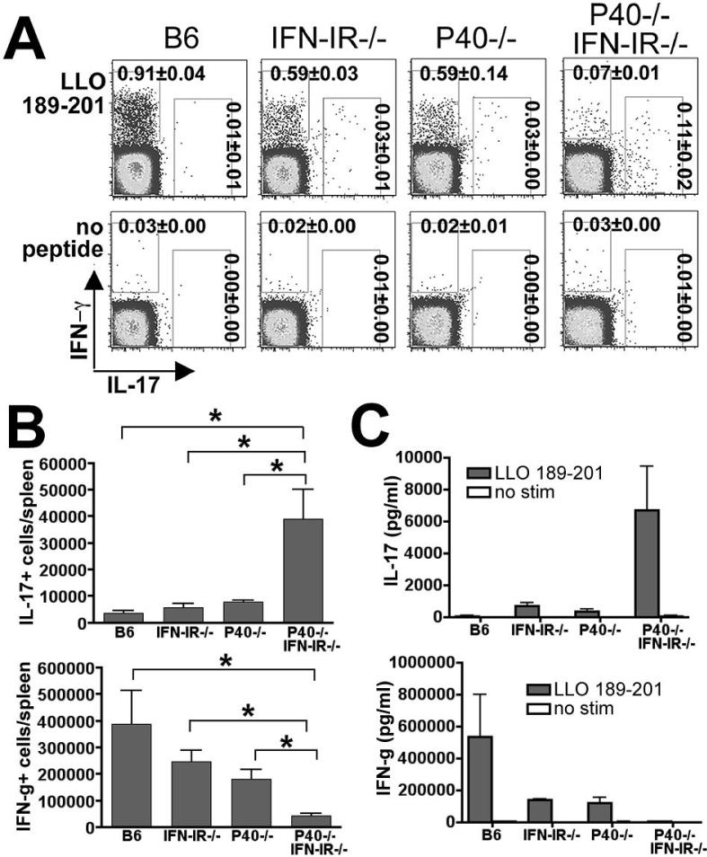
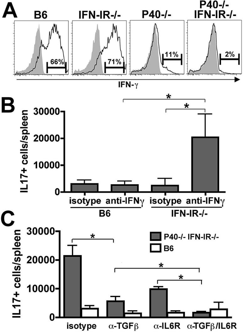
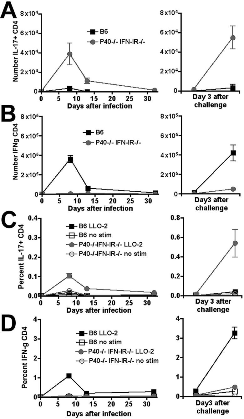
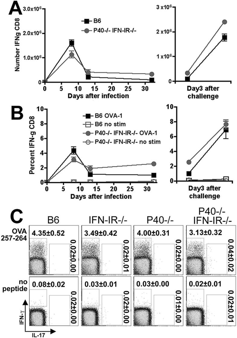
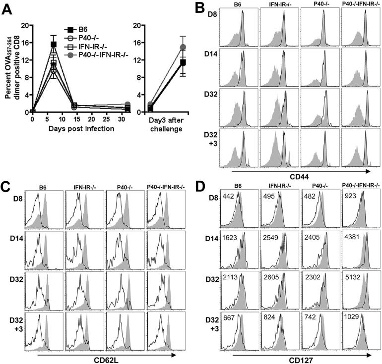
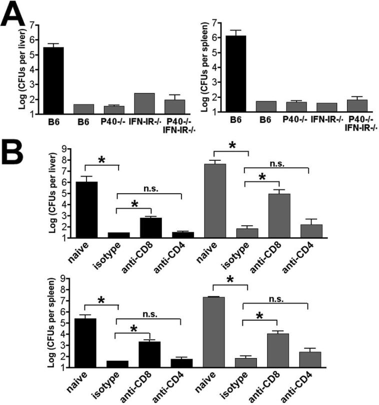
The differentiation of naive CD4 T cells into specific effector subsets is controlled in large part by the milieu of cytokines present during their initial encounter with Ag. Cytokines that drive differentiation of the newly described Th17 lineage have been characterized in vitro, but the cytokines that prime commitment to this lineage in response to infection in vivo are less clear. Listeria monocytogenes (Lm) induces a strong Th1 response in wild-type mice. By contrast, we demonstrate that in the absence of IL-12p40 (or IFN-gamma) and type I IFN receptor signaling, the Th1 Ag-specific CD4 T cell response is virtually abolished and replaced by a relatively low magnitude Th17-dominated response. This Th17 response was dependent on TGF-beta and IL-6. Despite this change in CD4 T cell response, neither the kinetics of the CD4 and CD8 T cell responses, the quality of the CD8 T cell response, nor the ability of CD8 T cells to mediate protection were affected. Thus, generation of protective CD8 T cell immunity was resilient to perturbations that replace a strong Th1-dominated to a reduced magnitude Th17-dominated Ag-specific CD4 T cell response.


Figures

Figure 1
Figure 1
Th17 response in the absence of both IL-12 and IFN-IR. A. FACS plot indicating IL-17 and IFN-γ production in a CD4+ cell gate among splenocytes from mice day 8 after infection with 106 Lm-OVA ΔactA and in vitro stimulation with the Lm-specific MHC class II peptide LLO189-201 or no peptide control. B. Total numbers of IL-17- or IFN-γ-producing, LLO189-201-specific CD4 T cells per mouse spleen as quantified by intracellular cytokine staining. C. Concentration of IL-17 and IFN-γ in splenocyte culture supernatants 72 hours after stimulation with the LLO189-201 peptide or no peptide. These data represent 6-10 mice for each experimental group combined from three independent experiments. Bar, standard error. * P < 0.05.
Figure 2
Figure 2
IFN-γ or IFN-I inhibits Th17 differentiation. A. Histogram plot indicating the percentage of NK1.1+ cells that produce IFN-γ 24 hours after infection with 106 Lm-OVA ΔactA (open histograms) or in the absence of infection (shaded histograms) for the indicated groups of mice. B. Total number of IL-17-producing CD4 T cells after stimulation with LLO189-201 peptide on day 8 after infection with 106 Lm-OVA ΔactA in B6 or IFN-IR-/- mice pretreated with anti-IFN-γ or isotype control antibody. C. Total numbers of IL-17-producing CD4 T cells after stimulation with LLO189-201 peptide on day 8 after infection with 106 Lm-OVA ΔactA as quantified by intracellular cytokine staining in P40-/-IFN-IR-/- (shaded bars) or B6 (open bars) mice pretreated with either rat IgG control (isotype), anti-Tgf-β, anti-IL-6 receptor, or both anti-Tgf-β and anti-IL-6 receptor antibodies. These data represent 6-8 mice for each experimental group combined from two independent experiments. Bar, standard error. * P < 0.05
Figure 3
Figure 3
Total number (A, B) and percentage (C, D) of IFN-γ- (B, D) producing CD4 T cells per spleen after stimulation with the MHC class II peptide LLO189-201 on days 8, 14, or 32 after primary infection with 106 Lm-OVA ΔactA (left sided panels), or on day 3 after challenge with 105 Lm-OVA in mice inoculated with Lm-OVA ΔactA 32 days prior to challenge (right sided panels). These represent 8-12 mice per time point combined from three independent experiments. Bar, standard error.
Figure 4
Figure 4
Total number (A) and percentage (B) of IFN-γ-producing CD8 T cells per spleen after stimulation with the MHC class I peptide OVA257-264 on days 8, 14, or 32 after primary infection with 106 Lm-OVA ΔactA (left sided panels), or on day 3 after challenge with 105 Lm-OVA in mice inoculated with Lm-OVA ΔactA 32 days prior to challenge (right sided panels). C. FACS plot indicating IL-17 and IFN-γ production in a CD8+ cell gate among splenocytes from mice day 8 after infection with 106 Lm-OVA ΔactA after in vitro stimulation with the MHC class I peptide OVA257-264 or no peptide control. These data represent 9-12 mice per time point combined from three independent experiments. Bar, standard error.
Figure 5
Figure 5
A. Percentage of antigen-specific CD8 T cells among splenocytes after primary infection with 106 Lm-OVA ΔactA (left sided panel), or on day 3 after challenge with 105 Lm-OVA in mice inoculated with Lm-OVA ΔactA 32 days prior to challenge (right sided panel) as determined by staining with OVA257-264 dimer. Bar, standard error. These data are from 6 mice per time point per group from two independent experiments. Expression of CD44 (B), CD62L (C), and CD127 (D) among OVA257-264 dimer positive antigen-specific (open histograms) compared with non-antigen specific (filled histograms) CD8 T cells at the indicated time points after primary infection with 106 Lm-OVA ΔactA (day 8, 14, and 32), and secondary challenge with 105 Lm-OVA (day 3). The mean fluorescent intensity for CD127 expression among dimer positive antigen-specific T cells is indicated.
Figure 6
Figure 6
Protective immunity in the absence of both IL-12 and IFN-I. A. Numbers of recoverable Lm CFUs in the liver and spleen day 3 after challenge with 105 Lm-OVA in either naïve B6 mice (black bar) or the indicated mice that had been primed with 106 Lm-OVA ΔactA 32 days prior to challenge. B. Numbers of recoverable Lm CFUs in the liver and spleen day 3 after challenge with 105 Lm-OVA in either B6 (black bar) or P40-/-IFN-IR-/- (shaded bar) mice not previously infected with Lm (naïve) or infected with 106 Lm-OVA ΔactA 32 days prior to challenge and treated with anti-CD8, anti-CD4, or isotype control antibody one day prior to challenge. These data represent 8-10 mice for each experimental group combined from two independent experiments. Bar, standard error. * P < 0.05, n.s., not significant.

References

    1. Bettelli E, Oukka M, Kuchroo VK. T(H)-17 cells in the circle of immunity and autoimmunity. Nat Immunol. 2007;8:345–350. - PubMed
    1. Weaver CT, Hatton RD, Mangan PR, Harrington LE. IL-17 family cytokines and the expanding diversity of effector T cell lineages. Annu Rev Immunol. 2007;25:821–852. - PubMed
    1. Mangan PR, Harrington LE, O'Quinn DB, Helms WS, Bullard DC, Elson CO, Hatton RD, Wahl SM, Schoeb TR, Weaver CT. Transforming growth factor-beta induces development of the T(H)17 lineage. Nature. 2006;441:231–234. - PubMed
    1. Ye P, Rodriguez FH, Kanaly S, Stocking KL, Schurr J, Schwarzenberger P, Oliver P, Huang W, Zhang P, Zhang J, Shellito JE, Bagby GJ, Nelson S, Charrier K, Peschon JJ, Kolls JK. Requirement of interleukin 17 receptor signaling for lung CXC chemokine and granulocyte colony-stimulating factor expression, neutrophil recruitment, and host defense. J Exp Med. 2001;194:519–527. - PMC - PubMed
    1. Happel KI, Zheng M, Young E, Quinton LJ, Lockhart E, Ramsay AJ, Shellito JE, Schurr JR, Bagby GJ, Nelson S, Kolls JK. Cutting edge: roles of Toll-like receptor 4 and IL-23 in IL-17 expression in response to Klebsiella pneumoniae infection. J Immunol. 2003;170:4432–4436. - PMC - PubMed

Publication types

MeSH terms