. 2008 Mar 7;319(5868):1405-8.
doi: 10.1126/science.1154470.

Coiled-coil irregularities and instabilities in group A Streptococcus M1 are required for virulence

Affiliations

Coiled-coil irregularities and instabilities in group A Streptococcus M1 are required for virulence

Case McNamara et al. Science. .

Abstract

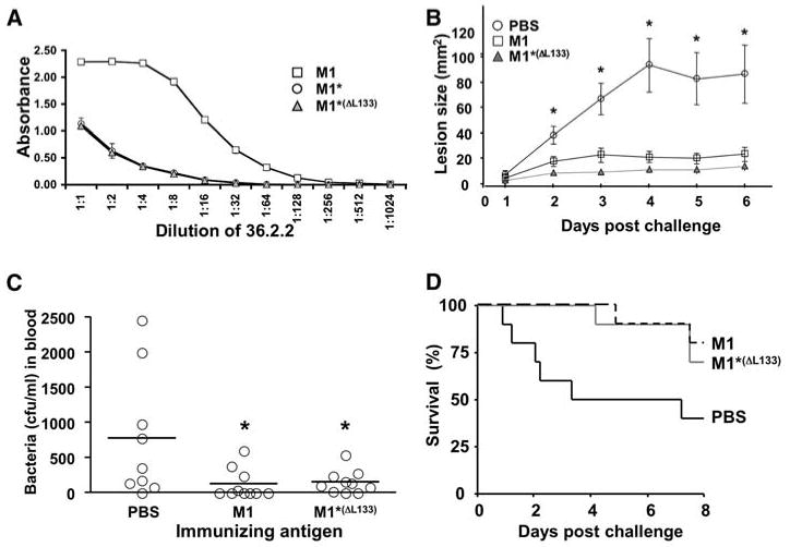
Antigenically variable M proteins are major virulence factors and immunogens of the human pathogen group A Streptococcus (GAS). Here, we report the approximately 3 angstrom resolution structure of a GAS M1 fragment containing the regions responsible for eliciting type-specific, protective immunity and for binding fibrinogen, which promotes M1 proinflammatory and antiphagocytic functions. The structure revealed substantial irregularities and instabilities throughout the coiled coil of the M1 fragment. Similar structural irregularities occur in myosin and tropomyosin, explaining the patterns of cross-reactivity seen in autoimmune sequelae of GAS infection. Sequence idealization of a large segment of the M1 coiled coil enhanced stability but diminished fibrinogen binding, proinflammatory effects, and antibody cross-reactivity, whereas it left protective immunogenicity undiminished. Idealized M proteins appear to have promise as vaccine immunogens.


Figures

Fig. 1
Fig. 1
(A) Mature M1 results from cleavage (arrowheads) of the N-terminal signal sequence and the C-terminal Leu-Pro-X-Thr-Gly motif (where X is any amino acid) and covalent attachment of the C terminus to the cell wall. Boundaries of the A region, B repeats, S region, C repeats, and D region are indicated. (B) (Top) a-a′ and d-d′ (prime refers to the opposing helix) packing in parallel dimeric coiled coils. Broken wedges indicate helices pointing the N to C termini into the page. (Bottom) Heptad register indicated above and below the sequence (a and d position residues boxed) of the M1 A region (blue) and B repeats (green). The circled residues are destabilizing to coiled coils, with relative instabilities ΔΔGu(Ala) ≤ 0 (11). Italicized residues form antiparallel coiled coils in the crystal. Residues highlighted in yellow and orange were substituted with Val and Leu, respectively, to create M1* and M1AB*. (C) Tail-to-tail packing of the two M1AB dimers in the asymmetric unit of the crystal (blue, A regions; green, B repeats).
Fig. 2
Fig. 2
(A) Structure of M1AB (blue, A region; green, B repeats) with boxed regions and labeling indicating irregularities. (B) Ala stagger shown by superposition of Cα traces of M1AB residues 70 to 97 (orange) with the ideal coiled coil of GCN4 (purple). (C) Conformation of individual helices from the two M1AB dimers in the asymmetric unit, superimposed on main-chain atoms of residues 60 to 77. The position of the Ala stagger is indicated by the arrowhead. (D) Conformation of Lys98 and Arg105 in the two M1AB dimers in the asymmetric unit, with heptad positions of residues indicated in parentheses and polar contacts in red dashed lines (with distances shown). (E) (Top) Schematic of a-d′ and d-a′ packing in antiparallel dimeric coiled coils. The broken wedge indicates the helix pointing the N to C termini into the page, and the solid wedge denotes out of the page. (Bottom) Antiparallel coiled coil of B repeats, with side chains of a and d position residues depicted and labeled.
Fig. 3
Fig. 3
CD spectra at 4 (triangles), 20 (squares), and 37°C (circles) of (A) M1AB (green), M1AB* (black), and M1AB*(ΔL133) (red) and (B) M1 (green), M1* (black), and M1*(ΔL133) (red). Mean residue 222:208 ellipticity (MRE) ratios are shown. (C) (Top) His6-tagged M1 (M1-H) and untagged M1 (M1) were coincubated at the indicated temperatures and coprecipitated at 4°C with Ni2+– nitrilotriacetic acid (NTA) agarose beads. (Middle) M1-H/M1 heterodimers were isolated, incubated, and coprecipitated at the indicated temperatures with Ni2+-NTA agarose beads. (Bottom) Only untagged M1 was incubated with beads. (A to C) Unbound protein (U) and protein bound to the beads (B) were visualized by Coomassie-stained, reducing SDS–polyacrylamide gel electrophoresis (PAGE).
Fig. 4
Fig. 4
(A) FgD incubated alone or with His6-tagged constructs of M1, M1*, or M1*(ΔL133) at 37°C and coprecipitated with Ni2+-NTA agarose beads. U and B proteins were visualized by Coomassie-stained, nonreducing SDS-PAGE. (B) Quantification of FgD binding in (A). Error bars indicate mean ± SD. (C) Western blot of HBP in supernatants from human neutrophils stimulated with M1 or M1*(ΔL133). rhHBP, recombinant human HBP. (D) Lung histopathology of Balb/c mice 30 min after intravenous injection of M1 or M1*(ΔL133). Representative histopathology (hematoxylin and eosin stain) with intra-alveolar edema (thick blue arrows) and macrovascular (asterisks) and microvascular (thin arrows) congestion is indicated. Magnification, ×100.
Fig. 5
Fig. 5
(A) Titer of mAb 36.2.2 versus M1, M1*, and M1*(ΔL133) by enzyme-linked immunosorbent assay. Error bars indicate mean ± SD. (B) Skin lesion size of mice immunized with M1 or M1*(ΔL133) after subcutaneous challenge with WT M1 GAS. Error bars indicate mean ± SEM (N = 10 mice per group). Analysis of variance (ANOVA) was significant (P < 0.002) on days 2 to 6; posthoc group comparisons (Tukey-Kramer multiple-comparison test) revealed significant protection of M1 or M1*(ΔL133) versus phosphate-buffered saline (PBS) on days 2 to 6 (asterisks denote P < 0.05). (C) Bacteremia of mice immunized with M1 or M1*(ΔL133) 4 hours after intraperitoneal challenge with WT M1 GAS. Mean (horizontal bars) and distribution are shown (N = 10 per group). ANOVA was significant at P = 0.02; posthoc group comparisons revealed significant protection of M1 or M1*(ΔL133) versus PBS control (asterisks denote P < 0.05). (D) Kaplan-Meyer survival curve of immunized mice from (C).

References

    1. Cunningham MW. Clin Microbiol Rev. 2000;13:470. - PMC - PubMed
    1. Fischetti VA. Clin Microbiol Rev. 1989;2:285. - PMC - PubMed
    1. Phillips GN, Jr, Flicker PF, Cohen C, Manjula BN, Fischetti VA. Proc Natl Acad Sci U S A. 1981;78:4689. - PMC - PubMed
    1. Ringdahl U, et al. Mol Microbiol. 2000;37:1318. - PubMed
    1. Sandin C, Carlsson F, Lindahl G. Mol Microbiol. 2006;59:20. - PubMed

Publication types

MeSH terms

Associated data