

Remodeling the brain: plastic structural brain changes produced by different motor therapies after stroke
- PMID: 18323492
- PMCID: PMC2574634
- DOI: 10.1161/STROKEAHA.107.502229
Remodeling the brain: plastic structural brain changes produced by different motor therapies after stroke
Abstract
Background and purpose: Studies on adult stroke patients have demonstrated functional changes in cortical excitability, metabolic rate, or blood flow after motor therapy, measures that can fluctuate rapidly over time. This study evaluated whether evidence could also be found for structural brain changes during an efficacious rehabilitation program.
Methods: Chronic stroke patients were randomly assigned to receive either constraint-induced movement therapy (n=16) or a comparison therapy (n=20). Longitudinal voxel-based morphometry was performed on structural MRI scans obtained immediately before and after patients received therapy.
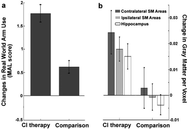
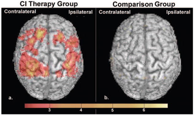
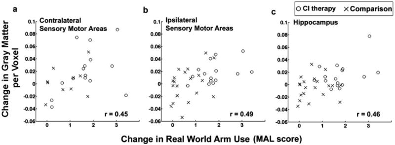
Results: The group receiving constraint-induced movement therapy exhibited far greater improvement in use of the more affected arm in the life situation than the comparison therapy group. Structural brain changes paralleled these improvements in spontaneous use of the more impaired arm for activities of daily living. There were profuse increases in gray matter in sensory and motor areas both contralateral and ipsilateral to the affected arm that were bilaterally symmetrical, as well as bilaterally in the hippocampus. In contrast, the comparison therapy group failed to show gray matter increases. Importantly, the magnitude of the observed gray matter increases was significantly correlated with amount of improvement in real-world arm use.
Conclusions: These findings suggest that a previously overlooked type of brain plasticity, structural remodeling of the human brain, is harnessed by constraint-induced movement therapy for a condition once thought to be refractory to treatment: motor deficit in chronic stroke patients.
Figures
References
-
- Merzenich MM, Nelson RJ, Stryker MP, Cynader MS, Schoppmann A, Zook JM. Somatosensory cortical map changes following digit amputation in adult monkeys. J Comp Neurol. 1984;224:591–605. - PubMed
-
- Kaas JH, Merzenich MM, Killackey HP. The reorganization of somatosensory cortex following peripheral nerve damage in adult and developing mammals. Annu Rev Neurosci. 1983;6:325–356. - PubMed
-
- Jenkins WM, Merzenich MM, Ochs MT, Allard T, Guic-Robles E. Functional reorganization of primary somatosensory cortex in adult owl monkeys after behaviorally controlled tactile stimulation. J Neurophysiol. 1990;63:82–104. - PubMed
-
- Pons TP, Garraghty PE, Ommaya AK, Kaas JH, Taub E, Mishkin M. Massive cortical reorganization after sensory deafferentation in adult macaques. Science. 1991;252:1857–1860. - PubMed
-
- Elbert T, Pantev C, Wienbruch C, Rockstroh B, Taub E. Increased cortical representation of the fingers of the left hand in string players. Science. 1995;270:305–307. - PubMed
Publication types
MeSH terms
Grants and funding
LinkOut - more resources
Full Text Sources
Medical
