. 2008 May;39(5):1569-74.
doi: 10.1161/STROKEAHA.107.502047. Epub 2008 Mar 6.

Dual-modality monitoring of targeted intraarterial delivery of mesenchymal stem cells after transient ischemia

Affiliations

Dual-modality monitoring of targeted intraarterial delivery of mesenchymal stem cells after transient ischemia

Piotr Walczak et al. Stroke. 2008 May.

Abstract

Background and purpose: In animal models of stroke, functional improvement has been obtained after stem cell transplantation. Successful therapy depends largely on achieving a robust and targeted cell engraftment, with intraarterial (IA) injection being a potentially attractive route of administration. We assessed the suitability of laser Doppler flow (LDF) signal measurements and magnetic resonance (MR) imaging for noninvasive dual monitoring of targeted IA cell delivery.

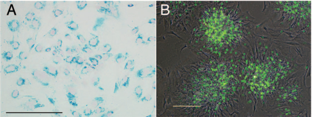
Methods: Transient cerebral ischemia was induced in adult Wistar rats (n=25) followed by IA or intravenous (IV) injection of mesenchymal stem cells (MSCs) labeled with superparamagnetic iron oxide. Cell infusion was monitored in real time with transcranial laser Doppler flowmetry while cellular delivery was assessed with MRI in vivo (4.7 T) and ex vivo (9.4 T).

Results: Successful delivery of magnetically labeled MSCs could be readily visualized with MRI after IA but not IV injection. IA stem cell injection during acute stroke resulted in a high variability of cerebral engraftment. The amount of LDF reduction during cell infusion (up to 80%) was found to correlate well with the degree of intracerebral engraftment, with low LDF values being associated with significant morbidity.

Conclusions: High cerebral engraftment rates are associated with impeded cerebral blood flow. Noninvasive dual-modality imaging enables monitoring of targeted cell delivery, and through interactive adjustment may improve the safety and efficacy of stem cell therapy.


Figures

Figure 1
Figure 1
In vitro characterization of labeled rat MSCs. A, Prussian blue staining of magnetically labeled cells. Nearly 100% of the cells are efficiently labeled with a characteristic perinuclear, endosomal distribution of the Feridex iron (blue precipitate). B, Fluorescent photomicrograph of anti-BrdU immunostaining, merged with phase-contrast image. BrdU-positive nuclei (green) are indicative of proliferative cells in the form of colonies, whereas peripheral cells are nondividing. Scale bars in A and B equal 200 µm.
Figure 2
Figure 2
LDF measurements of cerebral blood flow during ischemia induction and IA cell delivery compared to baseline values. Intraluminal occlusion of MCA initiated 150 minutes before cell infusion (I) resulted in a rapid and dramatic drop in LDF signal to 20% to 40% of baseline values which was maintained throughout the 2-hour ischemia period. After induction of ischemia, the occluding suture was removed, resulting in a quick restoration of cerebral blood flow (II). After intracarotid infusion of MSCs (III), the LDF signal decreases proportional to the intracapillary lodging of stem cells as detected by MRI (red - high engraftment; blue - moderate engraftment; black - no engraftment). The initial drop of LDF signal could be partially reversed when cell infusion was interrupted for 10 seconds (IV). A similar but variable restoration of LDF was observed at the completion of cell infusion (V). Whereas a reduction of LDF signal up to 50% did not seem to affect morbidity attributable to the intraarterial injection procedure (black and blue lines), a further reduction of cerebral blood flow was usually fatal (red line). The black, blue, and red lines represent mean ± SD for n = 3, n = 8, and n = 6 of animals, respectively.
Figure 3
Figure 3
MR images of intracerebral cell engraftment after IA (A–F, H) and IV (G) injection. MSCs appear as hypointense spots on T2*- weighted images. A, In vivo and (B) ex vivo MR images of an animal corresponding to the black line in Figure 2. No brain engraftment can be observed. In vivo (C) and ex vivo (D) MR images of an animal corresponding to the blue line in Figure 2. A moderate brain engraftment can be seen with some cells entering the other hemisphere. In vivo (E) and ex vivo (F) MR images of an animal corresponding to the red line in Figure 2. A massive engraftment can be observed, but the cell distribution is limited to the right hemisphere perfused by the right carotid artery used for injection. G, No engraftment could be detected after IV injection. H, 3D reconstruction of cellular distribution in the brain of example shown in E and F. Cells distributed widely throughout the right brain hemisphere, with a prevalence for the area directly fed by the right internal carotid artery; the territory of the anterior and posterior cerebral arteries also exhibit signficant engraftment. Very few cells were detected in the contralateral (left) hemisphere, suggesting that cells did not circulate systemically before arriving at their final location.
Figure 4
Figure 4
Histopathologic detection of engrafted MSCs. Prussian blue staining (A) and immunohistochemistry anti-BrdU (B) at early time points (1 day) demonstrate single cells found within the capillaries (arrow heads) throughout the ipsilateral brain hemisphere. Ten days after transplantation BrdU-positive cells were found within the brain parenchyma, indicating active migration across vessel walls (C, arrows). Scale bar = 40 µm.
Figure 5
Figure 5
T2-weighted MRI 24 hours after Feridex-labeled MSC injection. The presence of the cellular contrast agent does not hamper the detection of the ischemic lesion (asterisk). Inset shows 3D reconstruction with the area of ischemia in red and the ventricular system in green.

References

    1. Bjorklund A, Stenevi U. Reconstruction of the nigrostriatal dopamine pathway by intracerebral nigral transplants. Brain Res. 1979;177:555–560. - PubMed
    1. Borlongan CV, Tajima Y, Trojanowski JQ, Lee VM, Sanberg PR. Transplantation of cryopreserved human embryonal carcinoma-derived neurons (nt2n cells) promotes functional recovery in ischemic rats. Exp Neurol. 1998;149:310–321. - PubMed
    1. Kordower JH, Freeman TB, Snow BJ, Vingerhoets FJ, Mufson EJ, Sanberg PR, Hauser RA, Smith DA, Nauert GM, Perl DP, Olanow CW. Neuropathological evidence of graft survival and striatal reinnervation after the transplantation of fetal mesencephalic tissue in a patient with parkinson’s disease. N Engl J Med. 1995;332:1118–1124. - PubMed
    1. Kondziolka D, Wechsler L, Goldstein S, Meltzer C, Thulborn KR, Gebel J, Jannetta P, DeCesare S, Elder EM, McGrogan M, Reitman MA, Bynum L. Transplantation of cultured human neuronal cells for patients with stroke. Neurology. 2000;55:565–569. - PubMed
    1. Bang OY, Lee JS, Lee PH, Lee G. Autologous mesenchymal stem cell transplantation in stroke patients. Ann Neurol. 2005;57:874–882. - PubMed

Publication types

MeSH terms