

A case of postoperative Sphingomonas paucimobilis endophthalmitis after cataract extraction
- PMID: 18323709
- PMCID: PMC2629956
- DOI: 10.3341/kjo.2008.22.1.63
A case of postoperative Sphingomonas paucimobilis endophthalmitis after cataract extraction
Abstract
Purpose: To report a case of an acute onset of delayed postoperative endophthalmitis that was caused by Sphingomonas paucimobilis.
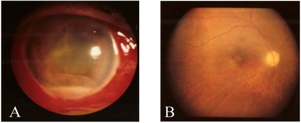
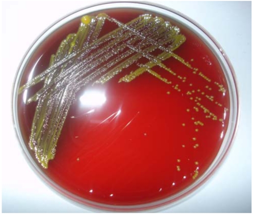
Methods: This case demonstrates an acute onset of delayed postoperative endophthalmitis at 3 months after uneventful cataract extraction and posterior chamber intraocular lens implantation. We performed vitrectomy, intraocular lens and capsular bag removal, and intravitreal antibiotics injection. On the smear stains from the aspirated vitreous humor, gram-negative bacilli were detected and S. paucimobilis was found in culture.
Results: At three months after vitrectomy, the best corrected visual acuity was 20/300. Fundus examination showed mild pale color of optic disc and macular degeneration.
Conclusions: Vitrectomy with intravitreal ceftazidime injection had contributed to the favorable result in case of an acute onset of delayed postoperatire endophthalmitis caused by S. paucimobilis.
Figures

References
-
- Dennis P. Hans and the Endophthalimitis Vitrectomy Study Group: Microbiologic factors and visual outcomes in the endophthalmitis vitrectomy group. Am J Ophthalmol. 1996;122:830–846. - PubMed
-
- Perola O, Nousiainen T, Soumalainen S, et al. Recurrent Sphingomonas paucimobilis bacteraemia associated with a multi bacterial water born epidemic among neutropenic patients. J Hosp Infect. 2002;50:196–201. - PubMed
-
- Liang C, Peyman GA, Molinari LC, Hegazy HM. Prophylaxis of experimental Pseudomonas aeruginosa endophthalmitis after vitrectomy using ceftazidime in the irrigating solution. Int Ophthalmol. 1998-1999;22:163–167. - PubMed
-
- Yabuuchi E, Yano I, Oyaizu H, et al. Proposals of Sphingomonas paucimobilis gen. nov. and comb. nov., Sphingomonas parapaucimobilis sp. nov., Sphingomonas yanoikuyae sp. nov., Sphingomonas adhaesiva sp. nov., Sphingomonas capsulata comb. nov., and two genospecies of the genus Sphingomonas. Microbiol Immunol. 1990;34:99–119. - PubMed
Publication types
MeSH terms
Substances
LinkOut - more resources
Full Text Sources
Medical
