. 2008 Apr 9;27(7):956-69.
doi: 10.1038/emboj.2008.38. Epub 2008 Mar 6.

Subversion of CtBP1-controlled macropinocytosis by human adenovirus serotype 3

Affiliations

Subversion of CtBP1-controlled macropinocytosis by human adenovirus serotype 3

Beat Amstutz et al. EMBO J. .

Abstract

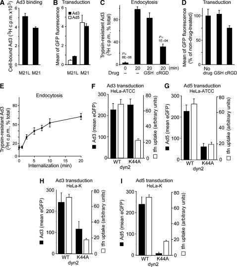
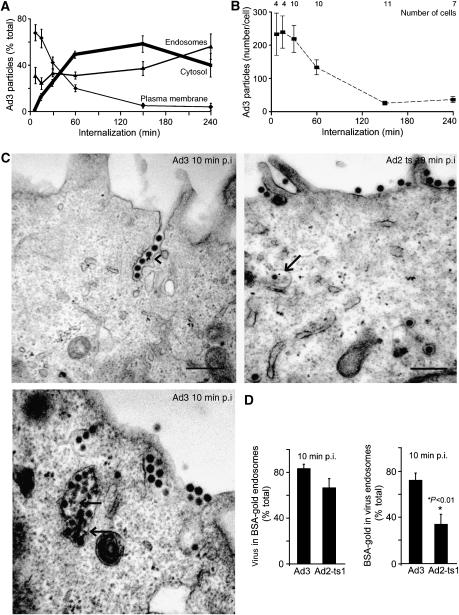
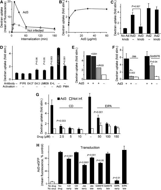
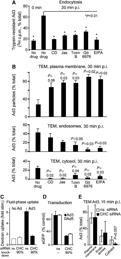
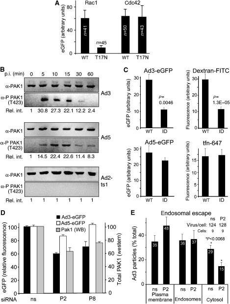
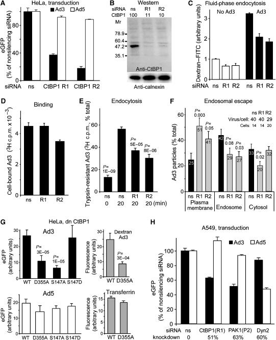
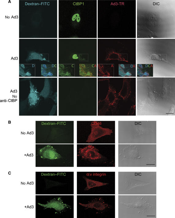
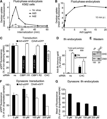
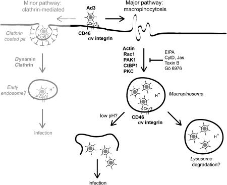
Endocytosis supports cell communication, growth, and pathogen infection. The species B human adenovirus serotype 3 (Ad3) is associated with epidemic conjunctivitis, and fatal respiratory and systemic disease. Here we show that Ad3 uses dynamin-independent endocytosis for rapid infectious entry into epithelial and haematopoietic cells. Unlike Ad5, which uses dynamin-dependent endocytosis, Ad3 endocytosis spatially and temporally coincided with enhanced fluid-phase uptake. It was sensitive to macropinocytosis inhibitors targeting F-actin, protein kinase C, the sodium-proton exchanger, and Rac1 but not Cdc42. Infectious Ad3 macropinocytosis required viral activation of p21-activated kinase 1 (PAK1) and the C-terminal binding protein 1 of E1A (CtBP1), recruited to macropinosomes. These macropinosomes also contained the Ad3 receptors CD46 and alpha v integrins. CtBP1 is a phosphorylation target of PAK1, and is bifunctionally involved in membrane traffic and transcriptional repression of cell cycle, cancer, and innate immunity pathways. Phosphorylation-defective S147A-CtBP1 blocked Ad3 but not Ad5 infection, providing a direct link between PAK1 and CtBP1. The data show that viruses induce macropinocytosis for infectious entry, a pathway used in antigen presentation and cell migration.


Figures

Figure 1
Figure 1
Infectious Ad3 endocytosis of HeLa cells requires αv integrins and to a low extent dynamin. (A) Human melanoma M21 or M21L cells were incubated with [3H]thymidine-labelled Ad3 in the cold and analysed for cell-associated radioactivity (106 cells, 0.75 μg Ad3). (B) M21 or M21L cells were transduced with Ad3-eGFP or Ad5-eGFP (MOI 5) and analysed by flow cytometry 8 h p.i. (C, D) [3H]thymidine-labelled Ad3 (8 × 10⁵/ml; 50 000 c.p.m.) was cold bound to HeLa-ATCC cells (2000 c.p.m. bound) and warmed for 0 or 20 min in the presence or absence of glutathione (GSH) or cyclic RGD peptide (cRGD, 0.2 mM), trypsinized in the cold, and analysed for cell-associated (internalized) or released [3H]thymidine-labelled Ad3 by liquid scintillation counting. Cells were treated with GSH or cRGD, infected with Ad3-eGFP (MOI 5), and analysed for GFP expression by flow cytometry 6 h p.i. (E) Kinetics of Ad3 endocytosis measured by trypsin resistance as described in panel C (100% equivalent to 2000 c.p.m.). Means of triplicate dishes of one representative experiment are shown (A–E). (FI) Ad3-eGFP and Ad5-eGFP transduction (6 h) and transferrin–Alexa647 internalization (10 μg/ml transferrin in the last 30 min of infection, open bars) in HeLa-ATCC or HeLa-K cells transfected with WT dyn2 or K44A dyn2 for 48 h. Single-cell analysis by confocal microscopy (MOI 5), showing the mean of at least 40 blindly selected cells, is shown. One representative experiment is shown (F–I).
Figure 2
Figure 2
Quantitative EM analyses of Ad3 endocytosis and endosomal escape. (A) Distribution of cold-bound Ad3 (5 × 10⁵ viral particles per cell, 4°C, 60 min) on the plasma membrane, endosomes, and the cytosol (bold line) upon internalization at 37°C. (B) Analyses of the total number of particles and cells. (C, D) Enrichment of Ad3 in fluid-phase-positive endosomes. HeLa cells were incubated with Ad3 or Ad2-ts1 in the cold (MOI as in (A)), washed, pulsed with BSA-gold for 10 min, fixed for ultrathin-section EM analyses, and quantified for viral particles in either gold-positive endosomes (arrows) or gold particles in endosomes that contain Ad3 or Ad2-ts1, respectively. Viruses in plasma membrane invaginations are indicated by an arrowhead.
Figure 3
Figure 3
CD46-, integrin-, F-actin-, PKC-, and EIPA-dependent stimulation of fluid-phase endocytosis by Ad3. (A) Ad3 transiently stimulates dextran uptake. HeLa-ATCC cells were incubated with Ad3 (5 μg/ml, equivalent to 2000 particles bound per cell) in the cold for 60 min, or noninfected, warmed for 0, 5, 10, 30, 60, 120, or 180 min, pulsed with dextran—FITC, and analysed by flow cytometry as described in Materials and methods. One out of three representative experiments is shown. (B) Dose dependence of fluid-phase uptake stimulation expressed as fold stimulation over noninfected cells. HeLa cells were incubated with different amounts of Ad3 (0, 0.5, 1, 10, 30, and 60 μg/ml) in the cold, warmed for 5 min, pulsed with dextran–FITC for 5 min, washed, and analysed by flow cytometry. One out of two similar experiments is shown. (C) Specificity of Ad3-stimulated fluid-phase endocytosis. HeLa cells were incubated with Ad3 or Ad2 (5 μg/ml) in the presence or absence of Ad3 fibre knob (5 μg/ml) and analysed for dextran–FITC stimulation as described above. (D) Fluid-phase stimulation by anti-CD46 and anti-integrin antibodies. HeLa cells were incubated with 4 μg/ml anti-CD46 antibody E4.3, or 0.8 μg/ml E4.3 (E4.3*), anti-αv β5 integrin (P1F6), anti-αv β3 (LM609), or a combination of E4.3 plus LM609 (E+L, 4 μg/ml each) in cold RPMI medium for 1 h, washed, and incubated with goat anti-mouse IgG antibodies (10 μg/ml), Ad3, or the phorbol ester PMA (Meier et al, 2002) on ice for 30 min. They were then warmed in the presence of dextran–FITC for 10 min and analysed by flow cytometry. The experiment was performed in triplicate and repeated once. (EG) Measurement of Ad3-induced dextran–FITC uptake 10 min p.i. in the presence or absence of cyclic RGD peptides (0.1 mM; Meier et al, 2002), jasplakinolide (Jas, 40 nM), the PKC inhibitor Gö6976 (1 μM), or different concentrations of cytochalasin D (CD) or EIPA. (H) Ad3-eGFP transduction (1000 viral particles/cell) of HeLa cells pretreated with CD, Jas, Gö6976, or EIPA. Gö6976 and EIPA were present during 1 h of warm infection and were then washed off (early), or present till 120–180 min p.i. (late), followed by flow cytometry of eGFP 6 h p.i. CD and Jas were present during the entire incubation time (early) or added 1 h after warming (late). Experiments were performed at least twice with triplicate samples.
Figure 4
Figure 4
Ad3 endocytosis and endosomal escape require F-actin, Rho GTPases, PKC, the sodium–proton exchanger 1, and clathrin. (A) Endocytosis of Ad3 measured by trypsin sensitivity of cell-surface-localized virus. HeLa-ATCC cells were pretreated with cytochalasin D (CD, 5 μM), jasplakinolide (Jas, 0.3 μM), Clostridium difficile toxin B (toxB, 0.3 μg/ml) (Aktories, 1997), Gö6976 (1 μM), or EIPA (100 μM) in growth medium for 30 min, incubated with [3H]thymidine-labelled Ad3 (50 000 c.p.m.) in the cold for 1 h, washed and internalized at 37°C for 20 min, washed with cold medium, and treated with trypsin (2 mg/ml) at 4°C for 1 h. Cells were pelleted by centrifugation at 500 g and the supernatants and cell pellets were analysed by liquid scintillation counting (fraction of total, 100% equivalent to 2000 c.p.m.). (B) Analysis of subcellular localization of Ad3 particles by transmission EM. HeLa cells were pretreated with drugs as described in panel A, incubated with Ad3 (30 μg/ml) in the cold, washed with binding medium, internalized in drug-containing medium for 30 min, and fixed for ultrathin-section EM analyses. Viral particles were quantified at the plasma membrane, in endosomes, and in the cytosol as described (Meier et al, 2005). The total number of particles blindly analysed for each condition was 200–300 in 6–9 different cells. For representative images, see Supplementary Figure 2. (CE) Ad3-stimulated dextran uptake and infection required CHC. Uptake measurements of dextran–FITC in normal HeLa-ATCC cells or cells transfected with nonsilencing siRNA (ns) or siRNA against CHC (CHC, double transfection, 72 h), eGFP transduction measurements, as well as EM analyses were performed as described.
Figure 5
Figure 5
Rac1 and PAK1 are required for Ad3 but not Ad5 endocytosis and infection. (A) HeLa-ATCC cells were transfected with plasmids encoding CFP–Rac1, CFP–Rac1 T17N, CFP–Cdc42, or CFP–Cdc42 T17N for 30 h, infected with Ad3-eGFP for 15 h, fixed, and analysed by confocal laser scanning microscopy. The eGFP intensity of at least 40 CFP-positive cells per condition was quantified by NIH image J with means and standard errors of the mean. The experiment was performed twice with similar results. Representative images are shown in Supplementary Figure 4. (B) PAK1 is activated by Ad3 and Ad5. Nonstarved HeLa-ATCC cells were incubated with Ad3, Ad5, or ts1 in the cold, washed, warmed for different times, and analysed for PAK1 and phosphorylated PAK1 (T423) using western blotting. One of three representative experiments is shown including the relative intensities of quantified phospho-PAK1 (rel. int.). (C) Cells expressing WT or dn PAK1 (inhibitory domain ID) were transduced with Ad3-eGFP or Ad5-eGFP and assessed for uptake of dextran–FITC or transferrin–Alexa647 upon Ad3 infection. (D) Cells were transfected with siRNAs P2 and P8 against PAK1 or nonsilencing (ns) siRNA for 72 h (double transfection, 20 pmol/ml siRNA), infected with Ad3-eGFP or Ad5-eGFP for 6 h, and analysed for eGFP expression by flow cytometry. Transfected cells (1 × 10⁵) were analysed by western blotting (WB) for PAK1 (grey panels). (E) Endosomal escape of Ad3 measured by thin-section EM in HeLa cells transfected with anti-PAK1 siRNA P2 and ns siRNA. Virions were counted at the plasma membrane, in endosomes, and in the cytosol.
Figure 6
Figure 6
CtBP1 is required for endosomal uptake and infection of Ad3 but not Ad5. (A) HeLa-ATCC cells transfected with CtBP1 siRNA R1 or R2 were infected with Ad3-eGFP or Ad5-eGFP and analysed for GFP expression by flow cytometry. (B) Western blot analysis of CtBP1 knockdown by siRNA R1 and R2, or ns siRNA, and normalization against calnexin. (C) Fluid-phase endocytosis of HeLa cells transfected with R1, R2, or ns siRNA. Cells were infected with Ad3 or not infected by cold binding and warming for 10 min in the presence of dextran–FITC, fixed, and analysed for dextran uptake by flow cytometry (10 000 cells in triplicate). (DF) Binding and endocytosis of [3H]thymidine-labelled Ad3 were determined by scintillation counting (106 cells, 0.75 μg Ad3) and endosomal escape of Ad3 was determined by EM in cells transfected with anti-CtBP1 siRNA R1, R2, or nonsilencing siRNA. (G) HeLa cells were transfected with myc-tagged CtBP1-S WT, CtBP1-S D355A mutant, or the S147A or S147D mutants for 30 h, infected with Ad3-eGFP or Ad5-eGFP at MOI 5 for 16 h, fixed, stained with an anti-myc antibody, and analysed for eGFP fluorescence by confocal laser scanning microscopy and NIH image J analysis of merged set of complete optical sections. CtBP1-S WT- or D355A-transfected cells were pulsed with dextran-TR (0.5 mg/ml) or transferrin–Alexa647 (10 μg/ml) for 30 min, fixed, and analysed for dextran and transferrin uptake by confocal microscopy, respectively (grey panels). (H) Human lung epithelial A549 cells were transfected with siRNA against CtBP1 (R1), PAK1 (P2), dyn2, or nonsilencing (ns), transduced with Ad3-eGFP or Ad5-eGFP (MOI 2.5), and analysed for GFP expression by flow cytometry. The levels of knockdown were determined by western blotting using calnexin as a reference.
Figure 7
Figure 7
Ad3-induced macropinosomes contain CtBP1, CD46, and αv β5 integrins. Ad3-TR (2 × 10⁴ viral particles/cell, MOI 50)-infected or not infected HeLa-ATCC cells were pulsed with 0.5 mg/ml dextran–FITC at 37°C for 10 min, washed extensively, fixed, immunostained against CtBP1 (0.5 μg/ml), CD46 (mab E4.3, 1 μg/ml), and αv β5 integrins (mab P1F6, 1 μg/ml), and analysed by confocal microscopy (AC). The enlarged boxes in panel A show dextran-filled macropinosomes (D) containing both CtBP1 (C) and Ad3-TR (A). Control stainings in the absence of primary anti-CtBP1 antibody are shown in panel A together with the differential interference contrast images (DIC). Scale bars=20 μm.
Figure 8
Figure 8
Infectious Ad3 entry into haematopoietic cells involves fluid-phase endocytosis and CtBP1. (A) Haematopoietic K562 cells were infected with Ad3, Ad2 (5 μg/ml virus, equivalent to 2000 viral particles bound per cell), or no virus for different times, pulsed with dextran–FITC (1 mg/ml) for 5 min, and subjected to flow cytometry. (B) Dose dependence of fluid-phase uptake stimulation by Ad3 with the indicated amounts of Ad3 in the cold, followed by warming for 5 min and dextran–FITC pulse for 5 min. (C) Cells were double transfected with CtBP1 siRNA R1, R2, nonsilencing siRNA (ns), or CHC (CHC) siRNA (20 pmol/ml) for 48 h, infected with Ad3-eGFP (1 μg/ml) or Ad5-eGFP (2 μg/ml) for 16 h, and analysed for eGFP expression by flow cytometry. (D) Transferrin (tfn) endocytosis by immuno-EM in CHC siRNA- or control siRNA-treated cells. (E) Western blot analyses of CHC siRNA-treated cells (1 × 10⁵ cells per lane, normalization against calnexin, CNX). (F, G) Dynasore block of Ad3-eGFP (1 μg/ml) or Ad5-eGFP (2 μg/ml) infection for 12 h and transferrin–Alexa488 (10 μg/ml) uptake for 30 min, analysed by flow cytometry.
Figure 9
Figure 9
Macropinocytosis is a major infectious uptake pathway of Ad3 in epithelial cells. Ad3 binds the membrane cofactor CD46 and is endocytosed into macropinocytic vesicles depending on αv β3 or β5 integrin coreceptors, F-actin, the Rac1 GTPase, which activates PAK1, and CtBP1 (C-terminal binding protein 1), a target of PAK1. Protein kinase C (PKC) and the sodium–proton exchanger 1 (sensitive to EIPA) are also required for the formation of Ad3-carrying macropinosomes. Ad3-bearing macropinosomes contain CD46 and αv β5 integrins. Low pH and additional triggers probably lead to virus release from macropinosomes and noninfectious virions are degraded in late endosomes and lysosomes. Besides the major macropinocytic pathway, there is a minor clathrin- and dynamin-dependent pathway, which is cell type-dependent and not found in K562 haematopoietic cells, for example. This pathway remains to be characterized.

References

    1. Aktories K (1997) Rho proteins: targets for bacterial toxins. Trends Microbiol 5: 282–288 - PubMed
    1. Barnes CJ, Vadlamudi RK, Mishra SK, Jacobson RH, Li F, Kumar R (2003) Functional inactivation of a transcriptional corepressor by a signaling kinase. Nat Struct Biol 10: 622–628 - PubMed
    1. Baumgartner M, Patel H, Barber DL (2004) Na(+)/H(+) exchanger NHE1 as plasma membrane scaffold in the assembly of signaling complexes. Am J Physiol Cell Physiol 287: C844–C850 - PubMed
    1. Benmerah A, Bayrou M, Cerf-Bensussan N, Dautry-Varsat A (1999) Inhibition of clathrin-coated pit assembly by an Eps15 mutant. J Cell Sci 112: 1303–1311 - PubMed
    1. Berk AJ (2005) Recent lessons in gene expression, cell cycle control, and cell biology from adenovirus. Oncogene 24: 7673–7685 - PubMed

Publication types

MeSH terms