. 2008 Apr 9;27(7):948-55.
doi: 10.1038/emboj.2008.36. Epub 2008 Mar 6.

Spatial separation of Golgi and ER during mitosis protects SREBP from unregulated activation

Affiliations

Spatial separation of Golgi and ER during mitosis protects SREBP from unregulated activation

René Bartz et al. EMBO J. .

Abstract

Sterol regulatory element-binding proteins (SREBPs) are membrane-bound transcription factors that reside as inactive precursors in the endoplasmic reticulum (ER) membrane. After sterol depletion, the proteins are transported to the Golgi apparatus, where they are cleaved by site-1 protease (S1P). Cleavage releases the active transcription factors, which then enter the nucleus to induce genes that regulate cellular levels of cholesterol and phospholipids. This regulation depends on the spatial separation of the Golgi and the ER, as mixing of the compartments induces unregulated activation of SREBPs. Here, we show that S1P is localized to the Golgi, but cycles continuously through the ER and becomes trapped when ER exit is inhibited. During mitosis, S1P is associated with mitotic Golgi clusters, which remain distinct from the ER. In mitotic cells, S1P is active, but SREBP is not cleaved as S1P and SREBP reside in different compartments. Together, these results indicate that the spatial separation of the Golgi and the ER is maintained during mitosis, which is essential to protect the S1P substrate SREBP from unregulated activation during mitosis.


Figures
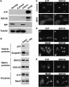
Figure 1
Figure 1
S1P associates with Golgi membranes and recycles through the ER. (A) S1P is enriched on Golgi membranes. Rat liver homogenate, cytosol, intermediate membrane fraction and Golgi membranes were separated by 10% SDS–PAGE (40 μg protein for homogenate (homog.), cytosol and intermediate (interm.); 10 μg for Golgi fraction) and analysed by immunoblotting with antibodies against S1P, GM130, Grp78/BiP and tubulin. (B) S1P is found in the Triton X-100-insoluble Golgi matrix fraction. Golgi membranes ( 300 μg) were extracted with Triton X-100 and pelleted by centrifugation. Equal amounts of protein were subjected to 10% SDS–PAGE and immunoblotted with the indicated antibodies. (C) BFA separates S1P from GM130. NRK cells were incubated for 45 min with 5 μg/ml BFA or carrier control. Cells were then fixed, permeabilized and stained with mouse anti-GM130 and rabbit anti-S1P antibodies followed by fluorescently conjugated secondary antibodies (anti-mouse AlexaFluor 594 and anti-rabbit AlexaFluor 488). (D) S1P is retained in the ER after Sar1DN microinjection. NRK cells were injected with 1 mg/ml purified Sar1DN protein, fixed after 3 h incubation, permeabilized and stained with an antibody against GM130 together with polyclonal antibodies against S1P or ManII. Injected cells were identified by coinjected cascade-blue-conjugated BSA (asterisks). (E) The Golgi apparatus re-forms without S1P. NRK cells were incubated for 45 min with 5 μg/ml BFA and injected with recombinant Sar1DN protein as in (D). BFA was washed out and after 2 h the cells were fixed, permeabilized and stained with an antibody against GM130 together with antibodies against S1P and ManII. Scale bars, 15 μm.
Figure 2
Figure 2
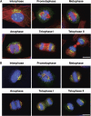
S1P colocalizes with Golgi proteins during mitosis. (A) Localization of S1P during mitosis. Exponentially growing NRK cells were fixed, permeabilized and triple stained with antibodies for S1P (green), tubulin (red) and DNA (blue). The different mitotic stages were identified by the organization of the microtubule network and the chromatin. (B) GM130 and S1P colocalize during mitosis. Cells were prepared and mitotic cells were identified as in (A) after staining with anti-GM130 and anti-S1P antibodies. Scale bars, 10 μm.
Figure 3
Figure 3
S1P is distinct from the ER during mitosis. Gradient fractionation of mitotic membranes. CHO cells treated with or without 3 μg/ml BFA were mitotically arrested with 2 mM S-trityl-L-cysteine. Mitotic cells were collected by shake-off and post-chromosomal supernatants were fractionated on a 5–30% glycerol gradient. Fractions were collected, separated by 12% SDS–PAGE and analysed by immunoblotting for S1P, GM130 and GRP78/BiP.
Figure 4
Figure 4
SREBP remains uncleaved in mitosis. (A) SREBP is not localized in the nuclei of post-mitotic cells. CHO cells were grown in sterol-depleting medium in the absence or presence of 1 μg/ml 25-hydroxycholesterol for 16 h. Cells were fixed, permeabilized and stained for SREBP and tubulin. DNA was labelled with Hoechst. Scale bar, 10 μm. (B) Biochemical analysis reveals SREBP processing in mitotic and interphase cells only after the addition of BFA. CHO cells treated with 3 μg/ml BFA or carrier control were arrested in mitosis for 5 h in the presence of 2 mM S-trityl-L-cysteine. Mitotic cells (M) were harvested by shake-off and the remaining cells (interphase, I) were collected by trypsinization. Equal amounts of protein (30 μg) were separated by 10% SDS–PAGE and immunoblotted with the indicated antibodies.
Figure 5
Figure 5
S1P remains active in mitotic cells. Stably transfected CHO cells were induced to express ATF6–GFP by induction with 5 μM ponasterone A for 16 h. Cells were then arrested in mitosis with 0.2 μg/ml nocodazole in the presence or absence of 3 μg/ml BFA. The arrested cells were then treated for 45 min with 2 mM DTT or control. The drugs were removed and the cells were followed by time-lapse microscopy (t=0 min) until mitosis was complete (t=80 min). The cells were fixed and stained with antibodies to GFP to detect ATF6–GFP. Note that only cells that entered mitosis in the presence of BFA that were further treated with DTT in mitosis showed cleaved ATF–GFP in the nucleus (+BFA +DTT), whereas in control cells treated with only BFA (+BFA) or DTT (+DTT), ATF6–GFP was restricted to the ER. Scale bar, 10 μm.

References

    1. Altan-Bonnet N, Sougrat R, Liu W, Snapp EL, Ward T, Lippincott-Schwartz J (2006) Golgi inheritance in mammalian cells is mediated through endoplasmic reticulum export activities. Mol Biol Cell 17: 990–1005 - PMC - PubMed
    1. Axelsson MA, Warren G (2004) Rapid, endoplasmic reticulum-independent diffusion of the mitotic Golgi haze. Mol Biol Cell 15: 1843–1852 - PMC - PubMed
    1. Brown MS, Ye J, Rawson RB, Goldstein JL (2000) Regulated intramembrane proteolysis: a control mechanism conserved from bacteria to humans. Cell 100: 391–398 - PubMed
    1. Cole NB, Sciaky N, Marotta A, Song J, Lippincott-Schwartz J (1996) Golgi dispersal during microtubule disruption: regeneration of Golgi stacks at peripheral endoplasmic reticulum exit sites. Mol Biol Cell 7: 631–650 - PMC - PubMed
    1. Collins RN, Warren G (1992) Sphingolipid transport in mitotic HeLa cells. J Biol Chem 267: 24906–24911 - PubMed

Substances