. 2008 Apr 9;27(7):1154-60.
doi: 10.1038/emboj.2008.35. Epub 2008 Mar 6.

Dimer ribbons of ATP synthase shape the inner mitochondrial membrane

Affiliations

Dimer ribbons of ATP synthase shape the inner mitochondrial membrane

Mike Strauss et al. EMBO J. .

Abstract

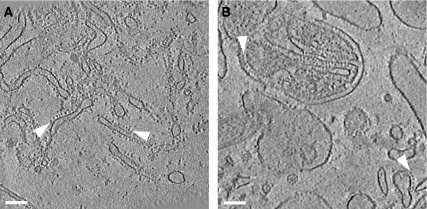
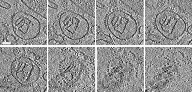
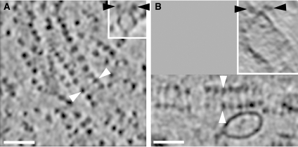
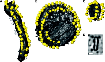
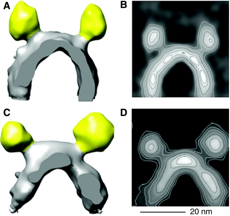
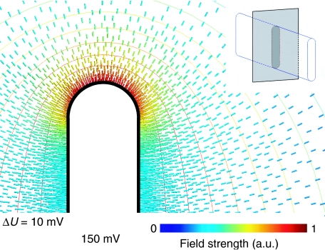
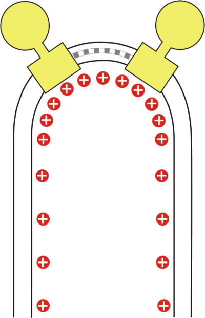
ATP synthase converts the electrochemical potential at the inner mitochondrial membrane into chemical energy, producing the ATP that powers the cell. Using electron cryo-tomography we show that the ATP synthase of mammalian mitochondria is arranged in long approximately 1-microm rows of dimeric supercomplexes, located at the apex of cristae membranes. The dimer ribbons enforce a strong local curvature on the membrane with a 17-nm outer radius. Calculations of the electrostatic field strength indicate a significant increase in charge density, and thus in the local pH gradient of approximately 0.5 units in regions of high membrane curvature. We conclude that the mitochondrial cristae act as proton traps, and that the proton sink of the ATP synthase at the apex of the compartment favours effective ATP synthesis under proton-limited conditions. We propose that the mitochondrial ATP synthase organises itself into dimer ribbons to optimise its own performance.


Figures

Figure 1
Figure 1
Activity of ATP synthase oligomers. Native gel of whole, digitonin-solubilised beef heart mitochondria stained for ATPase activity. The lowest band corresponds to the ATPase monomer, with higher bands indicating active oligomers up to the hexamer (Wittig and Schägger, 2005).
Figure 2
Figure 2
ATP synthase is arranged in rows. Slices of typical tomograms of membrane vesicles from rat liver (A) and bovine heart (B) mitochondria. Near the centre in panel B, a fragment of a mitochondrion with folded cristae and outer membrane is visible. Arrowheads indicate rows of ∼10-nm F1 ATP synthase heads. Slice thickness is 2.8 nm in panel A and 4.5 nm in panel B. Scale bars, 100 nm.
Figure 3
Figure 3
Tomographic series of ATP synthase dimer rows. Slices in xy show a disk-shaped crista vesicle from bovine heart mitochondria. Each slice has a thickness of 3 nm, and the spacing between slices is 7 nm. At given xy positions on consecutive sections from upper left to lower right, a circular row of F1 heads appears and then disappears, and then a second, parallel row becomes visible. Scale bar, 50 nm.
Figure 4
Figure 4
Dimer rows in bovine and rat mitochondria. Ribbons of ATP synthase dimers in tomograms of rat liver (A) and bovine heart (B) mitochondrial membranes. Black or white arrowheads indicate the same pairs of ATP synthase heads in (A) or (B), respectively. White arrowheads indicate the direction of perpendicular sections in the inserts. Slab thickness is 2.8 nm in panel A and 4.5 nm in panel B. Scale bars, 50 nm.
Figure 5
Figure 5
Tomographic volumes of dimer ribbons. Surface views of dimer ribbons in a tubular crista vesicle from rat liver mitochondria (A) and in round vesicles from bovine heart mitochondria (B, C). (D) A cross section through the tomographic volume of the small vesicle in panel C. F1 heads are yellow, while the membrane is grey. Particles not assigned to dimer ribbons are shown in lighter grey. The length of the tube in panel A is 280 nm, and the diameter of the vesicles in panels B and C is 180 and 51 nm, respectively. Scale bar in panel D, 20 nm.
Figure 6
Figure 6
Averaged dimer volumes. Averaged 3D volumes of ATP synthase dimers from rat liver (A) and bovine heart membranes (C) in situ. F1 heads are coloured as in Figure 5. In panel A, 235 dimer volumes were averaged. In panel C, 90 volumes of the largest of several classes with different ribbon curvature and, hence, dimer angles were averaged. Contour plots (B, D) of volumes in panels A, C projected in the viewing direction. Scale bar, 20 nm.
Figure 7
Figure 7
Simulation of electric field on membrane surface. Electric field strength around a cristae-shaped membrane surface calculated for a constant potential of 150 mV. The isopotential surfaces (ΔU=10 mV) are more closely spaced around the curved edges than along the planar sides, indicating that the potential gradient is strongest in the curved regions. The corresponding charge density on the inner membrane surface (solid black line) is directly proportional to the field strength. The numerical values indicate a 3.5-fold increase in surface charge density at the apex (red field lines), compared with the flat sides (blue field lines). The insert illustrates the geometry used to solve the electrostatic Poisson equation. The apical field strength is higher for larger axial ratios, but does not vary strongly with this parameter.
Figure 8
Figure 8
Proton density at position of dimers in the membrane. Schematic drawing of an F1–F0 ATP synthase dimer (yellow) in the inner mitochondrial membrane. Monomers are connected by dimer-specific subunits (grey). The dimers associate into ribbons, which induce a tight bend in the membrane, with an ∼17-nm outer radius of curvature. The higher surface density of protons (red) in the curved membrane regions would result in a local pH difference of ∼0.5 units.

References

    1. Abrahams JP, Leslie AG, Lutter R, Walker JE (1994) Structure at 2.8 Å resolution of F1–ATPase from bovine heart mitochondria. Nature 370: 621–628 - PubMed
    1. Abramoff M, Magelhaes P, Ram S (2004) Image processing with ImageJ. Biophotonics International 11: 36–42
    1. Allen RD, Schroeder CC, Fok AK (1989) An investigation of mitochondrial inner membranes by rapid-freeze deep-etch techniques. J Cell Biol 108: 2233–2240 - PMC - PubMed
    1. Arnold I, Pfeiffer K, Neupert W, Stuart RA, Schägger H (1998) Yeast mitochondrial F1F0–ATP synthase exists as a dimer: identification of three dimer-specific subunits. EMBO J 17: 7170–7178 - PMC - PubMed
    1. Boekema EJ, Braun HP (2007) Supramolecular structure of the mitochondrial oxidative phosphorylation system. J Biol Chem 282: 1–4 - PubMed

MeSH terms

Substances