

doi: 10.1038/nprot.2007.537.
A protocol for phenotypic detection and characterization of vascular cells of different origins in a lung neovascularization model in rodents
Affiliations
- PMID: 18323810
- PMCID: PMC2676340
- DOI: 10.1038/nprot.2007.537
Item in Clipboard
A protocol for phenotypic detection and characterization of vascular cells of different origins in a lung neovascularization model in rodents
Nat Protoc.
2008.
Abstract
The goal of many current studies of neovascularization is to define the phenotype of vascular cell populations of different origins and to determine how such cells promote assembly of vascular channel. Here, we describe a protocol to immunophenotype vascular cells by high-resolution imaging and by fluorescence-activated flow cytometry in an in vivo rodent model of pulmonary microvascular remodeling. Analysis of cells by this combined approach will characterize their phenotype, quantify their number and identify their role in the assembly of vascular channels.
Figures
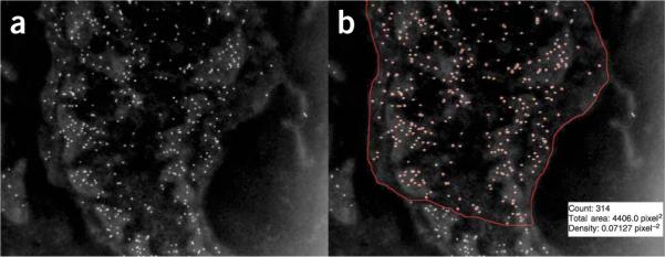
Quantification of gold particle density using image processing and analysis. In the inverted digital image collected by TEM (a) antigenic sites labeled with gold particles appear over the cell surface as bright dots of uniform size. The macro analysis of this image (b) detects, counts and outlines the particles, and also outputs the manually circumscribed area (red line enclosing the cell area). As shown for the fibroblast illustrated here (with pA-AU-labeled antigenic sites to PDGF-Rβ): the gold particle count is 314; the area analyzed is 4406.0 pixel2 and the density is 0.07127 pixel-2, which translates (from the number of nanometers per pixel) to an area of 3.90 μm2 and a calculated density of 80.0 μm-2.
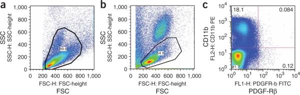
Representative flow cytometry plots using FlowJo software to analyze cells from lung tissue or from rat peripheral blood. (a) FSC-SSC plot of lung cells-gate set on cell size events; (b) FSC-SSC plot of blood cells-gate set on mononuclear cells, excluding neutrophils, red blood cells, platelets and cell fragments; (c) Analysis of PDGF-Rβ and CD11b expression on cells from rat peripheral blood.
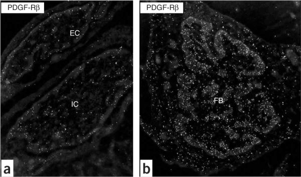
Inverted images of PDGF-Rβ+ vascular-associated cells in the rat lung after breathing high oxygen. (a) Endothelial cell (EC) and intermediate cell (IC) at D7; (b) mesenchymal cell, that is, fibroblast (FB) at D7. Note the condensation of heterochromatin at the edge of the nucleus. Original magnification: (a) ×19,636; (b) ×12,480.
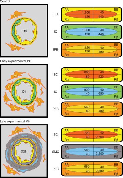
Schematic of vessel wall thickening by precursor cells in the rat after high oxygen (left); values for gold-labeled antigenic sites quantified by macro for each cell population at each time point (right). Note the distribution of sites in the normal lung (Control, D0) and the shift early (D4) and late (D28) in the hypertensive lung; although PDGF-AA expression predominates in vascular cells of the normal lung, a shift to PDGF-Rβ expression begins in the early stage of pulmonary hypertension and is pronounced in the late stage (reprinted with permission from ref. 4, Jones et al., 2006).
References
-
- Jones RC, Jacobson M. Angiogenesis in the hypertensive lung: response to ambient oxygen tension. Cell Tissue Res. 2000;300:263–284. - PubMed
-
- Jones R, Capen D, Jacobson M, Munn L. PDGF and microvessel wall remodeling in adult rat lung: imaging PDGF-AA and PDGF-Rα molecules in progenitor smooth muscle cells developing in experimental pulmonary hypertension. Cell Tissue Res. 2006;326:759–769. - PubMed
-
- Jones R, Capen D, Jacobson M. PDGF and microvessel wall remodeling in adult lung: imaging PDGF-Rβ and PDGF-BB molecules in progenitor smooth muscle cells developing in pulmonary hypertension. Ultrastruct. Pathol. 2006;30:267–281. - PubMed
Publication types
MeSH terms
Substances
Grants and funding
LinkOut - more resources
Full Text Sources
