. 2008 Apr;118(4):1511-8.
doi: 10.1172/JCI33764.

Mitotic spindle destabilization and genomic instability in Shwachman-Diamond syndrome

Affiliations

Mitotic spindle destabilization and genomic instability in Shwachman-Diamond syndrome

Karyn M Austin et al. J Clin Invest. 2008 Apr.

Abstract

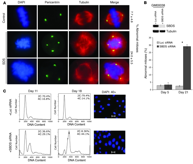
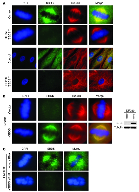
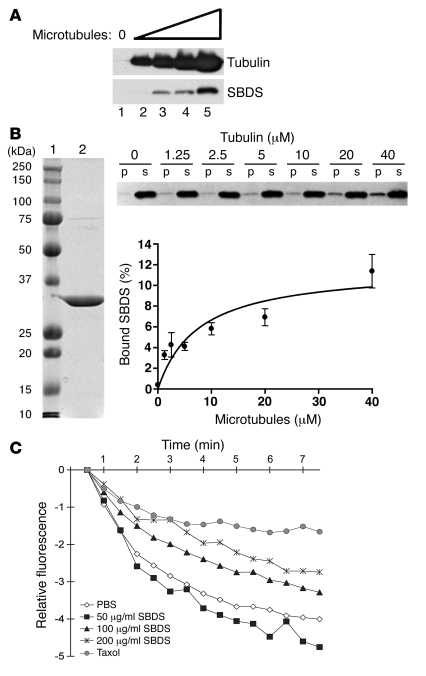
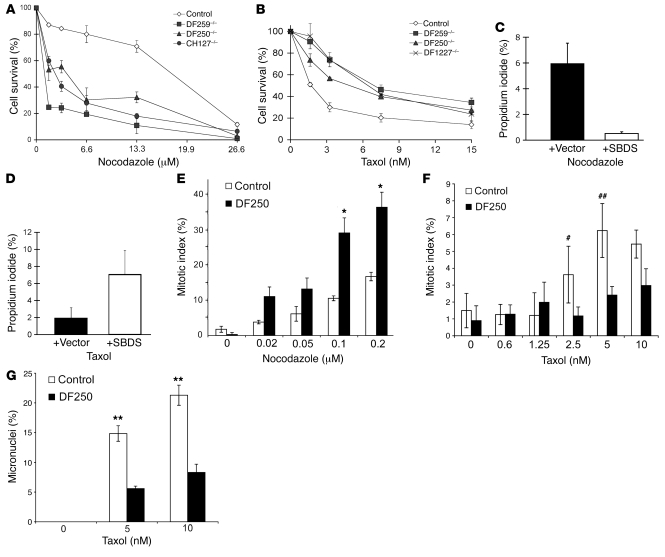
Deficiencies in the SBDS gene result in Shwachman-Diamond syndrome (SDS), an inherited bone marrow failure syndrome associated with leukemia predisposition. SBDS encodes a highly conserved protein previously implicated in ribosome biogenesis. Using human primary bone marrow stromal cells (BMSCs), lymphoblasts, and skin fibroblasts, we show that SBDS stabilized the mitotic spindle to prevent genomic instability. SBDS colocalized with the mitotic spindle in control primary BMSCs, lymphoblasts, and skin fibroblasts and bound to purified microtubules. Recombinant SBDS protein stabilized microtubules in vitro. We observed that primary BMSCs and lymphoblasts from SDS patients exhibited an increased incidence of abnormal mitoses. Similarly, depletion of SBDS by siRNA in human skin fibroblasts resulted in increased mitotic abnormalities and aneuploidy that accumulated over time. Treatment of primary BMSCs and lymphoblasts from SDS patients with nocodazole, a microtubule destabilizing agent, led to increased mitotic arrest and apoptosis, consistent with spindle destabilization. Conversely, SDS patient cells were resistant to taxol, a microtubule stabilizing agent. These findings suggest that spindle instability in SDS contributes to bone marrow failure and leukemogenesis.


Figures

Figure 1
Figure 1. SBDS loss promotes mitotic abnormalities.
(A) SBDS–/– cells exhibit mitotic abnormalities. Primary BMSCs from at least 3 different SDS patients were fixed and stained with antibodies against pericentrin (green) and α-tubulin (red) and with DAPI (blue). The top row illustrates normal control metaphase staining, while the middle and bottom rows illustrate the aberrant mitotic figures observed in SDS patient cells. Note multiple centrosomes, multipolar spindles, and broad DNA distribution. The percentage of abnormal mitotic cells in controls versus SBDS–/– cells is noted on the right (P < 0.01). A minimum of 200 cells were counted per sample in a blinded fashion in 3 independent experiments. Original magnification, ×60. (B) Targeted SBDS loss results in aberrant mitosis. GM00038 or GM00637 skin fibroblast cell lines immortalized with SV40 T antigen were infected with dual expression cassette lentiviral constructs encoding both GFP and siRNA sequences, the latter targeted against either SBDS or a LUC control. GFP-positive cells were sorted and analyzed for SBDS expression by western blot. SBDS protein expression was markedly reduced by 3 days following infection (upper panel). On day 5 and day 21 following infection, the cells were fixed and stained with antibodies against pericentrin and tubulin. At least 150 cells per sample were scored for abnormal mitoses (centrosomal amplification and multipolar spindles as illustrated in A) in a blinded fashion in at least 3 independent experiments, and the percentage of abnormal mitoses were tabulated in the histogram. *P = 0.01, LUC siRNA compared with SBDS siRNA on day 21. (C) SBDS loss results in aneuploidy. GM00038 cells from B were infected with lentivirus vectors as described in B. These immortalized GM00038 cells failed to exhibit p53-dependent p21 upregulation following exposure to ionizing radiation. GFP-positive cells were gated and analyzed for DNA content by flow cytometry on the indicated days following infection. DAPI staining shows enlargement of nuclei for cells lacking SBDS. Cells were visualized under ×40 magnification, and a scale bar (arbitrary units) is shown in C for comparison of the top and bottom panels.
Figure 2
Figure 2. SBDS localizes to a region corresponding to that of the mitotic spindle.
(A) SBDS colocalizes with the spindle by immunofluorescence. Primary BMSCs in metaphase (upper 2 panels) or in interphase (lower 2 panels) from normal control or SBDS–/– patients were fixed and stained with antibodies against SBDS (green), α-tubulin (red), and DAPI (blue). SBDS spindle staining was not detectable in any of the 6 SDS patients exhibiting low SBDS protein expression as analyzed by western blot. (B) The spindle-staining pattern is SBDS dependent. DF259 primary BMSCs were infected with a retroviral vector carrying SBDS cDNA or an empty vector control, as indicated. Cells were lysed or fixed for immunofluorescence studies 48–72 h after infection. Western blot analysis for SBDS expression is shown on the right. Tubulin was stained to control for equal sample loading. Fixed cells were stained for SBDS (green), α-tubulin (red), or DAPI (blue) and visualized by fluorescence microscopy. (C) Loss of SBDS abrogates the spindle-staining pattern. GM00038 skin fibroblasts were infected with lentiviral siRNA vectors against either SBDS or a LUC control and analyzed by immunofluorescence for SBDS (green).
Figure 3
Figure 3. SBDS binds to and stabilizes microtubules in vitro.
(A) Endogenous SBDS co-pellets with microtubules from cell lysates. Increasing concentrations of purified, taxol-stabilized bovine microtubules were incubated with HeLa cell extract, layered over a glycerol cushion, and centrifuged to pellet the microtubules and associated proteins. The pellets were analyzed by western blot for tubulin and SBDS. (B) Recombinant SBDS co-sediments with microtubules. Left: Coomassie-stained gel loaded with standard molecular markers (lane 1) or 3 μg purified recombinant SBDS protein (lane 2). Upper right: The fraction of 500 nM SBDS remaining in the supernatant (s) or co-pelleting (p) with increasing amounts of taxol-stabilized microtubules after centrifugation through a glycerol cushion was monitored by SBDS immunoblotting. Lower right: The average percentage bound from 3 experiments. The 1-site binding isotherm and Kd = 7.6 ± 3.0 μM were obtained by best-fit nonlinear regression using Graphpad Prism software. Approximately 14% of purified SBDS was co-pelleted with microtubules. Error bars represent SEM. (C) SBDS stabilizes microtubules in vitro. Preformed fluorescence-associated microtubules were diluted into PBS buffer alone or into buffer containing either taxol or increasing concentrations of purified, bacterially expressed recombinant SBDS protein. SBDS concentrations of 1.74 μM, 3.5 μM, and 7 μM were assayed. Dilution-induced depolymerization was monitored by the loss of microtubule-stimulated fluorescence signal. Three independent experiments yielded similar results. A representative experiment is shown. As a negative control, purified bovine serum albumin protein had no effect on microtubule stability (Supplemental Figure 4).
Figure 4
Figure 4. SBDS stabilizes microtubules in vivo.
(A and B) SDS patient cells are sensitive to nocodazole and resistant to taxol. Lymphoblast cell lines originating from SDS patients (DF259, DF250, CH127, and DF1227) or normal control lymphoblasts were treated with increasing concentrations of nocodazole for 24 h (A) or taxol for 72 h (B). Cell survival was determined using AqueousOne colorimetric assay reagent. Each cell survival assay was performed in triplicate for each experiment for a total of 3 independent experiments. SEM is denoted with vertical bars. (C and D) Nocodazole sensitivity and taxol resistance are SBDS dependent. DF259 lymphoblasts were transiently infected with a dual expression lentiviral vector containing either GFP and SBDS cDNA or GFP alone (vector). Two days following infection, cells were treated with 6.6 μM nocodazole (C) or 50 nM taxol (D) for 18 h. Cell death was assayed by flow cytometry for propidium iodide uptake. Three independent experiments were performed for each assay. P < 0.25 (C) and P < 0.05 (D). (E) SBDS–/– cells exhibit an increased mitotic arrest with nocodazole. Normal control or SDS patient primary BMSCs were treated with increasing doses of nocodazole for 18 h. Cells were stained with DAPI, and the percentage of cells in metaphase (expressed as the mitotic index) was visually scored for at least 100 cells in triplicate for 3 independent experiments. Samples were quantitated in a blinded fashion. The mitotic index was calculated from the total number of mitotic cells divided by the total cell number counted. BMSCs from 2 different SDS patients (DF250 and DF259) yielded similar results. *P < 0.01. (F) SBDS–/– cells are resistant to taxol-induced mitotic arrest. Normal control or SBDS–/– patient (DF250) primary BMSCs were treated with increasing doses of taxol for 10 h, and the mitotic index was quantitated as in E. BMSCs from 2 different SDS patients (DF250 and DF259) yielded similar results. #P = 0.11; ##P = 0.09. (G) SBDS–/– cells are resistant to micronuclei formation following taxol treatment. Normal control or SDS patient (DF250) primary BMSCs were treated with increasing concentrations of taxol for 10 h. Cells were stained with DAPI, and the percentage of cells with micronuclei was quantitated for at least 100 cells per experiment for 3 independent experiments. Samples were quantitated in a blinded fashion. Similar results were obtained with BMSCs from at least 2 different SDS patients. **P < 0.05.

References

    1. Dror Y. Shwachman-Diamond syndrome. Pediatr. Blood Cancer. 2005;45:892–901. doi: 10.1002/pbc.20478. - DOI - PubMed
    1. Shimamura A. Shwachman-Diamond syndrome. Semin. Hematol. 2006;43:178–188. doi: 10.1053/j.seminhematol.2006.04.006. - DOI - PubMed
    1. Boocock G.R., et al. Mutations in SBDS are associated with Shwachman-Diamond syndrome. Nat. Genet. 2003;33:97–101. doi: 10.1038/ng1062. - DOI - PubMed
    1. Zhang S., Shi M., Hui C.C., Rommens J.M. Loss of the mouse ortholog of the shwachman-diamond syndrome gene (sbds) results in early embryonic lethality. Mol. Cell. Biol. 2006;26:6656–6663. doi: 10.1128/MCB.00091-06. - DOI - PMC - PubMed
    1. Woloszynek J.R., et al. Mutations of the SBDS gene are present in most patients with Shwachman-Diamond syndrome. Blood. 2004;104:3588–3590. doi: 10.1182/blood-2004-04-1516. - DOI - PubMed

Publication types