Review
. 2008 Nov;35(4):711-8.
doi: 10.1007/s00726-008-0053-6. Epub 2008 Mar 7.

Direct linking of metabolism and gene expression in the proline utilization A protein from Escherichia coli

Affiliations
Review

Direct linking of metabolism and gene expression in the proline utilization A protein from Escherichia coli

Yuzhen Zhou et al. Amino Acids. 2008 Nov.

Abstract

The control of gene expression by enzymes provides a direct pathway for cells to respond to fluctuations in metabolites and nutrients. One example is the proline utilization A (PutA) protein from Escherichia coli. PutA is a membrane-associated enzyme that catalyzes the oxidation of L: -proline to glutamate using a flavin containing proline dehydrogenase domain and a NAD(+) dependent Delta(1)-pyrroline-5-carboxylate dehydrogenase domain. In some Gram-negative bacteria such as E. coli, PutA is also endowed with a ribbon-helix-helix DNA-binding domain and acts as a transcriptional repressor of the proline utilization genes. PutA switches between transcriptional repressor and enzymatic functions in response to proline availability. Molecular insights into the redox-based mechanism of PutA functional switching from recent studies are reviewed. In addition, new results from cell-based transcription assays are presented which correlate PutA membrane localization with put gene expression levels. General membrane localization of PutA, however, is not sufficient to activate the put genes.


Figures
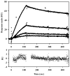
Fig. 1
Fig. 1
SPR sensorgrams of the association and dissociation kinetics of PutA-DNA interactionsin the presence of proline. From bottom to top, increasing concentrations of PutA (12.5, 25, 50, and 100 nM) in the presence of 5 mM proline were injected onto a SA sensor chip coated with E. coli put control DNA. The association phase (a) corresponds to the injection of PutA at 60 μl/min for 120 s and the dissociation phase (b) corresponds to the flow of HEPES-N buffer (pH 7.4, 150 mM NaCl) at 60 μl/min for 300 s. For immobilization of the put control DNA, put control DNA was 5′-end labeled by PCR using biotinylated primers. Biotinylated put control DNA was then immobilized onto the surface using a streptavidin derivatized chip (SA chip) as previously described (Buckle, 2001). PutA for these experiments was purified as previously described (Zhang et al., 2004). The data were fit by global analysis to a 1:1 Langmuir binding isotherm as described (Zhang et al., 2004). Signals from the control surface have been subtracted.
Fig. 2
Fig. 2
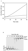
Functional membrane association and DNA-binding assays with PutA1294. A, Full-length PutA (100 μg, solid line) and PutA1294 (100 μg, dashed line) were incubated with 60 mM proline, 4 mM o-aminobenzaldehyde, and inverted membrane vesicles from E. coli strain JT31 putA- (0.1 mg/mL membrane protein) in 20 mM Mops buffer (pH 7.5) at 23 °C. The reactions were monitored at 443 nm as previously described (Becker and Thomas, 2001). The calculated specific activities for full-length PutA and PutA1294 are 81 and 8.0 mU mg−1 of membrane protein, respectively. B, Binding mixtures of TRdyed-700 labeled put control DNA (2 nM) and PutA1294 (0, 5, 10, 20, 50, 100, 200, 400, and 800 nM) were incubated at 20 °C for 20 min in 50 mM Tris buffer (pH 7.5, 50 mM NaCl). The PutA1294-DNA complexes were then separated by native polyacrylamide gel (4 %) electrophoresis at 4 °C as previously described (Gu et al., 2004).
Fig. 3
Fig. 3
Thermostability of PutA. Full-length PutA (2 mg/ml) and PutA86–630 (2 mg/ml) were incubated in 50 mM potassium phosphate buffer (pH 7.5) containing 50 mM NaCl at 45 °C. At the indicated time points protein samples were removed from the heating mixture and assayed for PRODH activity using the proline:oxidoreductase DCPIP assay as previously described (Becker and Thomas, 2001). Assay results for PutA (closed circles) and PutA86–630 (open circles) are reported as relative percent activity based on the initial PRODH activity at 0 min. A rate constant of 0.18 ± 0.03 min−1 was estimated for the decay in PRODH activity with full-length PutA using best-fit analysis to a single exponential equation. PutA and PutA86–630 were purified as described (Zhu and Becker, 2005; Zhang et al., 2007).
Fig. 4
Fig. 4
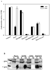
Correlation of PutA intracellular location and β-galactosidase activity from the lacZ reporter construct in cells containing various PutA mutant constructs. A, E. coli strain JT31 was grown in minimal medium at 37 °C containing the PputA:lacZ reporter construct and pUC18 constructs expressing wild-type PutA, PutAK9M, PutA1294, PutA-LacY, and PutA-CI-LacY. β-galactosidase activity is reported as mean ± standard errors of the mean of four independent experiments. aData for PutAR431M is from Zhang et al. (2007). Control cells have the PputA:lacZ reporter construct and pUC18 alone. B, Western blot analysis of different PutA proteins. The partitioning of wild-type PutA, PutAK9M, PutA1294 and PutAR431M proteins between the soluble (S) and membrane (M) fractions was analyzed using antibodies generated against purified PutA47 which contains the DNA-binding domain of PutA. PutA proteins were expressed from pUC18 constructs in E. coli strain JT31 grown in the absence and presence of 5 mM proline at 37 °C in minimal medium. Far left-hand lane shows purified wild-type PutA (~ 144 kDa band). Protein bands were visualized by chemiluminescence detection.

References

    1. Abrahamson JLA, Baker LG, Stephenson JT, Wood JM. Proline dehydrogenase from Escherichia K12, properties of the membrane-associated enzyme. Eur J Biochem. 1983;134:77–82. - PubMed
    1. Becker DF, Thomas EA. Redox properties of the PutA protein from Escherichia coli and the influence of the flavin redox state on PutA-DNA interactions. Biochemistry. 2001;40:4714–4722. - PubMed
    1. Brown E, Wood JM. Redesigned purification yields a fully functional PutA protein dimer from Escherichia coli. J Biol Chem. 1992;267:13086–13092. - PubMed
    1. Brown ED, Wood JM. Conformational change and membrane association of the PutA protein are coincident with reduction of its FAD cofactor by proline. J Biol Chem. 1993;268:8972–8979. - PubMed
    1. Buckle M. Surface plasmon resonance applied to DNA-protein complexes. In: Moss T, editor. DNA-Protein Interactions: Principles and Protocols. Vol 148: Methods in Molecular Biology. Humana Press Inc.; Totowa, NJ: 2001. pp. 535–546. - PubMed

Publication types

MeSH terms

LinkOut - more resources