

doi: 10.3201/eid1403.070825.
Hemagglutinating encephalomyelitis coronavirus infection in pigs, Argentina
Affiliations
- PMID: 18325268
- PMCID: PMC2570804
- DOI: 10.3201/eid1403.070825
Item in Clipboard
Hemagglutinating encephalomyelitis coronavirus infection in pigs, Argentina
Emerg Infect Dis.
2008 Mar.
Abstract
We describe an outbreak of vomiting, wasting, and encephalomyelitis syndrome in piglets in Argentina, caused by porcine hemagglutinating encephalomyelitis coronavirus (PHE-CoV) infection. Diagnosis was made by epidemiologic factors, pathologic features, immunohistochemistry, reverse transcription-PCR, and genomic sequencing. This study documents PHE-CoV infection in South America.
Figures
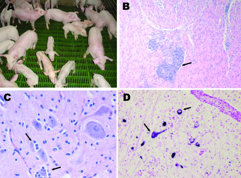
A) Nursery piglets showing clinical signs compatible with porcine hemagglutinating encephalomyelitis coronavirus (PHE-CoV). Nonaffected pigs of the same age are also shown. B) Muscle layer of stomach from affected piglet showing perivascular cuffing (arrow); hematoxylin-eosin stain, magnification ×100. C) Brainstem from affected piglet showing satellitosis (arrows) and gliosis; hematoxylin-eosin stain, magnification x400. D) Brainstem from affected piglet showing positive label of neuron perikarion (arrows); nitroblue-tetrazolium imunohistochemical stain, magnification x400.
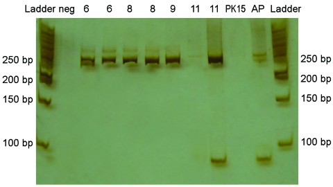
Polyacrylamide gel and silver staining of reverse transcription–PCR products from brains of piglets infected with porcine hemagglutinating encephalomyelitis coronavirus. Amplicons of ≈250 bp were found in brain samples from pigs 6, 8, 9, and 11 days of age. Neg, negative control (water + mastermix); PK15, amplification of PK15 cells inoculated with brain and tonsil from affected piglet; AP, asymptomatic piglet; and Ladder, 50-bp Fermentas.
References
-
- Chang GN, Chang TC, Lin SC, Tsai SS, Chern RS. Isolation and identification of hemagglutinating encephalomyelitis virus from pigs in Taiwan. J Chin Soc Vet Sci. 1993;19:147–58.
-
- Pensaert MB. Hemagglutinating encephalomyelitis virus. In: Straw BL, Zimmerman JJ, D’Allaire S, Taylor DJ, editors. Diseases of swine, 9th ed. Ames (IA): Blackwell Publishers; 2006. p. 353–8.
-
- Sasseville AM, Boutin M, Gélinas AM, Dea S. Sequence of the 3′-terminal end (8.1 kb) on the genome of porcine haemagglutinating encephalomyelitis virus: comparison with other hemagglutinating coronaviruses. J Gen Virol. 2002;83:2411–6. - PubMed
Publication types
MeSH terms
LinkOut - more resources
Full Text Sources