. 2009 Nov;30(11):1805-17.
doi: 10.1016/j.neurobiolaging.2008.01.001. Epub 2008 Mar 5.

Microglial responses to dopamine in a cell culture model of Parkinson's disease

Affiliations

Microglial responses to dopamine in a cell culture model of Parkinson's disease

Diego Mastroeni et al. Neurobiol Aging. 2009 Nov.

Abstract

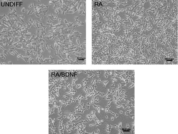
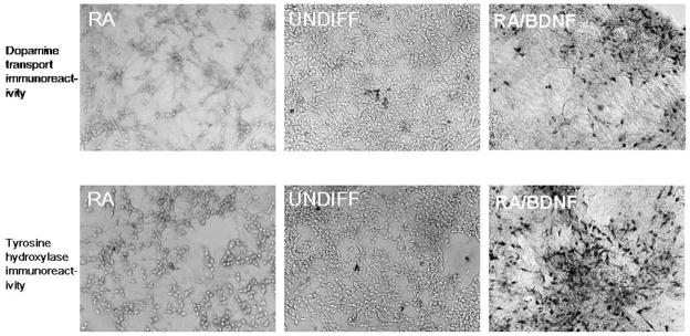
Activated microglia appear to selectively attack dopamine (DA) neurons in the Parkinson's disease (PD) substantia nigra. We investigated potential mechanisms using culture models. As targets, human SH-SY5Y cells were left undifferentiated (UNDIFF) or were differentiated with retinoic acid (RA) or RA plus brain-derived neurotrophic factor (RA/BDNF). RA/BDNF-treated cells were immunoreactive for tyrosine hydroxylase and the DA transporter, took up exogenous DA, and released DA after K(+) stimulation. Undifferentiated and RA-treated cells lacked these characteristics of a DA phenotype. Co-culture of target cells with human elderly microglia resulted in elevated toxicity in DA phenotype (RA/BDNF) cells. Lipopolysaccharide (LPS) plus K(+)-stimulated DA release enhanced toxicity by 500-fold. DA induced microglial chemotaxis in Boyden chambers. Spiperone inhibited this effect. Cultured human elderly microglia expressed mRNAs for D1-D4 but not D5 DA receptors. The microglia, as well as PD microglia in situ, were also immunoreactive for D1-D4 but not D5 DA receptors. These findings demonstrate that activated microglia express DA receptors, and suggest that this mechanism may play a role in the selective vulnerability of DA neurons in PD.


Conflict of interest statement

Disclosure Statement

There are no actual or potential conflicts of interest that the authors have.

Figures

Fig. 1
Fig. 1
Immunoreactivity for LN3 (A, B) (green fluorophore) and CD11b (C, D) (red fluorophore), classic markers for microglia and microglial activation in cultures of human elderly microglia (DAPI counterstain for DNA). Primary antibody deletion and/or prior absorption of antibodies with antigens gave uniformly negative immunohistochemical and immunocytochemical results both in this experiment and all others reported here. As was found in the present experiments, partial activation of human elderly microglia commonly occurs when the cells are placed into culture, presumably due to the rigors of cell isolation and maintenance in vitro, the prior state of the microglia in vivo, or other factors.
Fig. 2
Fig. 2
Morphology of SH-SY5Y cells 7 days after treatment with vehicle only (UNDIFF), RA only (RA), or RA followed by BDNF (RA/BDNF). (Phase contrast, 20×).
Fig. 3
Fig. 3
Immunoreactivity for the DA transporter (DAT) (upper panels) and tyrosine hydroxylase (TH) (bottom panels) in vehicle-treated (UNDIFF), RA-treated (RA), and RA/BDNF-treated (RA/BDNF) cells. (DAB chromagen, bright field, 10×).
Fig. 4
Fig. 4
DA uptake and release by the cultures. (Upper three panels) Fluorescence histochemistry (glyoxylic acid condensation method) for DA uptake by UNDIFF (vehicle-treated) (10×), RA-treated (10×), and RA/BDNF-treated (40×) cells after 30 min exposure to 50μM exogenous DA plus 1mM L-glutathione. (Bottom panel) DA release detected by HPLC-EC assays of culture supernatants. Cultures were exposed to 50 or 100 μg/ml exogenous DA for 30 min, washed 3×, then stimulated with 10% (v/v) high K+ Ringer’s solution. DA release was near background for UNDIFF and RA cells, and was significantly less than that for RA/BDNF cells (RA/BDNF vs UNDIFF at 50 and 100 μg/ml DA: t = 56.5, P < 0.001 and t = 42.6, P < 0.001, respectively; RA/BDNF vs RA at 50 and 100 μg/ml DA: t = 44.7, P < 0.001, and t = 34.8, P < 0.001, respectively). Comparisons were made over three independent HPLC-EC runs. A fourth run with materials that had been frozen and thawed twice was excluded from the analysis because of low values, although significance levels were still less than 0.001 when this run was included.
Fig. 5
Fig. 5
Effects of microglia co-culture with UNDIFF, RA, and RA/BDNF cells. (A) Toxicity, as measured by LDH release, after 96 hours co-incubation. (B) Nearly identical results were obtained using cell counts as the measure of toxicity, as suggested by the representative photomicrographs shown here of microglia and target cells after co-culture for 96 hours. Darker cells are LN3 immunoreactive, DAB-stained activated microglia. Lighter, unstained cells with a more flattened morphology are the target cells. (Phase contrast with LN3 immunocytochemistry, 10×).
Fig. 6
Fig. 6
Prior exposure of the target cells to 50mM DA, followed by stimulated release with high K+ Ringer’s, dramatically exacerbated toxicity in RA/BDNF-treated cells, especially when microglia were previously activated with LPS. This effect was again statistically significant in quantitative LDH release assays (A) and visually apparent in representative photomicrographs (B), where loss of the target cells (tan, neutral red-stained cells) and preservation of LN3 immunoreactive microglia (darker, DAB-stained cells) was especially obvious when RA/BDNF was used for differentiation to a DA phenotype and LPS exposure plus stimulated DA release were used as the treatment conditions (bright field, 20×).
Fig. 7
Fig. 7
Microglial chemotaxis in Boyden chambers in response to vehicle (serum free medium), 100nM MCP-1, 100 nM DA, or 100nM DA plus 10 nM spiperone. Significance levels after t-test comparisons are as shown in figure.
Fig. 8
Fig. 8
Dopamine receptor mRNA expression by cultured human elderly microglia. A) Representative gel image of D1 (DDR1) through D5 (DDR5) DA receptor mRNAs. B) Densitometry of bands in panel A. Replicating an earlier pilot experiment, D1–D4 DA receptor mRNAs (DRD1–DRD4) showed clear bands at appropriate amplicon sizes and were significantly elevated compared to D5 DA receptor mRNA (DRD5) (P < 0.001, all comparisons, by t-test). (Kb+: base pair standard; −ve: vehicle control; LML: Low Mass Ladder).
Fig. 9
Fig. 9
Photomicrographs of double immunostaining with the microglia marker LN3 and monoclonal antibodies to human D1 through D5 DA receptors. Confocal fluorescence images are of human elderly microglia cultures with a green fluorophor for the various DA receptors and a red fluorophor for LN3 (60×). Bright field images are of PD striatum with a purple chromagen (DAB plus nickel) for the various DA receptors and a brown chromagen (DAB alone) for LN3 (40×). Positive microglial immunoreactivity for D1, D2, D3, and D4 DA receptors was detected both in culture and in striatum sections, but D5 immunoreactivity was absent in microglia cultures and highly equivocal in striatum sections. Similar findings were obtained in the nigra and in NC cases (not shown). Notably, whereas the D5 DA receptor antibody did not uniquivocally label microglia, it did prominently stain neuronal/neuritic elements in cortex of the same patients (not shown), suggesting that the failure to label nigral and striatal microglia with the D5 antibody was not due to inadequate techniques or defective reagents.

Similar articles

Cited by

References

    1. Akiyama H, McGeer PL. Microglial response to 6-hydroxydopamine-induced substantia nigra lesions. Brain Res. 1989;489:247–253. - PubMed
    1. Akiyama H, McGeer PL. Brain microglia constitutively express beta-2 integrins. J Neuroimunol. 1990;30:81–93. - PubMed
    1. Arimoto T, Bing G. Up-regulation of inducible nitric oxide synthase in the substantia nigra by lipopolysaccharide causes microglial activation and neurodegeneration. Neurobiol Dis. 2003;12:35–45. - PubMed
    1. Chang RC, Hudson PM, Wilson BC, Liu B, Abel H, Hong JS. High concentrations of extracellular postassium enhance bacterial endotoxin lipopolysaccharide-induced neurotoxicity in glia-neuron mixed cultures. Neuroscience. 2000;97:757–764. - PubMed
    1. Chen H, Zhang SM, Hernan MA, Schwarzschild MA, Willet WC, Colditz GA, Speizer FE, Ascherio A. Nonsteroidal anti-inflammatory drugs and the risk of Parkinson disease. Arch Neurol. 2003;60:1059–1064. - PubMed

Publication types

MeSH terms