

Distinct horizontal patterns in the spatial organization of superficial zone chondrocytes of human joints
- PMID: 18325787
- PMCID: PMC2443102
- DOI: 10.1016/j.jsb.2008.01.010
Distinct horizontal patterns in the spatial organization of superficial zone chondrocytes of human joints
Abstract
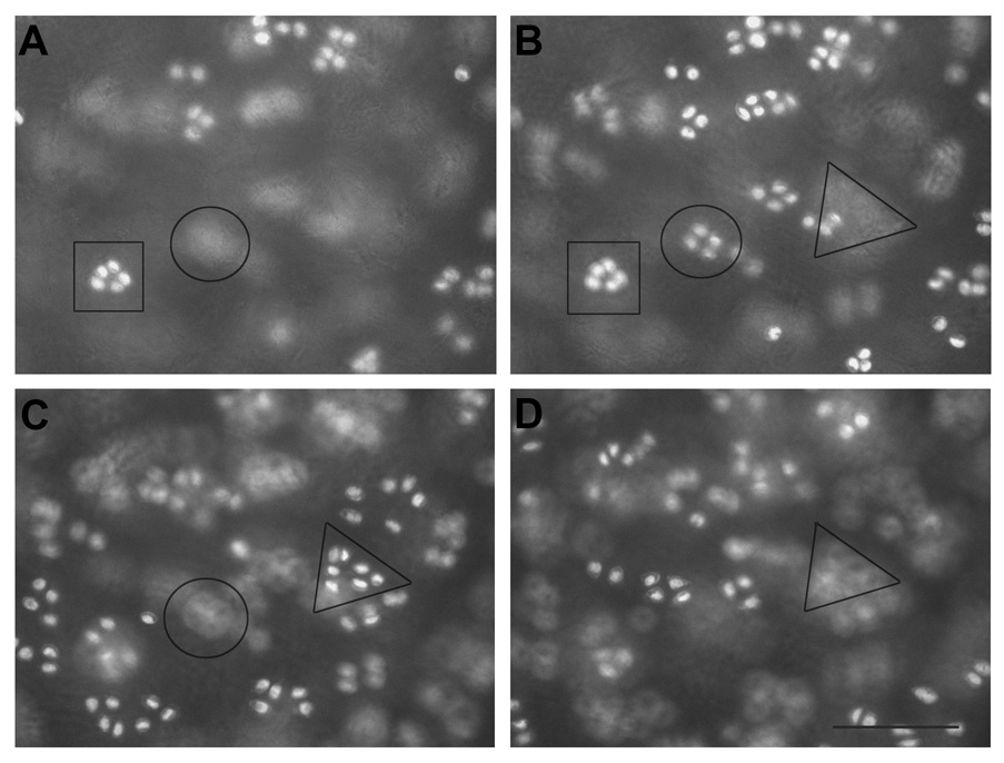
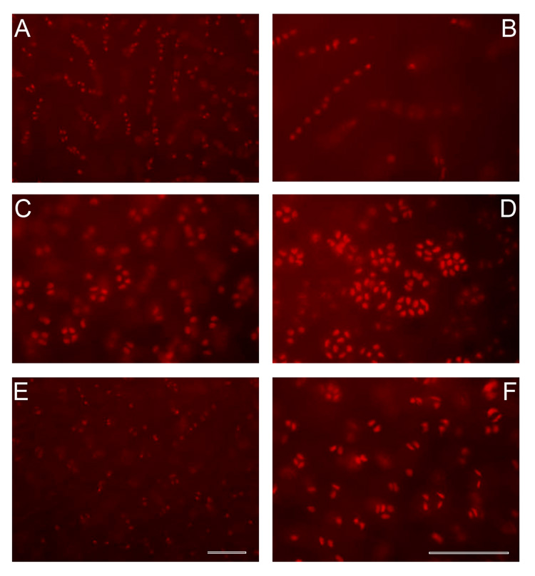
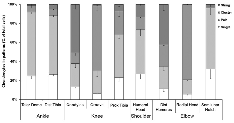
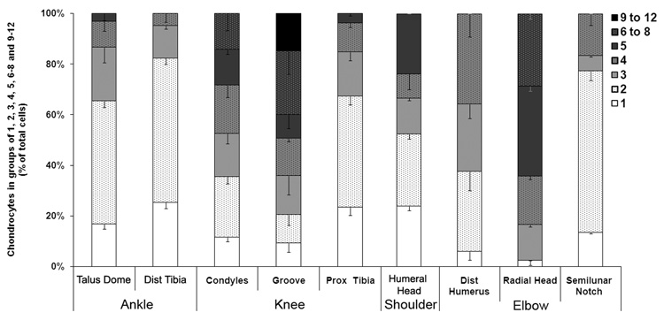
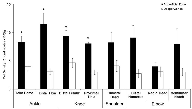
A better understanding of the unique cellular and functional properties of the superficial zone of articular cartilage may aid current strategies in tissue engineering which attempts a layered design for the repair of cartilage lesions to avert or postpone the onset of osteoarthritis. However, data pertaining to the cellular organization of non-degenerated superficial zone of articular cartilage is not available for most human joints. The present study analyzed the arrangement of chondrocytes of non-degenerated human joints (shoulder, elbow, knee, and ankle) by using fluorescence microscopy of the superficial zone in a top-down view. The resulting horizontal chondrocyte arrangements were tested for randomness, homogeneity or a significant grouping via point pattern analysis and were correlated with the joint type in which they occurred. The present study demonstrated that human superficial chondrocytes occurred in four distinct patterns of strings, clusters, pairs or single chondrocytes. Those patterns represented a significant grouping (p < 0.0001) with horizontal alignment. Each articular joint surface was dominated by only one of these four patterns (p < 0.001). Specific patterns correlated with specific diarthrodial joint types (p < 0.001). Further studies need to establish whether these organizational patterns are a consequence of their surrounding environment or whether they are linked to a functional purpose.
Figures
References
-
- Aydelotte MB, Kuettner KE. Differences between sub-populations of cultured bovine articular chondrocytes. I. Morphology and cartilage matrix production. Connect Tissue Res. 1988;18:205–222. - PubMed
-
- Below S, Arnoczky SP, Dodds J, Kooima C, Walter N. The split-line pattern of the distal femur: A consideration in the orientation of autologous cartilage grafts. Arthroscopy. 2002;18:613–617. - PubMed
-
- Brama PA, Tekoppele JM, Bank RA, Barneveld A, van Weeren PR. Functional adaptation of equine articular cartilage: the formation of regional biochemical characteristics up to age one year. Equine Vet J. 2000;32:217–221. - PubMed
-
- Brighton CT, Kitajima T, Hunt RM. Zonal analysis of cytoplasmic components of articular cartilage chondrocytes. Arthritis Rheum. 1984;27:1290–1299. - PubMed
-
- Burr DB. Anatomy and physiology of the mineralized tissues: role in the pathogenesis of osteoarthrosis. Osteoarthritis Cartilage. 2004;12 Suppl A:S20–S30. - PubMed
Publication types
MeSH terms
Grants and funding
LinkOut - more resources
Full Text Sources
Other Literature Sources
