. 2008 Jun;149(6):3196-205.
doi: 10.1210/en.2007-1655. Epub 2008 Mar 6.

Active intestinal calcium transport in the absence of transient receptor potential vanilloid type 6 and calbindin-D9k

Affiliations

Active intestinal calcium transport in the absence of transient receptor potential vanilloid type 6 and calbindin-D9k

Bryan S Benn et al. Endocrinology. 2008 Jun.

Abstract

To study the role of the epithelial calcium channel transient receptor potential vanilloid type 6 (TRPV6) and the calcium-binding protein calbindin-D9k in intestinal calcium absorption, TRPV6 knockout (KO), calbindin-D9k KO, and TRPV6/calbindin-D(9k) double-KO (DKO) mice were generated. TRPV6 KO, calbindin-D9k KO, and TRPV6/calbindin-D9k DKO mice have serum calcium levels similar to those of wild-type (WT) mice ( approximately 10 mg Ca2+/dl). In the TRPV6 KO and the DKO mice, however, there is a 1.8-fold increase in serum PTH levels (P < 0.05 compared with WT). Active intestinal calcium transport was measured using the everted gut sac method. Under low dietary calcium conditions there was a 4.1-, 2.9-, and 3.9-fold increase in calcium transport in the duodenum of WT, TRPV6 KO, and calbindin-D9k KO mice, respectively (n = 8-22 per group; P > 0.1, WT vs. calbindin-D9k KO, and P < 0.05, WT vs. TRPV6 KO on the low-calcium diet). Duodenal calcium transport was increased 2.1-fold in the TRPV6/calbindin-D9k DKO mice fed the low-calcium diet (P < 0.05, WT vs. DKO). Active calcium transport was not stimulated by low dietary calcium in the ileum of the WT or KO mice. 1,25-Dihydroxyvitamin D3 administration to vitamin D-deficient null mutant and WT mice also resulted in a significant increase in duodenal calcium transport (1.4- to 2.0-fold, P < 0.05 compared with vitamin D-deficient mice). This study provides evidence for the first time using null mutant mice that significant active intestinal calcium transport occurs in the absence of TRPV6 and calbindin-D9k, thus challenging the dogma that TRPV6 and calbindin-D9k are essential for vitamin D-induced active intestinal calcium transport.


Figures

Figure 1
Figure 1
Concentration of calcium and PTH in the serum of WT, TRPV6 KO, calbindin-D9k KO, and TRPV6/calbindin-D9k DKO mice at 3 months of age. Each value represents the mean ± sem for male mice (n = 7–23 mice per group; *, P < 0.05 compared with WT). Similar findings were observed using female mice (data not shown). Body weights for the mice at 3 months of age were as follows: WT, 25.4 ± 0.7 g; TRPV6 KO, 24.6 ± 1.1 g; calbindin-D9k KO, 24.7 ± 1.5 g; DKO, 26.3 ± 1.2 g (n = 7–23 mice per group; P > 0.1 compared with WT).
Figure 2
Figure 2
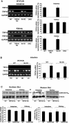
Possible compensatory or reciprocal mechanisms involving vitamin D target genes/proteins in the null mutant mice. A, Left panel, Representative RT-PCR analysis of TRPV6 and TRPV5 mRNA expression in intestine and kidney of WT and null mutant male mice; right panel, quantitation of TRPV5 and TRPV6 mRNA expression (note no induction of TRPV5 expression in the intestine and no change in renal TRPV5 expression in null mutant mice; n = 5–9 per group). B, Left panel, Representative RT-PCR analysis of TRPV5 and TRPV6 mRNA expression in WT and calbindin-D9k KO male mice; right panel, quantitative analysis. Note the induction of TRPV6 mRNA in the intestine under low dietary calcium conditions (0.02%, LC) and no difference in the levels of TRPV6 mRNA under high dietary calcium (1%, HC) or low dietary calcium conditions between WT and calbindin-D9k KO mice. All values are reported as the mean ± sem (n = 5–6 per group). *, Significantly different from the respective HC group at P < 0.05. C, Left, Representative Northern blot of calbindin-D9k mRNA (upper panel) and summary of densitometric scans of Northern blot analysis of calbindin-D9k mRNA levels in intestine and kidney of WT and TRPV6 KO mice (lower panel); right, representative Western blot (upper panel) and summary of densitometric scans of Western blots (lower panel) of calbindin-D9k in intestine and kidney of WT and TRPV6 KO mice. Note in the TRPV6 KO mice, there is no compensatory increase in calbindin-D9k. All values are reported as mean ± sem (n = 8–9 per group; WT vs. TRPV6 KO, calbindin-D9k mRNA, or calbindin-D9k protein in intestine and kidney, P > 0.1).
Figure 3
Figure 3
Active intestinal calcium transport in the duodenum of mice fed high- (1%) or low- (0.02%) calcium diet. A, Calcium transport was measured using everted intestinal sacs formed from the duodenum of 2-month-old mice that had been fed a high-calcium (HC) or low-calcium (LC) diet from 4 wk of age. Data are expressed relative to levels seen in WT high-calcium mice (HC WT = 1). Values represent the mean ± sem (n = 8–22 per group; *, significantly different from the respective high-calcium group at P < 0.05,; P > 0.1, WT vs. calbindin-D9k KO; and +, P < 0.05 WT vs. TRPV6 KO and TRPV6/calbindin-D9k DKO on the low-calcium diet). Right panel, Representative RT-PCR analysis of TRPV6 mRNA expression and Northern blot analysis for calbindin-D9k mRNA. Under low dietary calcium conditions, serum PTH was similarly elevated in all groups of mice (WT, 77.6 ± 11; TRPV6 KO, 85 ± 12; calbindin-D9k KO, 100 ± 12; DKO, 100.5 ± 20 pg/ml; P > 0.1 compared with WT; P > 0.1, comparison of multiple group means). 1,25(OH)2D3 serum levels were also similarly elevated in all groups of mice under low dietary calcium conditions (WT, 427 ± 77; TRPV6 KO, 414 ± 62; calbindin-D9k KO, 403 ± 65; DKO, 457 ± 46 pg/ml; P > 0.1 compared with WT; P > 0.1, comparison of multiple group means). Also, under low dietary calcium conditions, serum calcium levels were similar in all groups of mice (WT, 10.2 ± 0.2; TRPV6 KO, 9.8 ± 0.2; calbindin-D9k KO, 10.1 ± 0.2; DKO, 10.4 ± 0.4 mg/dl; P > 0.1 compared with WT; P > 0.1, comparison of multiple group means). B, Calcium transport was measured using everted intestinal sacs from the duodenum of 12- to 14-month-old mice that had been fed a high- or low-calcium diet for 4 wk. Data are relative to WT 12- to 14-month-old mice fed the high-calcium diet (aged high-calcium WT = 1; n = 6–18 per group).
Figure 4
Figure 4
1,25(OH)2D3-stimulated calcium transport in the duodenum of WT and null mutant mice. Calcium transport was measured using everted intestinal sacs from the duodenum of 12-wk-old mice made 1,25(OH)2D3 deplete by feeding a 0.8% strontium diet for 7 d. Mice were then injected with 1,25(OH)2D3 (+D3) or vehicle (−D3) 48, 24, and 6 h before termination (ip, 100 ng/100 g body weight per injection). Values represent the mean ± sem [n = 6–16 per group; *, P < 0.05 for 1,25(OH)2D3-treated (+D3) compared with the respective-deficient (−D3) mice; +, P < 0.05 compared with WT +D3]. This graph (as well as Fig. 3) represents data from both male and female mice. The number of male and female mice was balanced to increase the power to detect genotype vs. treatment differences. Right panel, Representative RT-PCR analysis of TRPV6 mRNA expression and Northern blot analysis for calbindin-D9k mRNA.

References

    1. Christakos S, Dhawan P, Liu Y, Peng X, Porta A 2003 New insights into the mechanisms of vitamin D action. J Cell Biochem 88:695–705 - PubMed
    1. Wasserman RH, Fullmer CS 1995 Vitamin D and intestinal calcium transport: facts, speculations and hypotheses. J Nutr 125:1971S–1979S - PubMed
    1. Raval-Pandya M, Porta AR, Christakos S 1998 Mechanism of action of 1,25 dihydroxyvitamin D3 on intestinal calcium absorption and renal calcium transportation. In: Holick MF, ed. Vitamin D: physiology, molecular biology, and clinical applications. Totowa, NJ: Humana Press; 163–174
    1. Feher JJ 1983 Facilitated calcium diffusion by intestinal calcium-binding protein. Am J Physiol Cell Physiol 244:C303–C307 - PubMed
    1. Bronner F, Pansu D, Stein WD 1986 An analysis of intestinal calcium transport across the rat intestine. Am J Physiol 250:G561–G569 - PubMed

Publication types