. 2008 May 9;283(19):13116-23.
doi: 10.1074/jbc.M707697200. Epub 2008 Mar 7.

Mechanism of ceftriaxone induction of excitatory amino acid transporter-2 expression and glutamate uptake in primary human astrocytes

Affiliations

Mechanism of ceftriaxone induction of excitatory amino acid transporter-2 expression and glutamate uptake in primary human astrocytes

Seok-Geun Lee et al. J Biol Chem. .

Abstract

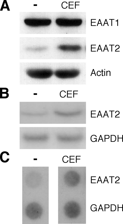
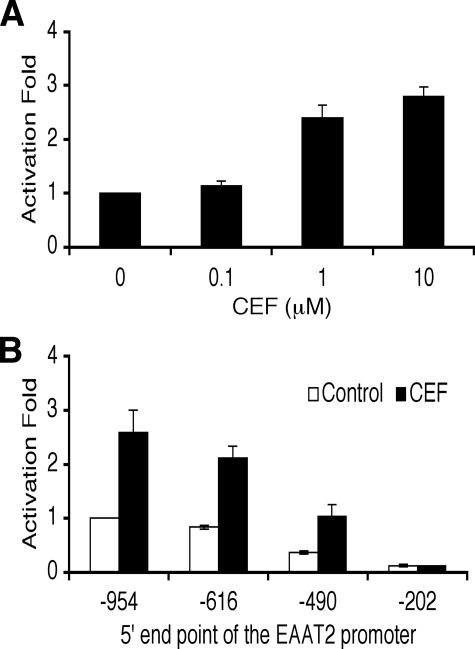
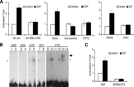
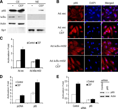
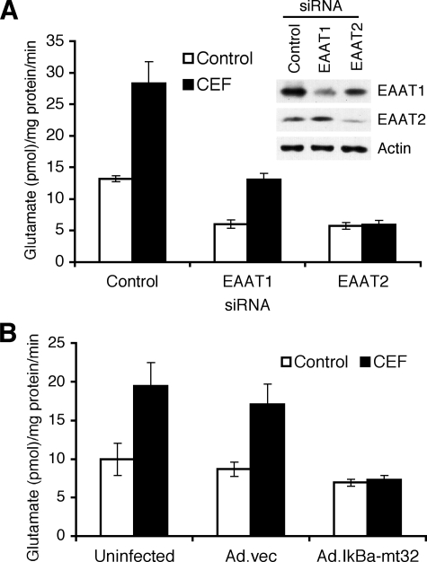
Glutamate is an essential neurotransmitter regulating brain functions. Excitatory amino acid transporter (EAAT)-2 is one of the major glutamate transporters primarily expressed in astroglial cells. Dysfunction of EAAT2 is implicated in acute and chronic neurological disorders, including stroke/ischemia, temporal lobe epilepsy, amyotrophic lateral sclerosis, Alzheimer disease, human immunodeficiency virus 1-associated dementia, and growth of malignant gliomas. Ceftriaxone, one of the beta-lactam antibiotics, is a stimulator of EAAT2 expression with neuroprotective effects in both in vitro and in vivo models based in part on its ability to inhibit neuronal cell death by glutamate excitotoxicity. Based on this consideration and its lack of toxicity, ceftriaxone has potential to manipulate glutamate transmission and ameliorate neurotoxicity. We investigated the mechanism by which ceftriaxone enhances EAAT2 expression in primary human fetal astrocytes (PHFA). Ceftriaxone elevated EAAT2 transcription in PHFA through the nuclear factor-kappaB (NF-kappaB) signaling pathway. The antibiotic promoted nuclear translocation of p65 and activation of NF-kappaB. The specific NF-kappaB binding site at the -272 position of the EAAT2 promoter was responsible for ceftriaxone-mediated EAAT2 induction. In addition, ceftriaxone increased glutamate uptake, a primary function of EAAT2, and EAAT2 small interference RNA completely inhibited ceftriaxone-induced glutamate uptake activity in PHFA. Taken together, our data indicate that ceftriaxone is a potent modulator of glutamate transport in PHFA through NF-kappaB-mediated EAAT2 promoter activation. These findings suggest a mechanism for ceftriaxone modulation of glutamate transport and for its potential effects on ameliorating specific neurodegenerative diseases through modulation of extracellular glutamate.


Figures

FIGURE 1.
FIGURE 1.
CEF induces EAAT2 expression in primary human fetal astrocytes. PHFA cells were cultured in antibiotic-free medium for at least 7 days prior to all experiments reported in this study. Cells were treated with 10 μm CEF for 2 days. A, cell lysates were prepared, and EAAT1 and EAAT2 protein expressions were analyzed. Actin was used as an internal control. B, total cellular RNA was extracted and subjected to Northern blotting for EAAT2 mRNA expression. C, nuclei were extracted, and the isolated nuclei were used to label preinitiated RNA transcription with [α-32P]UTP in vitro, and the purified RNA was hybridized to a dot blot carrying an equivalent amount of panel DNA probes. The transcript rate of glyceraldehyde-3-phosphate dehydrogenase (GAPDH) served as a control.
FIGURE 2.
FIGURE 2.
CEF activates the EAAT2 promoter. A, PHFA cells were transfected with the EAAT2Pro-954 together with a pSV-β-galactosidase plasmid as an internal control. One day after transfection, cells were treated with various concentrations of CEF for 2 days as indicated. Values are presented as -fold-normalized activity relative to that of the untreated cells taken as 1. B, PHFA cells were transfected with different EAAT2 promoter deletion-reporter constructs with pSV-β-galactosidase as an internal control. One day after transfection, cells were treated with 10 μm CEF for 2 days. Values are presented as -fold-normalized activity relative to that of the EAAT2Pro-954 in untreated cells taken as 1. Error bars indicate S.D.
FIGURE 3.
FIGURE 3.
NF-κB is crucial for CEF-induced EAAT2 expression. A, PHFA were infected with Ad.vec or Ad.IκBα-mt32. The infected cells (left) or uninfected PHFA (middle and right) were transfected with EAAT2Pro-954 together with pSV-β-galactosidase as an internal control. One day after transfection, cells were treated with 10 μm CEF together with the indicated inhibitors (middle, 5 μm quinalzoline or 20 μm pyrrolidine dithiocarbamate; right, 5 μm SN50M or SN50) for 2 days. Values are presented as –fold-normalized activity relative to that of the EAAT2Pro-954 in Ad.vec-infected (left), mock-treated (middle), or SN50M-treated (right) cells. B, PHFA were treated with 10 μm CEF for 4 days, and nuclear extracts were prepared. Nuclear extracts were mixed with each radiolabeled oligonucleotide containing the NF-κB binding site located at -583, -334, -272, or -251 of the EAAT2 promoter as follows: lanes 1, 3, 5, 7, and 9, untreated nuclear extracts; lanes 2, 4, 6, 8, and 10–13, CEF-treated nuclear extracts; lane 14, no extracts added. For competition assays, a 100-fold excess of corresponding unlabeled probe (lane 11) or unlabeled probe encompassing the mutated NF-κB site (lane 12) was added. Supershift analysis was performed with an anti-p65 antibody (lane 13). The asterisk and arrow indicate, respectively, NF-κB and a supershift band. C, PHFA were transfected with either the EAAT2Pro-954 or NF-κBmt-272 construct together with pSV-β-galactosidase as an internal control. One day after transfection, cells were treated with 10 μm CEF for 2 days. Values are represented as –fold-normalized activity to that of the EAAT2Pro-954 in untreated cells taken as 1. Error bars indicate S.D.
FIGURE 4.
FIGURE 4.
CEF activates NF-κB. A, PHFA were treated with 10 μm CEF for 4 days. Nuclear extracts (NE) and cytoplasmic extracts (CE) were prepared and immunoblotted with the indicated antibodies. Actin and Sp1 were used as the internal control for CE and NE, respectively. B, PHFA cells were infected with Ad.vec or Ad.IκBα-mt32 and then incubated with 10 μm CEF for 2 days. The p65 immunostaining and 4′,6-diamidino-2-phenylindole (DAPI) staining were evaluated by confocal microscopy. C, PHFA were infected with Ad.vec or Ad.IκBα-mt32. The infected cells were transfected with 3κB-Luc, containing three tandem NF-κB binding sites, together with pSV-β-galactosidase as an internal control. One day after transfection, the cells were treated with 10 μm CEF for 2 days. Values are presented as -fold-normalized activity relative to that of 3κB-Luc in Ad.vec-infected cells untreated with CEF taken as 1. D, PHFA were transfected with the EAAT2Pro-954 and pcDNA or p65 expression vector together with a pSV-β-galactosidase plasmid as an internal control. One day after transfection, cells were treated with 10 μm CEF for 2 days. Values are presented as -fold-normalized activity relative to that of the pcDNA-transfected and untreated cells taken as 1. E, PHFA were transfected with the EAAT2Pro-954 and control siRNA or p65 siRNA together with a pSV-β-galactosidase plasmid as an internal control. One day after transfection, cells were treated with 10 μm CEF for 2 days. Values are presented as -fold-normalized activity relative to that of the control siRNA-transfected and untreated cells taken as 1. The cell lysates treated with control and p65 siRNA were used for Western blotting. Error bars indicate S.D.
FIGURE 5.
FIGURE 5.
CEF induces glutamate uptake in PHFA. A, PHFA were transfected with control, EAAT1, or EAAT2 siRNA, and then the transfected cells were treated with 10 μm CEF. Two days later, glutamate uptake levels were measured as described under “Experimental Procedures.” Accordingly, cell lysates from PHFA transfected with control, EAAT1, or EAAT2 siRNA were prepared and EAAT1 and EAAT2 expressions were analyzed. B, PHFA were infected with Ad.vec or Ad.IκBα-mt32, and then the uninfected or infected cells were treated with 10 μm CEF. Two days later, glutamate uptake levels were measured. Error bars indicate S.D.

References

    1. Fonnum, F. (1984) J. Neurochem. 42 1-11 - PubMed
    1. Headley, P. M., and Grillner, S. (1990) Trends Pharmacol. Sci. 11 205-211 - PubMed
    1. Anderson, C. M., and Swanson, R. A. (2000) Glia 32 1-14 - PubMed
    1. Rothstein, J. D., Martin, L., Levey, A. I., Dykes-Hoberg, M., Jin, L., Wu, D., Nash, N., and Kuncl, R. W. (1994) Neuron 13 713-725 - PubMed
    1. Plaitakis, A., and Caroscio, J. T. (1987) Ann. Neurol. 22 575-579 - PubMed

Publication types

MeSH terms

LinkOut - more resources