. 2008 Mar 18;105(11):4453-8.
doi: 10.1073/pnas.0800897105. Epub 2008 Mar 7.

Organization of the core structure of the postsynaptic density

Affiliations

Organization of the core structure of the postsynaptic density

Xiaobing Chen et al. Proc Natl Acad Sci U S A. .

Erratum in

  • Proc Natl Acad Sci U S A. 2008 Jun;105(22):7893

Abstract

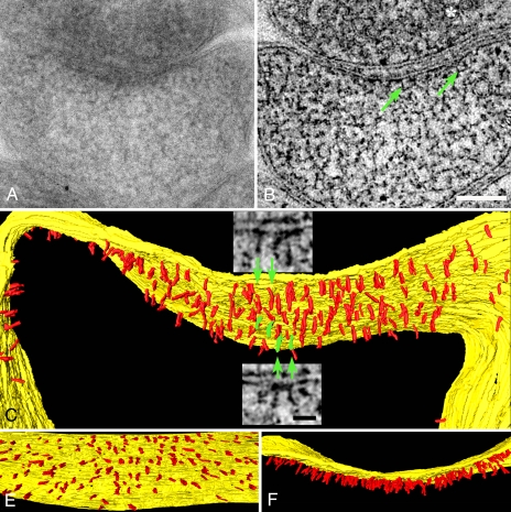
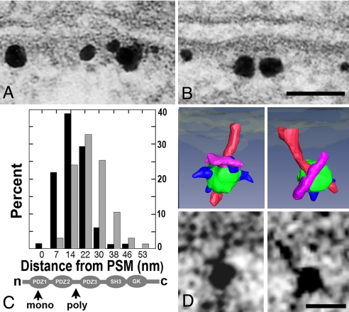
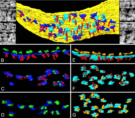
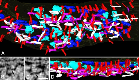
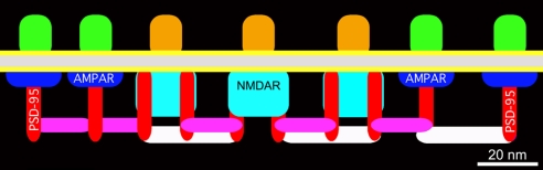
Much is known about the composition and function of the postsynaptic density (PSD), but less is known about its molecular organization. We use EM tomography to delineate the organization of PSDs at glutamatergic synapses in rat hippocampal cultures. The core of the PSD is dominated by vertically oriented filaments, and ImmunoGold labeling shows that PSD-95 is a component of these filaments. Vertical filaments contact two types of transmembrane structures whose sizes and positions match those of glutamate receptors and intermesh with two types of horizontally oriented filaments lying 10-20 nm from the postsynaptic membrane. The longer horizontal filaments link adjacent NMDAR-type structures, whereas the smaller filaments link both NMDA- and AMPAR-type structures. The orthogonal, interlinked scaffold of filaments at the core of the PSD provides a structural basis for understanding dynamic aspects of postsynaptic function.


Conflict of interest statement

The authors declare no conflict of interest.

Figures

Fig. 1.
Fig. 1.
Vertical filaments at the PSD. (A) EM of a PSD in a mushroom-shaped dendritic spine. Structural details are obscured by overlap within this 120-nm-thick section. (B) Vertical filaments (green arrows) are apparent in a 1.5-nm-thick virtual section derived from the tomographic reconstruction of the section in A. The synaptic vesicle is indicated by an asterisk. (Scale bar: 100 nm.) (C) Rendering of vertical filaments (red) from the tomographic reconstruction. Vertical filaments, 5 nm in diameter and 20 nm long, contact the postsynaptic membrane (yellow). (Insets) Virtual sections from which particular vertical filaments (green) are segmented. (Scale bar: 20 nm.) (D) En face view showing uniform distribution of vertical filaments at the PSD. (E) Overlap of vertical filaments contributes to the typical thickened appearance of a PSD viewed in cross-section.
Fig. 2.
Fig. 2.
Mapping and labeling of PSD-95. (A) ImmunoGold label for PSD-95 with monoclonal antibody to PDZ1 domain. (B) ImmunoGold label for PSD-95 with polyclonal antibody to the region between PDZ2 and PDZ3. Both labels are confined to the vicinity of the PSD. (Scale bar: 50 nm.) (C) Label with monoclonal antibody (black bars) is closer to the PSM than label with polyclonal antibody (gray bars). Percentage refers to the percentage of total gold label. Locations of epitopes on PSD-95 are indicated below (arrows). (D) Label associated with filaments in single virtual sections (below) and surface renderings (above). Vertical filaments are rendered in red, and silver-enhanced gold particles are rendered in green. Structures rendered in purple are the correct size to represent the primary antibody contacting the vertical filaments. Smaller structures rendered in blue may represent the secondary Fab fragment partially buried in the silver encrusted on the gold particle. (Scale bar: 10 nm.)
Fig. 3.
Fig. 3.
Transmembrane structures at the PSD. (A) Cytoplasmic surface of the membrane (yellow) at the PSD, showing AMPAR-type (blue) and NMDAR-type cytoplasmic domains (cyan), and vertical filaments (red). (Left Insets) AMPAR-type structures in virtual sections from the tomographic reconstruction. Arrow points to a transcleft filament. (Right Insets) NMDAR-type structures. Arrow points to a transcleft filament, and double arrow (lower right) indicates the extent of synaptic cleft. (Scale bar: 20 nm.) (B–D) AMPAR-type structures shown in cross-section (B, extracellular domain green), en face from inside the spine (C), and en face from outside the spine (D). Cytoplasmic domains of AMPAR-type structures are contacted by vertical filaments (C). (E–G) NMDAR-type structures shown in cross-section (E, extracellular domain gold), en face from inside the spine (F), and en face from outside the spine (G). Cytoplasmic domains of NMDAR-type structures are contacted by one or two vertical filaments (F).
Fig. 4.
Fig. 4.
Filament network at the PSD. (A) Meshwork of horizontal filaments at the core of the PSD. The shorter type (purple) is 4–5 nm in diameter and ≈20 nm long, and the longer type (white) is 5–6 nm in diameter and 30–35 nm long. The asterisks indicate sheet-like structures. (B and C) Shorter (B, arrow) and longer (C, arrowhead) types of horizontal filament in virtual sections. (Scale bar: 20 nm.) (D) Cross-sectional view of the PSD in A showing layering of horizontal filaments. The shorter filaments (B, purple) lie somewhat closer to the postsynaptic membrane than the longer filaments (C, white).
Fig. 5.
Fig. 5.
Core structure of the PSD based on tomographic reconstructions. Components and spacings between components are drawn approximately to scale. Dominant structure is an array of vertical filaments containing PSD-95 (red). NMDAR-type structures, differentiated by their large cytoplasmic extensions (cyan), concentrate in the center of the PSD. AMPAR-type structures, differentiated by their flattened cytoplasmic aspects (blue), surround the NMDAR structures. Virtually all NMDAR- and AMPAR-type structures are contacted by vertical filaments. Short horizontal filaments (purple) link vertical filaments associated with both NMDAR- and AMPAR-type structures. Longer horizontal filaments (white) concentrated under the NMDAR-type structures cross-link the vertical filament meshwork.

References

    1. Siekevitz P. The postsynaptic density: A possible role in long-lasting effects in the central nervous system. Proc Natl Acad Sci USA. 1985;82:3494–3498. - PMC - PubMed
    1. Ziff EB. Enlightening the postsynaptic density. Neuron. 1997;19:1163–1174. - PubMed
    1. Kennedy MB. Signal-processing machines at the postsynaptic density. Science. 2000;290:750–754. - PubMed
    1. Palay SL. Synapses in the central nervous system. J Biophys Biochem Cytol. 1956;2:193–202. - PMC - PubMed
    1. Malinow R, Malenka RC. AMPA receptor trafficking and synaptic plasticity. Annu Rev Neurosci. 2002;25:103–126. - PubMed

Publication types

MeSH terms

Substances

LinkOut - more resources