. 2008 Jun;94(12):4948-56.
doi: 10.1529/biophysj.107.124107. Epub 2008 Mar 7.

Label-free, high-throughput measurements of dynamic changes in cell nuclei using angle-resolved low coherence interferometry

Affiliations

Label-free, high-throughput measurements of dynamic changes in cell nuclei using angle-resolved low coherence interferometry

Kevin J Chalut et al. Biophys J. 2008 Jun.

Abstract

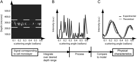
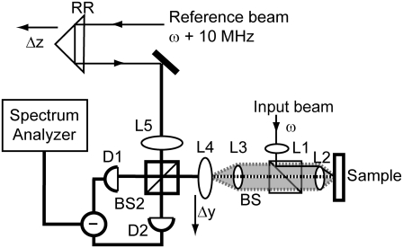
Accurate measurements of nuclear deformation, i.e., structural changes of the nucleus in response to environmental stimuli, are important for signal transduction studies. Traditionally, these measurements require labeling and imaging, and then nuclear measurement using image analysis. This approach is time-consuming, invasive, and unavoidably perturbs cellular systems. Light scattering, an emerging biophotonics technique for probing physical characteristics of living systems, offers a promising alternative. Angle-resolved low-coherence interferometry (a/LCI), a novel light scattering technique, was developed to quantify nuclear morphology for early cancer detection. In this study, a/LCI is used for the first time to noninvasively measure small changes in nuclear morphology in response to environmental stimuli. With this new application, we broaden the potential uses of a/LCI by demonstrating high-throughput measurements and by probing aspherical nuclei. To demonstrate the versatility of this approach, two distinct models relevant to current investigations in cell and tissue engineering research are used. Structural changes in cell nuclei due to subtle environmental stimuli, including substrate topography and osmotic pressure, are profiled rapidly without disrupting the cells or introducing artifacts associated with traditional measurements. Accuracy > or = 3% is obtained for the range of nuclear geometries examined here, with the greatest deviations occurring for the more complex geometries. Given the high-throughput nature of the measurements, this deviation may be acceptable for many biological applications that seek to establish connections between morphology and function.


Figures

FIGURE 1
FIGURE 1
Acquisition and processing of light scattering data from a cell monolayer using the a/LCI technique. In A, the optical depth corresponding to the cell monolayer is summed to obtain the intensity of the scattered light versus scattering angle (B). The scattering distribution is then low-pass filtered and a second-order polynomial is subtracted to isolate the nuclear scattering. Finally, the processed scattering distribution is compared to a database of known scattering distributions (calculated from an appropriate light scattering model) to ascertain the nuclear morphology (C). In this case, the sample comprised a monolayer of chondrocyte cells for which the a/LCI size determination was 6.5 μm.
FIGURE 2
FIGURE 2
a/LCI schematic in a Mach-Zehnder interferometer configuration (used with permission of Pyhtila and colleagues (5)). Axial translation of retroreflector permits optical sectioning of the sample by taking advantage of coherence properties of the source. Lenses L1–L4 are configured as shown so that 1), the beam is collimated when incident on the sample, and 2), L4 is in the image plane of the scattered light (the periphery of which is shaded). By scanning L4, the intensity of scattered light can be mapped as a function of the scattering angle.
FIGURE 3
FIGURE 3
Schematic of the four configurations used in the a/LCI setup for the experiment regarding the macrophage cells on a 2-μm PDMS grating. The two orientations determine whether the symmetry axis of the macrophage cell nuclei will present itself to the electric field vector or the magnetic field vector; the incident light polarization determines the direction of the two field vectors as presented to the sample.
FIGURE 4
FIGURE 4
Results of using the a/LCI analysis technique to measure size of spheroidal particles by using angular scattering distributions calculated using the T-matrix model as input into the analysis. Angular scattering distributions correspond to a particle with an equal volume radius of 3.75 μm, a background refractive index of 1.36, a target refractive index of 1.42, and a size distribution of 2.5%. The polarization and orientation of the light and particle respectively are (a) S11, α = 90°, β = 90°; (b) S22, α = 90°, β = 90°, (c) S11, α = 0°, β = 60°, and (d) S22, α = 0°, β = 90°, where α and β are Euler angles and S11 and S22 are parallel and perpendicular incident polarizations, respectively. The equatorial and polar manifolds are 1 wavelength (830 nm) thick, with the center corresponding to the equatorial and polar axes of the spheroid at that aspect ratio. These results indicate that the size measurement of a spheroidal particle as determined by the a/LCI analysis technique almost universally falls on either the equatorial or polar manifold; however, there is no method of consistently predicting which it will be.
FIGURE 5
FIGURE 5
Confocal images of stained chondrocyte cell nucleus equilibrated at (a) 500 mOsm and (b) 330 mOsm. At 500 mOsm, the cell nucleus is smaller and less rounded than the cell nucleus equilibrated at 330 mOsm, which is a typical observation in this experiment.
FIGURE 6
FIGURE 6
Results of measurements of porcine chondrocyte cell nuclei using (a) image analysis and (b) the a/LCI technique. The error bars correspond to mean ± SE (standard error of the mean in the 95% confidence interval). Both experiments demonstrate a statistically significant (p < 0.05) increase in nuclear size with decreasing osmolarity.
FIGURE 7
FIGURE 7
DAPI-stained images of murine macrophage cell nuclei in planar control configuration (a) and on 2-μm PDMS grating (b). Elongation of nuclei along the direction of the grating is clear compared to planar controls.
FIGURE 8
FIGURE 8
Results of measurements of murine macrophage cell nuclei using (a) image analysis and (b) a/LCI technique. The planar controls were determined by image analysis to be nonoriented spheroids with a small aspect ratio; it is reported as the average between the major and minor axes. For the a/LCI measurements, there was a bimodal distribution of sizes. One size was within the 95% confidence interval of the planar controls, and the other size distribution contained sizes at least four standard deviations larger than the planar controls. The short size was determined to correspond to the minor (equatorial) axis of the macrophage cell nuclei; the large size was determined to correspond to the major (polar) axis of the macrophage cell nuclei. Results of measurements using image analysis and a/LCI analysis are reported in the table. Nuclear elongation is statistically significant (p < 0.01) for both a/LCI and image analysis measurements.

References

    1. Guilak, F. 1995. Compression-induced changes in the shape and volume of the chondrocyte nucleus. J. Biomech. 28:1529–1541. - PubMed
    1. Chen, C. S., and D. E. Ingber. 1999. Tensegrity and mechanoregulation: from skeleton to cytoskeleton. Osteoarthritis Cartilage. 7:81–94. - PubMed
    1. Wax, A., J. W. Pyhtila, R. N. Graf, R. Nines, C. W. Boone, R. R. Dasari, M. S. Feld, V. E. Steele, and G. D. Stoner. 2005. Prospective grading of neoplastic change in rat esophagus epithelium using angle-resolved low-coherence interferometry. J. Biomed. Opt. 10:051604. - PubMed
    1. Chalut, K. J., L. A. Kresty, J. W. Pyhtila, R. Nines, M. Baird, V. E. Steele, and A. Wax. 2007. In situ assessment of intraepithelial neoplasia in hamster trachea epithelium using angle-resolved low-coherence interferometry. Cancer Epidem. Biomarkers Prev. 16:223–227. - PubMed
    1. Pyhtila, J. W., R. N. Graf, and A. Wax. 2003. Determining nuclear morphology using an improved angle-resolved low coherence interferometry system. Opt. Express. 11:3473–3484. - PubMed

Publication types

MeSH terms

LinkOut - more resources