

Mapping translocation breakpoints by next-generation sequencing
- PMID: 18326688
- PMCID: PMC2493403
- DOI: 10.1101/gr.076166.108
Mapping translocation breakpoints by next-generation sequencing
Abstract
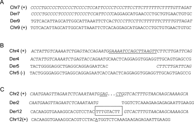
Balanced chromosome rearrangements (BCRs) can cause genetic diseases by disrupting or inactivating specific genes, and the characterization of breakpoints in disease-associated BCRs has been instrumental in the molecular elucidation of a wide variety of genetic disorders. However, mapping chromosome breakpoints using traditional methods, such as in situ hybridization with fluorescent dye-labeled bacterial artificial chromosome clones (BAC-FISH), is rather laborious and time-consuming. In addition, the resolution of BAC-FISH is often insufficient to unequivocally identify the disrupted gene. To overcome these limitations, we have performed shotgun sequencing of flow-sorted derivative chromosomes using "next-generation" (Illumina/Solexa) multiplex sequencing-by-synthesis technology. As shown here for three different disease-associated BCRs, the coverage attained by this platform is sufficient to bridge the breakpoints by PCR amplification, and this procedure allows the determination of their exact nucleotide positions within a few weeks. Its implementation will greatly facilitate large-scale breakpoint mapping and gene finding in patients with disease-associated balanced translocations.
Figures


References
-
- Abelson J.F., Kwan K.Y., O’Roak B.J., Baek D.Y., Stillman A.A., Morgan T.M., Mathews C.A., Pauls D.L., Rasin M.R., Gunel M., et al. Sequence variants in SLITRK1 are associated with Tourette’s syndrome. Science. 2005;310:317–320. - PubMed
-
- Alkuraya F.S., Saadi I., Lund J.J., Turbe-Doan A., Morton C.C., Maas R.L. SUMO1 haploinsufficiency leads to cleft lip and palate. Science. 2006;313:1751. - PubMed
-
- Arkesteijn G., Jumelet E., Hagenbeek A., Smit E., Slater R., Martens A. Reverse chromosome painting for the identification of marker chromosomes and complex translocations in leukemia. Cytometry. 1999;35:117–124. - PubMed
-
- Bache I., Hjorth M., Bugge M., Holstebroe S., Hilden J., Schmidt L., Brondum-Nielsen K., Bruun-Petersen G., Jensen P.K., Lundsteen C., et al. Systematic re-examination of carriers of balanced reciprocal translocations: A strategy to search for candidate regions for common and complex diseases. Eur. J. Hum. Genet. 2006;14:410–417. - PubMed
-
- Backx L., Van Esch H., Melotte C., Kosyakova N., Starke H., Frijns J.P., Liehr T., Vermeesch J.R. Array painting using microdissected chromosomes to map chromosomal breakpoints. Cytogenet. Genome Res. 2007;116:158–166. - PubMed
Publication types
MeSH terms
Associated data
- Actions
LinkOut - more resources
Full Text Sources
Other Literature Sources
Molecular Biology Databases