Rapid repair of UVA-induced oxidized purines and persistence of UVB-induced dipyrimidine lesions determine the mutagenicity of sunlight in mouse cells
- PMID: 18326785
- PMCID: PMC2714223
- DOI: 10.1096/fj.07-105437
Rapid repair of UVA-induced oxidized purines and persistence of UVB-induced dipyrimidine lesions determine the mutagenicity of sunlight in mouse cells
Abstract
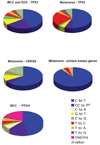
Despite the predominance of ultraviolet A (UVA) relative to UVB in terrestrial sunlight, solar mutagenesis in humans and rodents is characterized by mutations specific for UVB. We have investigated the kinetics of repair of UVA- and UVB-induced DNA lesions in relation to mutagenicity in transgenic mouse fibroblasts irradiated with equilethal doses of UVA and UVB in comparison to simulated-sunlight UV (SSL). We have also analyzed mutagenesis-derived carcinogenesis in sunlight-associated human skin cancers by compiling the published data on mutation types found in crucial genes in nonmelanoma and melanoma skin cancers. Here, we demonstrate a resistance to repair of UVB-induced cis-syn cyclobutane pyrimidine-dimers (CPDs) together with rapid removal of UVA-induced oxidized purines in the genome overall and in the cII transgene of SSL-irradiated cells. The spectra of mutation induced by both UVB and SSL irradiation in this experimental system are characterized by significant increases in relative frequency of C-->T transitions at dipyrimidines, which are the established signature mutation of CPDs. This type of mutation is also the predominant mutation found in human nonmelanoma and melanoma tumor samples in the TP53, CDKN2, PTCH, and protein kinase genes. The prevailing role of UVB over UVA in solar mutagenesis in our test system can be ascribed to different kinetics of repair for lesions induced by the respective UV irradiation.
Figures


Similar articles
-
Wavelength dependence of ultraviolet radiation-induced DNA damage as determined by laser irradiation suggests that cyclobutane pyrimidine dimers are the principal DNA lesions produced by terrestrial sunlight.FASEB J. 2011 Sep;25(9):3079-91. doi: 10.1096/fj.11-187336. Epub 2011 May 25. FASEB J. 2011. PMID: 21613571 Free PMC article.
-
Bipyrimidine photoproducts rather than oxidative lesions are the main type of DNA damage involved in the genotoxic effect of solar UVA radiation.Biochemistry. 2003 Aug 5;42(30):9221-6. doi: 10.1021/bi034593c. Biochemistry. 2003. PMID: 12885257
-
[Ultraviolet A-induced DNA damage: role in skin cancer].Bull Acad Natl Med. 2014 Feb;198(2):273-95. Bull Acad Natl Med. 2014. PMID: 26263704 French.
-
Mutations induced by ultraviolet light.Mutat Res. 2005 Apr 1;571(1-2):19-31. doi: 10.1016/j.mrfmmm.2004.06.057. Epub 2005 Jan 20. Mutat Res. 2005. PMID: 15748635 Review.
-
Mechanistic considerations on the wavelength-dependent variations of UVR genotoxicity and mutagenesis in skin: the discrimination of UVA-signature from UV-signature mutation.Photochem Photobiol Sci. 2018 Dec 5;17(12):1861-1871. doi: 10.1039/c7pp00360a. Photochem Photobiol Sci. 2018. PMID: 29850669 Review.
Cited by
-
Hydroxychloroquine induces oxidative DNA damage and mutation in mammalian cells.DNA Repair (Amst). 2021 Oct;106:103180. doi: 10.1016/j.dnarep.2021.103180. Epub 2021 Jul 16. DNA Repair (Amst). 2021. PMID: 34298488 Free PMC article.
-
Measuring the formation and repair of UV damage at the DNA sequence level by ligation-mediated PCR.Methods Mol Biol. 2012;920:189-202. doi: 10.1007/978-1-61779-998-3_14. Methods Mol Biol. 2012. PMID: 22941605 Free PMC article.
-
Mutation Analysis in Cultured Cells of Transgenic Rodents.Int J Mol Sci. 2018 Jan 16;19(1):262. doi: 10.3390/ijms19010262. Int J Mol Sci. 2018. PMID: 29337872 Free PMC article.
-
The interplay of DNA damage and repair, gene expression, and mutagenesis in mammalian cells during oxidative stress.Carcinogenesis. 2024 Nov 22;45(11):868-879. doi: 10.1093/carcin/bgae046. Carcinogenesis. 2024. PMID: 39023127 Free PMC article.
-
Wavelength dependence of ultraviolet radiation-induced DNA damage as determined by laser irradiation suggests that cyclobutane pyrimidine dimers are the principal DNA lesions produced by terrestrial sunlight.FASEB J. 2011 Sep;25(9):3079-91. doi: 10.1096/fj.11-187336. Epub 2011 May 25. FASEB J. 2011. PMID: 21613571 Free PMC article.
References
-
- de Gruijl FR. Photocarcinogenesis: UVA vs. UVB radiation. Skin Pharmacol Appl Skin Physiol. 2002;15:316–320. - PubMed
-
- Woodhead AD, Setlow RB, Tanaka M. Environmental factors in nonmelanoma and melanoma skin cancer. J Epidemiol. 1999;9:S102–S114. - PubMed
-
- Kelfkens G, de Gruijl FR, van der Leun JC. Ozone depletion and increase in annual carcinogenic ultraviolet dose. Photochem Photobiol. 1990;52:819–823. - PubMed
-
- Pfeifer GP, You YH, Besaratinia A. Mutations induced by ultraviolet light. Mutat Res. 2005;571:19–31. - PubMed
Publication types
MeSH terms
Substances
Grants and funding
LinkOut - more resources
Full Text Sources
Other Literature Sources
Research Materials
Miscellaneous

