. 2008 Feb 20;3(2):e1641.
doi: 10.1371/journal.pone.0001641.

Endogenous morphine in SH-SY5Y cells and the mouse cerebellum

Affiliations

Endogenous morphine in SH-SY5Y cells and the mouse cerebellum

Arnaud Muller et al. PLoS One. .

Abstract

Background: Morphine, the principal active agent in opium, is not restricted to plants, but is also present in different animal tissues and cell types, including the mammalian brain. In fact, its biosynthetic pathway has been elucidated in a human neural cell line. These data suggest a role for morphine in brain physiology (e.g., neurotransmission), but this hypothesis remains a matter of debate. Recently, using the adrenal neuroendocrine chromaffin cell model, we have shown the presence of morphine-6-glucuronide (M6G) in secretory granules and their secretion products, leading us to propose that these endogenous alkaloids might represent new neuroendocrine factors. Here, we investigate the potential function of endogenous alkaloids in the central nervous system.

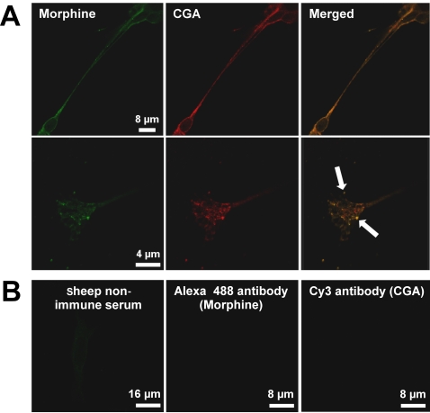
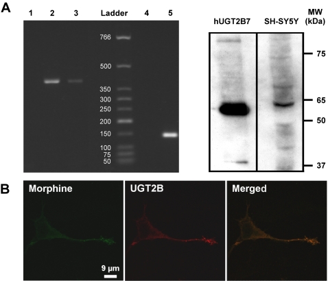
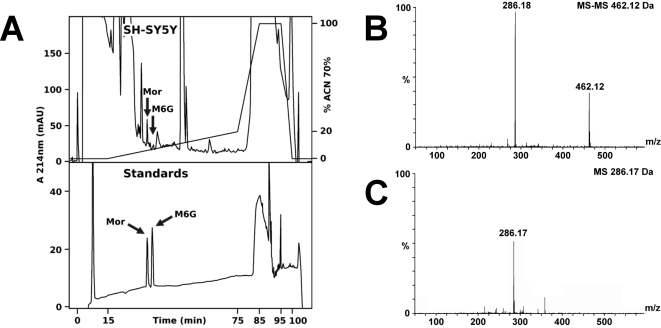
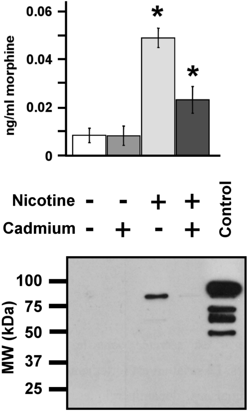
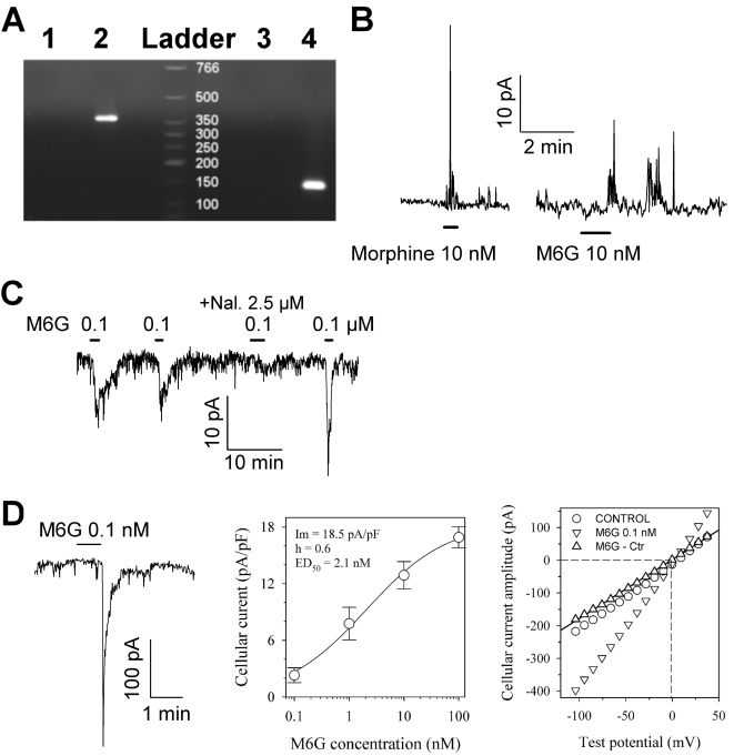
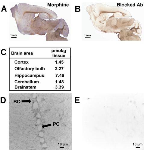
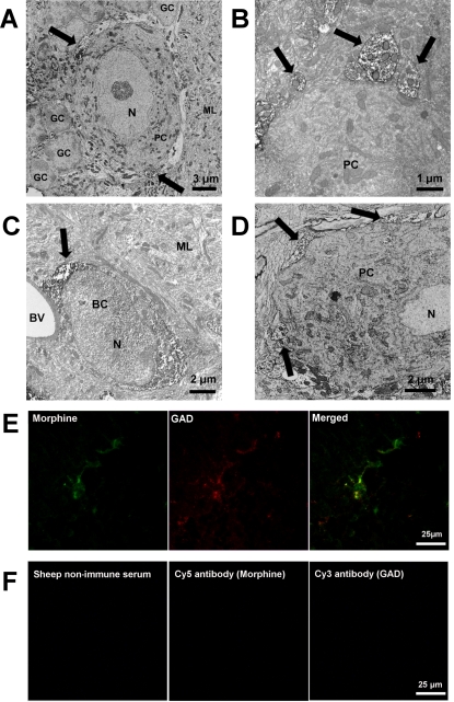
Methodology and principal findings: Microscopy, molecular biology, electrophysiology, and proteomic tools were applied to human neuroblastoma SH-SY5Y cells (i) to characterize morphine and M6G, and (ii) to demonstrate the presence of the UDP-glucuronyltransferase 2B7 enzyme, which is responsible for the formation of M6G from morphine. We show that morphine is secreted in response to nicotine stimulation via a Ca(2+)-dependent mechanism involving specific storage and release mechanisms. We also show that morphine and M6G at concentrations as low as 10(-10) M are able to evoke specific naloxone-reversible membrane currents, indicating possible autocrine/paracrine regulation in SH-SY5Y cells. Microscopy and proteomic approaches were employed to detect and quantify endogenous morphine in the mouse brain. Morphine is present in the hippocampus, cortex, olfactory bulb, and cerebellum at concentration ranging from 1.45 to 7.5 pmol/g. In the cerebellum, morphine immunoreactivity is localized to GABA basket cells and their termini, which form close contacts on Purkinje cell bodies.

Conclusions/significance: The presence of morphine in the brain and its localization in particular areas lead us to conclude that it has a specific function in neuromodulation and/or neurotransmission. Furthermore, its presence in cerebellar basket cell termini suggests that morphine has signaling functions in Purkinje cells that remain to be discovered.


Conflict of interest statement

Competing Interests: The authors have declared that no competing interests exist.

Figures

Figure 1
Figure 1. Evidence of the presence of morphine-like immunoreactivity in secretory granules.
A. Upper panel, double immunofluorescence confocal micrographs. Labeling was performed with a sheep anti-morphine antibody (visualized in green with an Alexa Fluor 488-conjugated IgG) and with an antibody against CGA (a specific intragranular marker) visualized in red with a mouse Cy3-conjugated IgG. Colocalized immunolabelling (merged window) appears as yellow staining. Lower panel, SH-SY5Y termini shown at higher magnification. Arrows indicate colocalization points. An identical pattern of labelling was obtained for a different mouse monoclonal anti-morphine antibody (data not shown). B. To assess the specificity of morphine immunolabelling, control experiments were performed using either sheep non-immune serum and an Alexa Fluor 488-conjugated IgG, or Alexa Fluor 488-conjugated IgG and Cy3-conjugated IgG without a primary antibody.
Figure 2
Figure 2. Evidence for UGT2B7 in SH-SY5Y cells.
A. Amplification of UGT2B7 RNA and Western blot analysis. Left panel, total SH-SY5Y RNA was submitted to RT-PCR using specific human UGT2B7 primers. Lane 1, control using water to test for contamination. Lane 2, total RNA from human hepatocyte extract was used as a positive control, showing a single band of 462 bp corresponding to the expected UGT2B7 PCR product. Lane 3, SH-SY5Y total RNA showing a single band of 462 bp. Lane 4, negative control of GAPDH amplification (water). Lane 5, SH-SY5Y total RNA (GAPDH, 142 bp). The size of standards are indicated in bp. Right panel, Western blot analysis of SH-SY5Y cell extracts was done using 1 µg of human recombinant UGT2B7 (positive control) and 50 µg of SH-SY5Y cell extract. Western blots using anti-UGT2B antibody show a band at 65 kDa . B. Localization of UGT2B immunoreactivity in SH-SY5Y cells. Double immunofluorescence confocal micrographs were obtained using an anti-UGT2B antibody (detected in red with an Alexa Fluor 568-conjugated IgG) and with an antibody against morphine visualized in green with an Alexa Fluor 488-conjugated IgG. Colocalized immunolabelling appears as yellow staining (merged window).
Figure 3
Figure 3. Characterization of morphine and M6G in SH-SY5Y extracts.
A. Purification of morphine and M6G. Upper panel, RP-HPLC chromatogram showing the purification of endogenous alkaloids from SH-SY5Y cells (375×106 cells). Lower panel, RP-HPLC purification of morphine (Mor) and M6G standards (500 pmol). B. Characterization of M6G. Q-TOF MS-MS analysis of the HPLC fraction showing M6G in the intragranular material (marked with an arrow in Fig. 3A, upper panel). The fragment at 462.12 Da corresponds to M6G, whereas the 286.18 Da fragment corresponds to morphine. C. Q-TOF MS analysis of the HPLC fraction showing morphine (286.17 Da) in the intragranular material (marked with an arrow in Fig. 3A, upper panel).
Figure 4
Figure 4. Characterization of morphine secretion from SH-SY5Y cells.
Upper panel. The amount of morphine secreted into the culture medium was determined after stimulating 4×10⁵ cells with 100 µM of nicotine with or without cadmium (200 µM) for 24 h. The basal secretion level was obtained from cells incubated without nicotine at the same time (n = 6). Amounts of secreted morphine in the nicotine and nicotine+cadmium groups were statistically different and were both different from the two control groups (no nicotine and no nicotine+cadmium; Mann-Whitney test, * p<0.01). Lower panel. The efficiency of secretion was assessed by monitoring the secretion of chromogranin B (CGB), an intra-LDC vesicle protein , by Western blot analysis. A positive control, intragranular protein matrix from bovine chromaffin cells (10 µg), was loaded in order to evaluate the molecular weight of the entire CGB (80 kDa).
Figure 5
Figure 5. Amplification of MOR1 RNA and characterization of the electrophysiological effects of a low concentration of morphine and M6G.
A. Total SH-SY5Y RNA was submitted to RT-PCR using specific human MOR1 primers. Lane 1, control using water as a PCR template to test for contamination. Lane 2, total RNA from SH-SY5Y, showing a single band of 376 bp. Lane 3, negative control of GAPDH amplification (water). Lane 4, SH-SY5Y total RNA (GAPDH, 142 bp). Size standards are indicated in bp. B and C. Typical responses of SH-SY5Y cells to low concentrations of morphine and M6G antagonized by naloxone, as measured by the patch clamp technique in cell-attached mode at a pipette potential of 60 mV and 80 mV (B panel), as well as at −135 mV (C panel). M6G was repeatedly applied in the absence or presence of naloxone (Nal.). D. Whole-cell patch clamp recording of the M6G response: the left-hand panel shows a typical trace recorded at a holding potential of −135 mV, the middle panel shows a dose-response curve obtained at a holding potential of −80 mV, and the right-hand panel shows an I-V plot of the peak amplitude response. The dose-response data were fitted with Hill's equation (continuous line) with optimal parameters as indicated in the Figure. The I-V plot of the steady state current for the control (circles) and at the peak of the response to 0.1 nM M6G (inverted triangles) was obtained using a ramp potential protocol from 80 to −140 mV lasting 800 ms. The specific current elicited in the presence of M6G (upright triangles) was linear, and the reversal potential calculated by linear regression gave a value of −0.5 mV (dashed line). Traces were filtered at 2 kHz and digitized at 5 kH. Bars indicate the period of drug application at the indicated concentrations.
Figure 6
Figure 6. Mapping of endogenous morphine in the mouse brain.
A. Immunodetection of morphine present in the mouse brain. Sagittal slices were incubated with a mouse monoclonal anti-morphine antibody and visualized with an HRP-conjugated donkey anti-mouse IgG, in order to detect endogenous morphine in specific brain areas. B. Control experiment using morphine immunoadsorbed mouse monoclonal antibody (same incubation time as in A). C. Quantification of the morphine present in different mouse brain areas using morphine-specific ELISA. The table shows the quantities of endogenous morphine (pmoles) present per gram of wet tissue. The values correspond to an average of the morphine amount determined for 5 brains (n = 5). D. Localization of morphine label in the cerebellum using mouse monoclonal anti-morphine antibody and an HRP-conjugated secondary antibody. Morphine labelling was observed around Purkinje cells and in basket cells. PC, Purkinje cell; BC, basket cell. E. Control for immunolabelling using morphine-immunoadsorbed mouse monoclonal antibody.
Figure 7
Figure 7. Characterization of morphine immunoreactivity in the mouse cerebellum.
A. Characterization of morphine immunolabelling around Purkinje cells. Electron microscopy using sheep anti-morphine antibody showed morphine-like immunoreactivity in nerve termini innervating Purkinje cell bodies (PC). Arrows indicate strong immunolabelling of morphine-containing termini around the Purkinje cell body. GC, granular cell. ML, molecular layer, N, nucleus. B. Higher magnification showing the presence of morphine immunoreactivity in nerve termini innervating Purkinje cell bodies. C. Electron microscopy showing morphine immunoreactivity in basket cells (BC). BV, blood vessel. ML, molecular layer. Control experiments using anti-sheep HRP-conjugated IgG showed the specificity of the immunolabelling. D. Electron microscopy showing PEBP-immunoreactivity in nerve termini covering Purkinje cell bodies (PC). Arrows indicate strong immunolabelling of PEBP-containing basket cell termini. E. Evidence of colocalization of morphine and glutamic acid decarboxylase (GAD). Double immunofluorescence confocal micrographs were obtained using a sheep anti-morphine antibody (detected with a CY5-conjugated IgG, green pseudocolor label) and an antibody against GAD visualized in red with a Cy3-conjugated IgG. Colocalized immunolabelling (arrows and merged window) appears as yellow staining. F. Control experiments were performed using sheep non-immune serum, or only secondary antibodies detected with Cy5- and Cy3-conjugated IgG (primary antibody omitted), to demonstrate the specificity of the immunolabelling.

Similar articles

Cited by

References

    1. Stefano GB, Goumon Y, Casares F, Cadet P, Fricchione GL, et al. Endogenous morphine. Trends Neurosci. 2000;23:436–442. - PubMed
    1. Hazum E, Sabatka JJ, Chang KJ, Brent DA, Findlay JW, et al. Morphine in cow and human milk: could dietary morphine constitute a ligand for specific morphine (mu) receptors? Science. 1981;213:1010–1012. - PubMed
    1. Gintzler AR, Gershon MD, Spector S. A nonpeptide morphine-like compound: immunocytochemical localization in the mouse brain. Science. 1978;199:447–448. - PubMed
    1. Goldstein A, Barrett RW, James IF, Lowney LI, Weitz CJ, et al. Morphine and other opiates from beef brain and adrenal. Proc Natl Acad Sci U S A. 1985;82:5203–5207. - PMC - PubMed
    1. Herbert RB, Venter H, Pos S. Do mammals make their own morphine? Nat Prod Rep. 2000;17:317–322. - PubMed

Publication types