. 2008 Jun;228(1):61-78.
doi: 10.1007/s00425-008-0719-z. Epub 2008 Mar 8.

Pepper pectin methylesterase inhibitor protein CaPMEI1 is required for antifungal activity, basal disease resistance and abiotic stress tolerance

Affiliations

Pepper pectin methylesterase inhibitor protein CaPMEI1 is required for antifungal activity, basal disease resistance and abiotic stress tolerance

Soo Hyun An et al. Planta. 2008 Jun.

Abstract

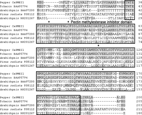
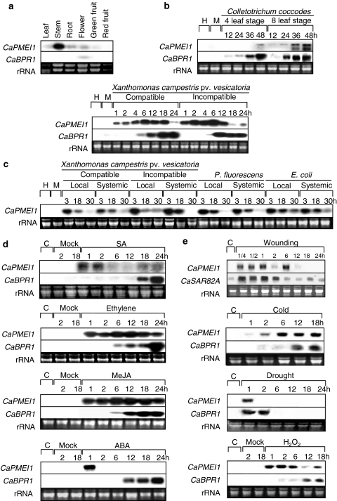
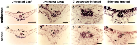
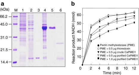
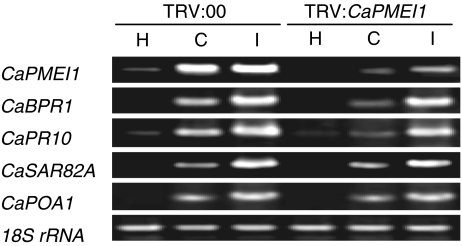
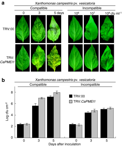
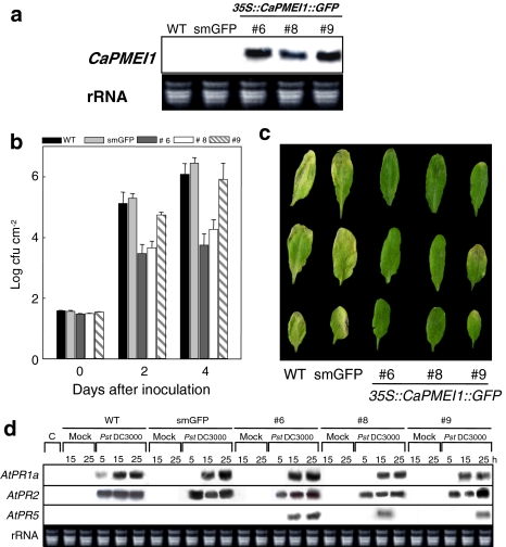
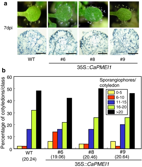
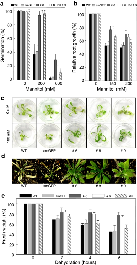
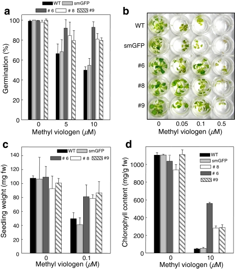
Pectin is one of the main components of the plant cell wall that functions as the primary barrier against pathogens. Among the extracellular pectinolytic enzymes, pectin methylesterase (PME) demethylesterifies pectin, which is secreted into the cell wall in a highly methylesterified form. Here, we isolated and functionally characterized the pepper (Capsicum annuum L.) gene CaPMEI1, which encodes a pectin methylesterase inhibitor protein (PMEI), in pepper leaves infected by Xanthomonas campestris pv. vesicatoria (Xcv). CaPMEI1 transcripts are localized in the xylem of vascular bundles in leaf tissues, and pathogens and abiotic stresses can induce differential expression of this gene. Purified recombinant CaPMEI1 protein not only inhibits PME, but also exhibits antifungal activity against some plant pathogenic fungi. Virus-induced gene silencing of CaPMEI1 in pepper confers enhanced susceptibility to Xcv, accompanied by suppressed expression of some defense-related genes. Transgenic Arabidopsis CaPMEI1-overexpression lines exhibit enhanced resistance to Pseudomonas syringae pv. tomato, mannitol and methyl viologen, but not to the biotrophic pathogen Hyaloperonospora parasitica. Together, these results suggest that CaPMEI1, an antifungal protein, may be involved in basal disease resistance, as well as in drought and oxidative stress tolerance in plants.


Figures

Fig. 1
Fig. 1
Amino acid sequence alignments of pepper CaPMEI1 with Nicotiana tabacum DC1.2 protein (accession no. BAA95794), A. thaliana ripening-related protein (accession no. BAA97200), A. thaliana invertase homolog (accession no. NP201267) and Pinus radiata pectinesterase homolog (accession no. T08112). The boxed amino acid sequences represent the pectin methylesterase inhibitor (PMEI) domain. The conserved cysteine residues are marked by asterisks and the shaded regions represent conserved amino acid residues
Fig. 2
Fig. 2
RNA gel blot analysis of expression of CaPMEI1, CaSAR82A and CaBPR1 in pepper plants. Membranes were hybridized with probes from the 3′ UTR region of pepper CaPMEI1 cDNA or full-length CaBPR1 cDNA. Equal loading (20 μg) was verified by visualizing RNA on a gel stained with ethidium bromide. H healthy, M mock-inoculated or mock-treated. aCaPMEI1 expression in various organs of pepper plants. b Expression of CaPMEI1 and CaBPR1 in pepper leaves at various time intervals after inoculation with C. coccodes or virulent strain Ds1 (compatible interactions with pepper: susceptible response) and avirulent strain Bv5-4a (incompatible interactions with pepper: resistant response) of Xcv.cCaPMEI1 expression in lower (local) infected and upper (systemic) uninfected leaves at various time intervals after inoculation with the virulent and avirulent strains Ds1 and Bv5-4a of Xcv, P.fluorescence ATCC13525 and E.coli JM109. The lower leaves of pepper plants were inoculated at the 6-leaf stage. For the mock-inoculation, the lower leaves were infiltrated with 10 mM MgSO4. d Expression of CaPMEI1 and CABPR1 in pepper leaves at various time intervals after treatment with salicylic acid (SA, 5 mM), methyl jasmonate (MeJA, 100 μM), ethylene (5 μl L−1) and abscisic acid (100 μM). e Expression of CaPMEI1 and CaSAR82A in pepper leaves at various time intervals after treatment with drought, wounding, cold and H2O2
Fig. 3
Fig. 3
In situ localization of CaPMEI1 transcripts in pepper leaf and stem tissues. Cross sections of leaf tissues were hybridized with CaPMEI1 antisense (a, c, e, g) and sense (b, d, f, h) DIG-labeled RNA probes, and then photographed under bright-field conditions. The transcript signal is purple. a, b Untreated leaf tissues. c, d Untreated stems. e, f Leaf tissues at 24 h after inoculation with C. coccodes. g, h Leaf tissues treated with 5 μl L−1 ethylene. P phloem, X xylem, UE upper epidermis, LE lower epidermis, Vs vascular bundle, C cortical cell
Fig. 4
Fig. 4
Inhibition of pectin methylesterase (PME) activity by the pepper pectin methylesterase inhibitor (CaPMEI). a Recombinant CaPMEI1 expression in E. coli BL21 (DE3) pLysS. Cells were grown in LB media, and recombinant protein expression was induced with 1 mM IPTG. M, molecular marker (kDa); Lane 1, Uninduced E. coli BL21 cell extracts; Lane 2, Soluble fraction of E. coli BL21 cell extracts encoding thioredoxin protein after IPTG induction; Lane 3, Purified thioredoxin; Lane 4, Crude protein extracts of E. coli cells producing the thioredoxin–CaPMEI1 fusion protein after IPTG induction; Lane 5, Purified thioredoxin–CaPMEI1 fusion protein; Lane 6, Cleaved CaPMEI1 and thioredoxin proteins following enterokinase digestion. Protein staining was performed using Coomassie brilliant blue. b Inhibition of PME activity following treatment with CaPMEI1. For the inhibition assay, crude CaPMEI1 (0.5 μg) was mixed with 7.8 mU of orange peel PME in a total volume of 11 μL, and then preincubated at 25°C for 15 min, followed by addition to the reaction solution
Fig. 5
Fig. 5
Assay of thioredoxin–CaPMEI1 fusion protein antimicrobial activity. a Inhibitory effects of the CaPMEI1–thioredoxin fusion protein on mycelial growth of the plant pathogenic fungi F. oxysporum f.sp. matthiolae, A. brassicicola and B. cinerea. In each plate, the upper wells (a) were treated with purified thioredoxin and the lower wells (b) were treated with the purified thioredoxin-CaPMEI1 fusion protein. b Inhibition of germination and hyphal growth of F.oxysporum f.sp. matthiolae. Fungal spores were allowed to germinate and grow in 100 μL potato dextrose broth medium alone (top), or with 500 μg mL−1 thioredoxin (middle) or 500 μg mL−1 thioredoxin–CaPMEI1 fusion protein (bottom). Photographs were taken after incubation for 10 h at 28°C. Bars 20 μm. c Inhibition of spore germination and hyphal growth of F.oxysporum f.sp. matthiolae by CaPMEI1. The percentage of germinated spores and the length of fungal hyphae were determined by light microscopy. Data represent means ± SD from three independent experiments
Fig. 6
Fig. 6
RT-PCR analysis of expression of CaPMEI1 and several defense-related genes in empty vector control (TRV:00) and CaPMEI1 gene-silenced (TRV:CaPMEI1) pepper plants 12 h after inoculation with the virulent (Ds1; C, compatible) and avirulent (Bv5-4a; I, incompatible) strains of Xcv (5 × 106 cfu mL−1). 18S rRNA levels were visualized as a loading control. This experiment was repeated three times with similar results. H healthy leaves, CaBPR1 basic pathogenesis-related protein 1, CaPR10 putative ribonuclease-like protein, CaPOA1 ascorbate peroxidase 1, and CaSAR82A SAR8.2
Fig. 7
Fig. 7
Enhanced disease susceptibility of CaPMEI1-silenced pepper plants to infection by the virulent Xcv strain Ds1, but not the avirulent Xcv strain Bv5-4a. a Disease symptoms developed on the leaves at different time points after inoculation with the virulent Xcv strain Ds1 (5 × 106 cfu mL−1) and the avirulent Xcv strain Bv5-4a (various bacterial concentrations). b Bacterial growth in leaves of empty vector control (TRV:00) or CaPMEI1-silenced (TRV:CaPMEI1) pepper plants at different time points after inoculation with the virulent Xcv strain Ds1 or the avirulent Xcv strain Bv5-4a (104 cfu mL−1). Data represent the mean ± SD from three independent experiments
Fig. 8
Fig. 8
Responses of wild-type (Col-0) Arabidopsis and CaPMEI1-OX transgenic plants to infection with P. syringae pv. tomato DC3000. a RNA gel blot analysis confirming CaPMEI1 overexpression (OX) in the transgenic Arabidopsis lines. Total RNA (10 μg) was loaded into each lane. The 3′ UTR region of pepper CaPMEI1 cDNA was used as a probe. b Growth of Pst DC3000 in the leaves of wild-type and transgenic plants. The mature leaves of the 6-week-old plants were infiltrated with a Pst Dc3000 suspension (105 cfu mL−1), and the degree of bacterial growth was rated at 0, 2 and 4 days after inoculation. c Disease symptoms on leaves of 6-week-old plants infiltrated with virulent Pst DC3000 (105 cfu mL−1). d Expression of pathogen-related (PR) genes in transgenic plants. Northern blot analyses were performed with 10 μg total RNA prepared from 5-week-old leaves of the wild-type (WT), vector control (smGFP) and transgenic (CaPMEI1::smGFP) plants. The samples were collected at 5, 15 and 25 h following pathogen infiltration with a suspension of the virulent strain Pst DC3000 (105 cfu mL−1)
Fig. 9
Fig. 9
Responses of wild-type (Col-0) Arabidopsis and CaPMEI1-OX transgenic plants to infection with H. parasitica isolate Noco2. a Disease symptoms and trypan blue-stained pathogen structures on 7-day-old cotyledons of wild-type and transgenic plants 7 days after inoculation; dpi days post-inoculation. Bars 0.5 mm. b Quantification of asexual sporangiophores per cotyledon for at least 50 cotyledons of wild-type and transgenic plants 7 days after inoculation. The average number of sporangiophores produced on the cotyledons of wild-type and transgenic lines are shown below each of the lines tested
Fig. 10
Fig. 10
Transgenic ArabidopsisCaPMEI1-OX lines exhibit enhanced tolerance to drought stress. a Seed germination in wild-type, smGFP and transgenic plants on the MS media containing 0, 200 and 600 mM mannitol. The data represent the mean ± SD of 100 seeds for each line tested. b Relative root length of wild-type, smGFP and transgenic lines in MS agar medium containing different concentrations of mannitol. Three independent experiments were performed with 40 seedlings of both wild-type and transgenic lines. c Drought tolerance test of transgenic seedlings. Wild-type, smGFP and transgenic lines were germinated and grown in 1× MS agar medium. Each seedling was transferred to liquid medium containing 100 mM mannitol. d Wild-type, smGFP and the CaPMEI1 transgenic Arabidopsis plants after 15 days without water. e Water loss from the excised leaves of wild-type, smGFP and transgenic plants. Data represent the mean ± SD from three independent experiments
Fig. 11
Fig. 11
Transgenic ArabidopsisCaPMEI1-OX lines exhibit tolerance to oxidative stress. a Effects of methyl viologen on the seed germination of transgenic lines. Seeds from wild-type, vector control and transgenic lines were plated on media with or without methyl viologen (MV, 5 and 10 μM) and incubated for 3 days. The data represent mean ± SD of 100 seeds for each line tested. b Phenotypes of wild-type, vector control and transgenic line seedlings treated with different concentrations of MV. c Fresh weights of seedlings grown in the indicated concentrations of MV for 2 weeks. The results are presented as the average fresh weight per seedling. Data represent mean ± SD from three independent experiments. d Chlorophyll content of MV-treated leaves of wild-type, vector control and transgenic plants, which were floated on 0, 0.05, 0.1 and 0.5 μM MV in MS medium and then incubated for 24 h in a growth chamber

References

    1. {'text': '', 'ref_index': 1, 'ids': [{'type': 'DOI', 'value': '10.1105/tpc.8.10.1683', 'is_inner': False, 'url': 'https://doi.org/10.1105/tpc.8.10.1683'}, {'type': 'PMC', 'value': 'PMC161307', 'is_inner': False, 'url': 'https://pmc.ncbi.nlm.nih.gov/articles/PMC161307/'}, {'type': 'PubMed', 'value': '12239358', 'is_inner': True, 'url': 'https://pubmed.ncbi.nlm.nih.gov/12239358/'}]}
    2. Alfano JR, Collmer A (1996) Bacterial pathogens in plants: life up against the wall. Plant Cell 8:1683–1698 - PMC - PubMed
    1. {'text': '', 'ref_index': 1, 'ids': [{'type': 'DOI', 'value': '10.1093/nar/25.17.3389', 'is_inner': False, 'url': 'https://doi.org/10.1093/nar/25.17.3389'}, {'type': 'PMC', 'value': 'PMC146917', 'is_inner': False, 'url': 'https://pmc.ncbi.nlm.nih.gov/articles/PMC146917/'}, {'type': 'PubMed', 'value': '9254694', 'is_inner': True, 'url': 'https://pubmed.ncbi.nlm.nih.gov/9254694/'}]}
    2. Altschul SF, Madden TL, Schaffer AA, Zhang J, Zhang Z, Miller W, Lipman DJ (1997) Gapped BLAST and PSI-BLAST: a new generation of protein database search programs. Nucleic Acids Res 25:3389–3402 - PMC - PubMed
    1. {'text': '', 'ref_index': 1, 'ids': [{'type': 'DOI', 'value': '10.1146/annurev.arplant.55.031903.141701', 'is_inner': False, 'url': 'https://doi.org/10.1146/annurev.arplant.55.031903.141701'}, {'type': 'PubMed', 'value': '15377225', 'is_inner': True, 'url': 'https://pubmed.ncbi.nlm.nih.gov/15377225/'}]}
    2. Apel K, Hirt H (2004) Reactive oxygen species: metabolism, oxidative stress, and signal transduction. Annu Rev Plant Biol 55:373–399 - PubMed
    1. None
    2. Asada K (1996) Radical production and scavenging in the chloroplasts. In: Baker NR (ed) Photosynthesis and the environment. Kluwer, Netherlands, pp. 123–150
    1. None
    2. Asoufi H, Hameed KM, Mahasneh A (2007) The cellulase and pectinase activities associated with the virulence of indigenous Sclerotinia sclerotiorum isolates in Jordan Valley. Plant Pathol J 23:233–238

Publication types

MeSH terms