Clinical Trial
. 2008 Aug;26(8):1046-52.
doi: 10.1002/jor.20550.

Quantifying tibial plafond fracture severity: absorbed energy and fragment displacement agree with clinical rank ordering

Affiliations
Clinical Trial

Quantifying tibial plafond fracture severity: absorbed energy and fragment displacement agree with clinical rank ordering

Donald D Anderson et al. J Orthop Res. 2008 Aug.

Abstract

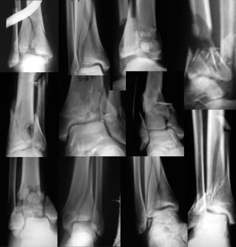
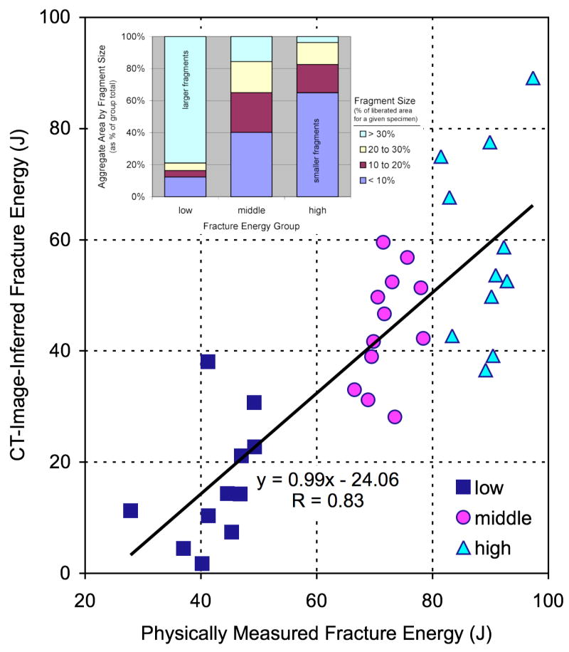
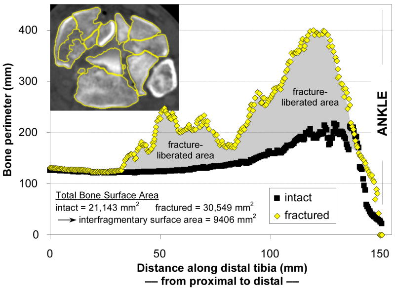
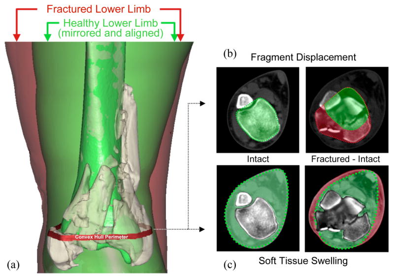
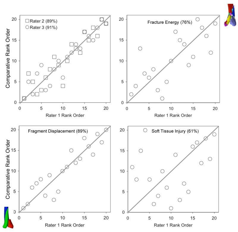
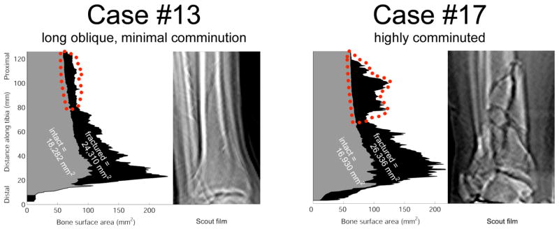
The energy of producing a fracture is considered one of the most important factors relating to injury severity, prognosis, and risk of complications, yet there has been no objective way to measure this important variable. The purpose of this study was to compare assessments of fracture severity made using new objective computed tomography (CT)-based methods versus the expert opinion of experienced orthopedic traumatologists. Fracture energy, fragment displacement, and soft tissue swelling were quantified in twenty tibial plafond fractures by analyzing injury CT scans. Three experienced orthopedic traumatologists then independently performed a rank order analysis of fracture severity, using plain radiographs. The concordance among the three clinicians ranged from 87% to 91%. Objective fracture severity measurements made with the CT algorithm agreed well with the clinical assessments (fracture energy concordance from 73% to 76%, fragment displacement from 82% to 89%, and soft tissue swelling from 61% to 65%). These are the first objective, CT-based measures of fracture severity. With further refinement, this conceptually novel method has the potential to serve as a valuable tool to provide objective measurement of fracture severity, allowing one to control for this previously confounding variable in large multicenter studies.


Figures

Figure 1
Figure 1
A montage of radiographs from comminuted tibial plafond fractures demonstrates the range of variation in both the number of fragments and in the degree of intra-articular extension.
Figure 2
Figure 2
Controlled fracture experiments performed in bovine bone showed a highly linear relationship (R=0.83) between CT-inferred fracture energy and the physically measured energy absorbed in fracturing the cortical bone specimens. Inset: The data also showed that fragment size distribution correlated with fracture energy, shifting from a relatively high proportion of large fragments at lower energies to a higher proportion of smaller fragments at higher energies.
Figure 3
Figure 3
Fragment surface area is calculated by identifying bone perimeters at each CT slice. Bone perimeters (matched intact and fractured) for each slice, plotted along the length of the distal tibia, help to clarify how the fracture energy measure is calculated. The interfragmentary surface area is calculated by summing the areas (for each slice, the product of perimeter and slice thickness) over the length of the fractured tibia, and then subtracting the areas of the intact contralateral tibia summed over the same length. Inset: CT slice from fracture case, with identified tibia bone edges highlighted.
Figure 4
Figure 4
Assessment of fragment displacement and soft tissue injury began with (a) alignment of the proximal base of the fractured tibia with a mirrored image of its healthy contralateral limb. (b) In each CT slice, a convex hull (the smallest convex polygon circumscribing an object) of both the mirrored intact, and of the composite aligned intact and fractured tibias was generated. The difference in circumscribed volumes between these convex hulls was used to quantify fragment dispersion and axial mal-alignment in a single aggregate displacement metric. (c) Fracture-associated soft tissue swelling was quantified by calculating the mathematical difference in volume of non-osseous regions in the fractured and intact limbs.
Figure 5
Figure 5
Agreement between injury severity rankings and CT-based metrics. Raters were blinded as to the CT-based data and to patient age. The graphs compare the rank ordering of rater 1 versus that of raters 2 and 3, and of the individual CT-based metrics. Concordance values are enclosed in parentheses following the rater/metric.
Figure 6
Figure 6
A plot of bone surface areas as they vary over the length of two fractured tibias (plotted regions shaded black), along with the areas of their contralateral intact tibias (shaded gray), shows the unique signatures of different fractures. The fractured surface areas plotted are the total over all fragments within a given CT slice. The region outlined in red shows characteristic differences in the degree of proximal comminution, confirmed on scout films.

References

    1. Wyrsch B, McFerran MA, McAndrew M, et al. Operative treatment of fractures of the tibial plafond. J Bone Joint Surg Am. 1996;78:1646–1657. - PubMed
    1. Crutchfield E, Seligson D, Henry S, Warnholtz A. Tibial pilon fractures: a comparative clinical study of management techniques and results. Orthopedics. 1995;18:613–617. - PubMed
    1. Evangelopoulos D, Anastasakis D, Samelis P, Vlachopoulos J. Articular fractures of the tibia (pilon) grade III. Treatment with external fixator and minimal internal fixation. J Bone Joint Surg Br. 1999;81(Suppl II):181.
    1. Hernandez JA, Rodriguez D, Garreta JL, et al. Fractures of the tibial plafond. Influence of fracture type and accuracy of articular reduction in their prognosis. J Bone Joint Surg Br. 1999;81(Suppl II):181.
    1. Stojkovska E, Saveski J, Todorov I, et al. Results of surgical treatment of displaced pilon fractures. J Bone Joint Surg Br. 1999;81(Suppl II):181.

Publication types