Review
. 2008 Jun;10(6):997-1030.
doi: 10.1089/ars.2007.1893.

Intracellular iron transport and storage: from molecular mechanisms to health implications

Affiliations
Review

Intracellular iron transport and storage: from molecular mechanisms to health implications

Elizabeth L MacKenzie et al. Antioxid Redox Signal. 2008 Jun.

Abstract

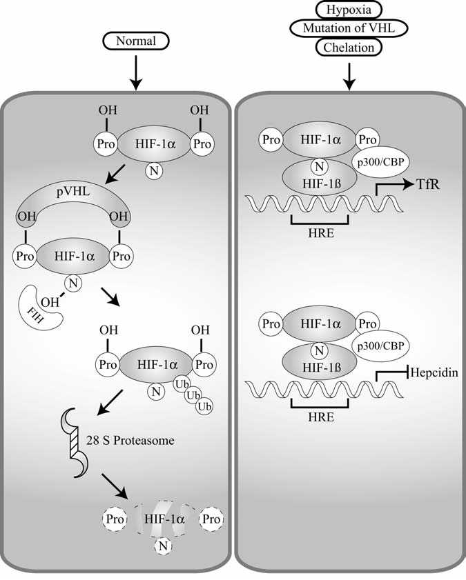
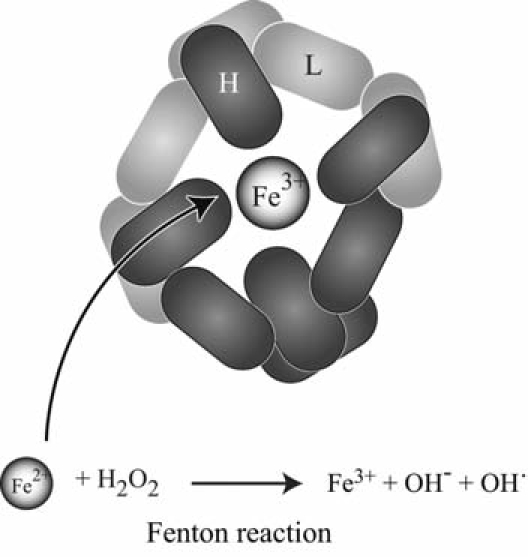
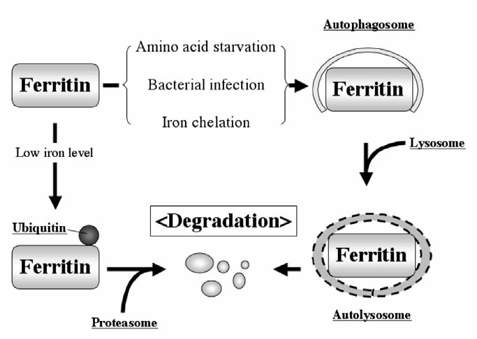
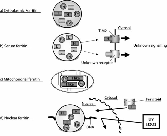
Maintenance of proper "labile iron" levels is a critical component in preserving homeostasis. Iron is a vital element that is a constituent of a number of important macromolecules, including those involved in energy production, respiration, DNA synthesis, and metabolism; however, excess "labile iron" is potentially detrimental to the cell or organism or both because of its propensity to participate in oxidation-reduction reactions that generate harmful free radicals. Because of this dual nature, elaborate systems tightly control the concentration of available iron. Perturbation of normal physiologic iron concentrations may be both a cause and a consequence of cellular damage and disease states. This review highlights the molecular mechanisms responsible for regulation of iron absorption, transport, and storage through the roles of key regulatory proteins, including ferroportin, hepcidin, ferritin, and frataxin. In addition, we present an overview of the relation between iron regulation and oxidative stress and we discuss the role of functional iron overload in the pathogenesis of hemochromatosis, neurodegeneration, and inflammation.


Figures

FIG. 1
FIG. 1
Cellular uptake of Tf-bound iron. Transferrin (Tf) is bound by two atoms of Fe3+ and circulates in plasma until reaching a target cell. It binds the transferrin receptor (TfR) on the plasma membrane of the cell, and then the Fe-Tf and TfR complex is endocytosed by the cell. In the acidic pH of the endosome, Fe3+ dissociates from Tf and is exported from the endosome by divalent metal transporter 1 (DMT1). The Tf-TfR complex is then recycled to the cell surface. Hereditary hemochromatosis protein (HFE) interacts with TfR in the Fe-Tf binding region and may thereby block the binding of Fe-Tf to TfR, preventing uptake of Fe-Tf, and negatively regulating cellular uptake of Tf-bound iron.
FIG. 2
FIG. 2
The IRP-IRE regulatory system. When iron regulatory proteins (IRPs) are activated by such conditions as low cellular iron, hypoxia, or H2O2, IRPs are able to bind to the iron-responsive element (IRE) of target mRNAs. When an IRP binds to an IRE located in the 5′-untranslated region (UTR) of target mRNA (ferritin H, ferritin L, eALAS, Fpn), it decreases translation by inhibiting the formation of the translation preinitiation complex (upper left panel). When an active IRP binds an IRE located in the 3′ UTR of target mRNA (TfR, DMT1), it increases mRNA stability (upper right panel). When the concentration of iron is high, IRP1 is deactivated by formation of an iron-sulfur cluster (ISC) and switches to cytosolic aconitase, whereas IRP2 is degraded. Under such conditions, IRP does not bind to IREs; therefore, mRNAs with IREs in the 5′ UTR are no longer blocked from translating (lower left panel), and those with IREs in the 3′ UTR of mRNA are destabilized (lower right panel).
FIG. 3
FIG. 3
Intestinal absorption of dietary iron. Heme is primarily obtained from meat sources and is taken up by enterocytes by heme carrier protein 1 (HCP1) (upper), whereas inorganic iron (Fe3+) is reduced to Fe2+ by duodenal cytochrome b1 (DCytb1) and is subsequently transported into the enterocyte by divalent metal transporter 1 (DMT1) (lower).
FIG. 4
FIG. 4
Iron transport in reticuloendocytic macrophages. Senescent red blood cells (RBCs) are phagocytosed and degraded. Their heme is broken down into biliverdin and Fe2+ by heme oxygenase 1 (HO-1), allowing Fe2+ to be exported from the macrophage by ferroportin1 (Fpn1). Once exported, Fe2+ is oxidized by ceruloplasmin (Cp) and binds to transferrin (Tf), where it may be delivered to target cells to fulfill cellular iron requirements for erythropoiesis or other processes.
FIG. 5
FIG. 5
Transport of intestinal iron. Once iron is taken up by intestinal enterocytes by divalent metal transporter 1 (DMT1), it is then transported out of the enterocyte and into plasma by ferroportin1 (Fpn1). Fpn1 couples with hephaestin (Heph) in intestinal cells (in other cell types, it couples with ceruloplasmin), whose ferroxidase activity converts Fe2+ into Fe3+. This oxidation is necessary to aid in the binding of Fe3+ to apo-Tf in the plasma.
FIG. 6
FIG. 6
Hepcidin negatively regulates ferroportin. A truncated version of hepcidin is unable to bind to ferroportin (Fpn). Fpn is coupled with anchored ceruloplasmin (Cp), and exported iron is released and oxidized (left). On binding of wild-type hepcidin, Fpn is phosphorylated and subsequently endocytosed, ubiquitinated, and degraded by the 28S proteasome (right).
FIG. 7.
FIG. 7.
HIF-1α is involved in transcriptional regulation of hepcidin and Tf. Under normal conditions, hypoxia-inducible factor 1α (HIF-1α) is negatively regulated (left panel). Normally, HIF-1α is hydroxylated on proline 402 and proline 564. Tight control of HIF-1α is mediated by the tumor-suppressor protein von Hippel–Lindau (VHL), which is an E3 ligase. VHL targets hydroxylated HIF-1α for degradation via the ubiquitin–proteasomal pathway. In addition, hydroxylation of asparagines (N) by FIH hydroxylase prevents binding of HIF-1α and p300. Under conditions of hypoxia, iron chelation, or mutation of VHL, HIF-1α is positively regulated (right panel). The hydroxylases are not active, and therefore it can bind to p300 and also not targeted for degradation. Because it is not targeted for degradation, it is capable of binding to the hypoxia-responsive element (HRE) of target genes in a heterodimer with HIF-1β. Once bound to the HRE, it may positively or negatively regulate the transcription of the target genes. In the case of transferrin receptor (TfR), it activates transcription; however, it represses transcription of hepcidin.
FIG. 8
FIG. 8
Ferritin and iron storage. Ferritin is composed of 24 subunits, heavy (H) and light (L), with varied ratios of H to L in different cell types and physiologic conditions. Ferritin H has ferroxidase activity to convert Fe2+ to Fe3+ inside ferritin shell. Iron is imported and exported through the channels constructed by ferritin H or L subunits.
FIG. 9
FIG. 9
Mechanisms of ferritin degradation. Ferritin degradation occurs through either lysosomal or proteasomal degradation pathways. Amino acid starvation, bacterial infection, or iron chelation triggers navigation of ferritin into the autophagy-lysosome pathway, in which ferritin is trapped by autophagosomes, followed by lysosome fusion, resulted in hydrolase-mediated degradation. In the proteasomal degradation pathway, lower iron concentration induces monoubiquitination of ferritin, resulting in proteasomal degradation. Polyubiquitination of ferritin has not been observed.
FIG. 10
FIG. 10
Different types of ferritin. (A) Cytoplasmic ferritin is composed of heteropolymers of H and L subunits and stores iron. The H subunit has ferroxidase activity to oxidize Fe2+ to Fe3+ for the iron storage. (B) Serum ferritin is L rich; however, only a T-cell immunoglobulin domain and mucin domain 2 (TIM-2) was identified as a receptor of ferritin H on B-cell surfaces. Downstream signaling after the association of ferritin H with TIM-2 is still largely unknown. (C) Mitochondrial ferritin is unique in that it is composed of ferritin H-like subunits. It has ferroxidase activity and maintains iron homeostasis in mitochondria. (D) Nuclear ferritin is composed of only H subunits and protects DNA from DNA-damaging agents, such as UV and H2O2. Ferritoid enhances ferritin translocation from cytosol to nucleus.
FIG. 11
FIG. 11
Transcriptional regulation of ferritin H. (A) The mouse and human ferritin genes have a similar 5′-region that regulates transcription in response to external stimuli. TNF-α activates the mouse ferritin H gene through 4.8 kb upstream from the start codon, in which NF-κB is involved in this activation mechanism. Human ferritin H gene is also activated by TNF-α, but the responsible region has not been identified, although NF-κB participation was observed. Chemical and oxidative stress (such as H2O2, t-BHQ, hemin) activate human and mouse ferritin H gene through an antioxidant-responsive element (ARE). The ARE activation is achieved by Nrf2 and AP-1 family transcription factors synergized with p300/CBP histone acetyl transferases. Among the AP-1 family transcription factors, ATF1 serves as a repressor of ferritin H gene. Hemin and cAMP were also shown to induce ferritin genes through the proximal region, termed A- or B-box, by NF-Y transcription factors, which recruit coactivator p300/CBP proteins. Adenovirus E1A oncogene represses ferritin H transcription by inhibiting p300/CBP function. (B) Both human and mouse ferritin H genes have bidirectional ARE sequences (AP1-like and AP1/NFE2). Ferritin L has a single ARE (AP1/NFE2). The core ferritin ARE sequences are completely conserved.
FIG. 12
FIG. 12
Maturation of frataxin proteins. Protein processing of human frataxin by mitochondrial protein peptidase-β (MPP-β) is summarized (top). The comparison of amino acid sequences between human and yeast frataxin and their cleavage sites by MPP-β is shown (bottom).
FIG. 13
FIG. 13
The role of frataxin in iron–sulfur cluster (ISC) formation and heme synthesis. Cytoplasmic iron is imported into mitochondria by Mrs3/4 (in yeast) or mitoferrin (in vertebrates). Frataxin interacts with a complex of the ISC scaffold protein Isu1and the cysteine desulfurase Nfs1, which release sulfur from cysteine. Frataxin also interacts with ferrochelatase and facilitates the heme synthesis through ferrochelatase-mediated insertion of ferrous iron into protoporphyrin IX. Frataxin protects aconitase activity by restoring the aconitase 4Fe-4S cluster. ISCs in mitochondria are exported into cytoplasm by Atm1 (ABC transporter of the mitochondrion 1 protein) in yeast or ABCB7, the human and mouse homologues of yeast Atm1.

References

    1. Abboud S. Haile DJ. A novel mammalian iron-regulated protein involved in intracellular iron metabolism. J Biol Chem. 2000;275:19906–19912. - PubMed
    1. Adamec J. Rusnak F. Owen WG. Naylor S. Benson LM. Gacy AM. Isaya G. Iron-dependent self-assembly of recombinant yeast frataxin: implications for Friedreich ataxia. Am J Hum Genet. 2000;67:549–562. - PMC - PubMed
    1. Adams PC. Powell LW. Halliday JW. Isolation of a human hepatic ferritin receptor. Hepatology. 1988;8:719–721. - PubMed
    1. Aisen P. Leibman A. Zweier J. Stoichiometric and site characteristics of the binding of iron to human transferrin. J Biol Chem. 1978;253:1930–1937. - PubMed
    1. Al-Mahdawi S. Pinto RM. Varshney D. Lawrence L. Lowrie MB. Hughes S. Webster Z. Blake J. Cooper JM. King R. Pook MA. GAA repeat expansion mutation mouse models of Friedreich ataxia exhibit oxidative stress leading to progressive neuronal and cardiac pathology. Genomics. 2006;88:580–590. - PMC - PubMed

Publication types