. 2008 Mar;13(3):206-20.
doi: 10.1016/j.ccr.2008.01.034.

HIF1alpha induces the recruitment of bone marrow-derived vascular modulatory cells to regulate tumor angiogenesis and invasion

Affiliations

HIF1alpha induces the recruitment of bone marrow-derived vascular modulatory cells to regulate tumor angiogenesis and invasion

Rose Du et al. Cancer Cell. 2008 Mar.

Abstract

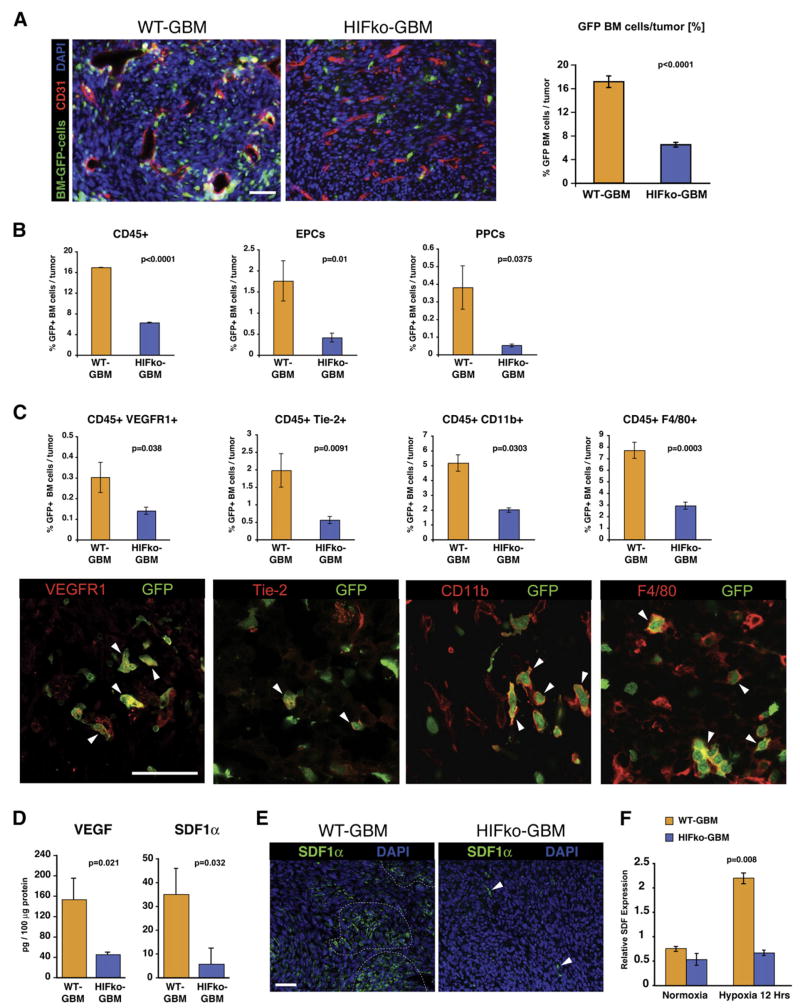
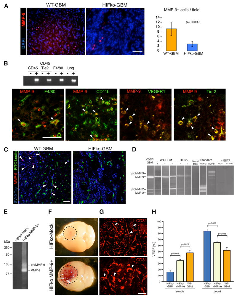
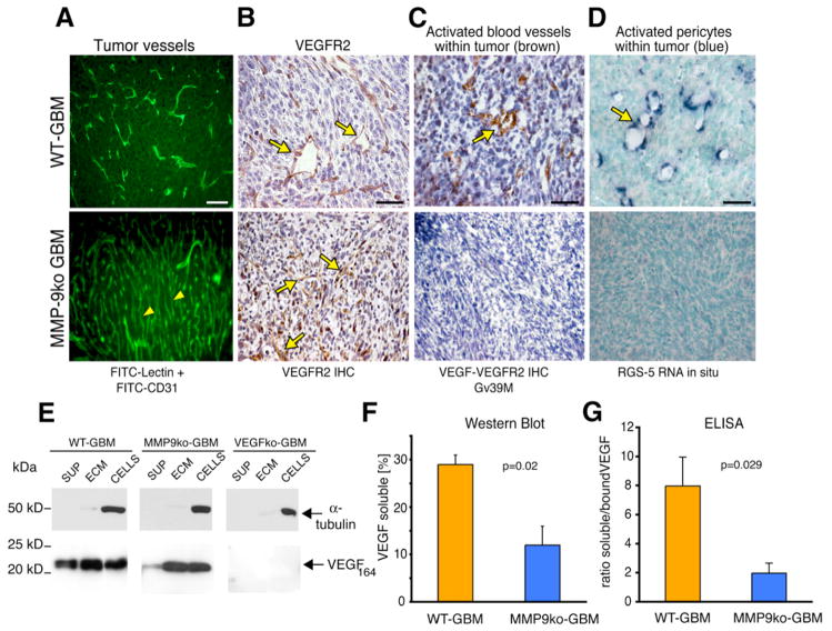
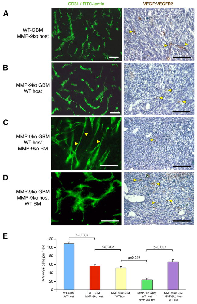
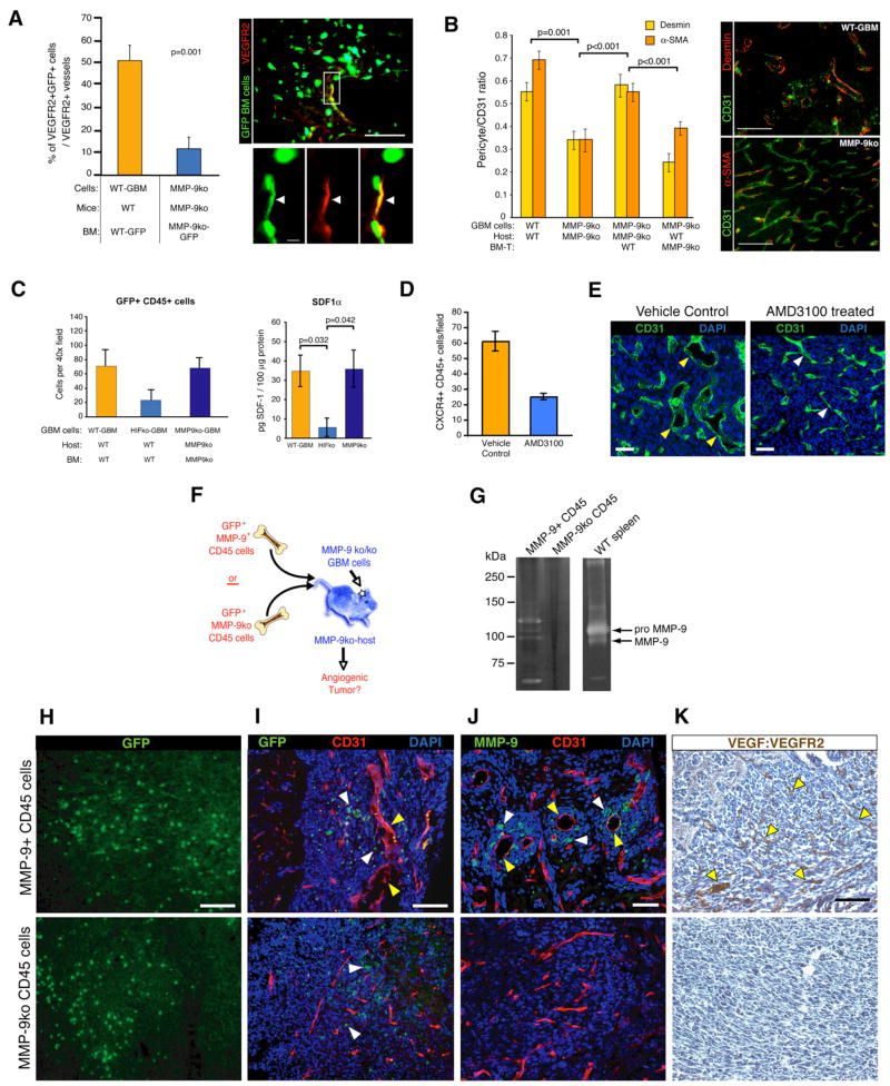
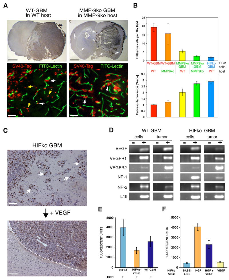
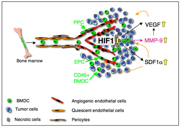
Development of hypoxic regions is an indicator of poor prognosis in many tumors. Here, we demonstrate that HIF1alpha, the direct effector of hypoxia, partly through increases in SDF1alpha, induces recruitment of bone marrow-derived CD45+ myeloid cells containing Tie2+, VEGFR1+, CD11b+, and F4/80+ subpopulations, as well as endothelial and pericyte progenitor cells to promote neovascularization in glioblastoma. MMP-9 activity of bone marrow-derived CD45+ cells is essential and sufficient to initiate angiogenesis by increasing VEGF bioavailability. In the absence of HIF1alpha, SDF1alpha levels decrease, and fewer BM-derived cells are recruited to the tumors, decreasing MMP-9 and mobilization of VEGF. VEGF also directly regulates tumor cell invasiveness. When VEGF activity is impaired, tumor cells invade deep into the brain in the perivascular compartment.


Figures

Figure 1
Figure 1. HIF1 Recruits Bone Marrow-Derived Cells to Orthotopic Glioblastomas
(A) Immunofluorescent analysis of GBM tumors. HIFkoGBM contained ~4 times less GFP+ BMDCs (green) than WT-GBM (n = 4 to 5 fields per tumor of 7 tumors/group). Angiogenic vasculature (red) in WT-GBMs was tortuous, hyperdilated, and distorted, whereas vessels in nonangiogenic HIFko GBMs appeared slim and more regular. Scale bar, 50 μm. (B) Fluorescence-activated cell sorting (FACS) analysis of GBM tumors. HIF1 recruited three distinct GFP+ BMDC populations to the tumor site: CD45+ monocytes, endothelial progenitors (EPC) (VE-Cadherin+/VEGFR2+), and pericyte progenitors (PPC) (Sca1+/PDGFRβ+) (n = 4 to 5/group). Error bars indicate ± SEM. (C) FACS and immunohistochemical analyses. Subsets of the GFP+ BMDC population of CD45+ monocytes were VEGFR1+, Tie2+, CD11b+ myeloid cells, and F4/80+ macrophages (all labeled in red, white arrowheads). Scale bar, 50 μm. All CD45+ subpopulations were reduced 2- to 4-fold in HIFko GBM. (n = 3 to 4/group). Error bars indicate ± SEM. (D) ELISA assay. Intratumoral VEGF and SDF1α was ~4 times higher in WT-GBM than HIFko GBM (n = 3 to 4/group). (E) Immunohistochemical analysis of SDF1α. WT-GBM contained clusters of SDF1α+ cells (green, encircled), whereas SDF1α expression in HIFko GBM was limited to a few single cells (white arrows). Scale bar, 50 μm. (F) Real-time PCR analysis. HIF1 induced SDF1α in GBM tumor cells. WT-GBM and HIFko GBM cells were cultured under normoxic (20% pO2) or hypoxic conditions (1% pO2) for 12 hr and harvested, and transcription levels of SDF1α were determined. Error bars indicate ± SEM.
Figure 2
Figure 2. MMP-9 Is Expressed in Various Subpopulations of CD45+ BMDC and in Infiltrating Tumor Cells
(A) Immunohistochemical analysis. WT-GBMs contained about 3.5 times more MMP-9+ cells than HIFko GBMs (n = 4 fields per tumor of 5 tumors/group). Scale bar, 50 μm. Error bars indicate ± SEM. (B) RT-PCR analyses of fractionated cell populations from the tumor and immunohistochemical analyses. MMP-9 was expressed in F4/80+ macrophages, CD11b+ myeloid cells, VEGFR1+ hemangiocytes, and in Tie2+ monocytes. Scale bar, 50 μm. (C) MMP-9 was expressed in a small subset of tumor cells at the invading edge of WT-GBM, but not in the perivascular invasive areas of HIFko GBMs. (MMP-9+ cells, red; SV40Tag+ tumor cells, blue; blood vessels, green). Scale bar, 50 μm. (D) Gelatin zymography. WT-GBM tumors contained predominantly active MMP-9 and, to a much lesser extent, MMP-2. In HIFko GBM, MMP-9 activity was reduced. As controls, recombinant pro-MMP-9 and -2 as well as active MMP-9 and -2 were loaded, and MMP-activity was obliterated in the presence of the MMP-inhibitor EDTA. (E) Gelatin zymography. HIFko MMP-9+ cells that ectopically express MMP-9 had substantially higher MMP-9 activity than mock-infected control. (F) HIFko MMP-9+ GBM, but not HIFko Mock control tumors, were hemorrhagic by gross morphology. (G) Blood vessels in HIFko Mock control tumors were slim and elongated, whereas vessels in HIFko MMP-9+ tumors were rather distorted and hyperdilated. (H) ELISA assay. Soluble VEGF was measured in GBM in the supernatant. Sequestered (bound) VEGF was determined both in the ECM and in GBM cells. Error bars indicate SEM.
Figure 3
Figure 3. MMP-9-Deficient GBM Do Not Undergo Vascular Remodeling and Neovascularization
(A) FITC-lectin perfusion of the tumor vasculature and anti-CD31 immunohistochemical analysis (green). In the complete absence of MMP-9, tumor vessels did not undergo vascular remodeling but contained a regular, slim, and dense vascular network (yellow arrowheads). All scale bars, 50 μm. (B) Immunohistochemical analysis. VEGFR2 expression on tumor endothelial cells was apparent in WT-GBM and MMP-9ko GBM (yellow arrows). (C) Immunohistochemical analysis. Activated tumor vessels were detected with the Gv39M antibody that recognizes the VEGF:VEGFR2 complex (brown) in WT-GBM, but not in MMP-9ko tumors. (D) In situ RNA hybridization. RGS-5 (blue), a marker for activated pericytes, was expressed in pericytes of WT-GBM, but not in MMP-9ko GBM. (E) VEGF western blot analysis of tumor cells. In WT-GBM, soluble VEGF was detected in the supernatant of GBM. In MMP-9ko GBM, VEGF-164 was predominantly sequestered in the ECM and bound to cell surfaces. VEGFko GBM served as negative controls. (F) Quantification of soluble VEGF levels determined by densitometry of VEGF western blots shown in (E). Soluble VEGF was significantly decreased in the absence of MMP-9. Error bars indicate SEM. (G) ELISA. Ratio of soluble VEGF (in supernatant) and sequestered VEGF (ECM and cells) was determined in WT-GBM and MMP-9ko GBM. Soluble, mobilized VEGF was significantly reduced in MMP-9ko GBM. Error bars indicate SEM.
Figure 4
Figure 4. MMP-9+ BMDCs Contribute to the Angiogenic Switch in GBM
Immunohistochemical analysis and FITC-lectin perfusion of mice (A–D). The activated tumor vasculature was identified by the presence of VEGF:VEGFR2 complexes on endothelial cells. Cohorts of 6–13 mice were used. White bars, 50 μm. Black bars, 75 μm. (A) WT-GBM cells in MMP-9ko mice. Vascular remodeling of tumor vessels was indicated by their irregular shape and the presence of a few activated tumor vessels (yellow arrowheads). (B) MMP-9ko GBM cells in wild-type mice. Vascular remodeling of tumor vessels was indicated by their irregular shape and the presence of a few activated tumor vessels (yellow arrowheads). (C) Wild-type mice, reconstituted with MMP-9ko bone marrow, were injected with MMP-9ko GBM cells. Tumor vessels contained areas of elongated and slim vessels mimicking the phenotype of MMP-9ko tumor vessels and did not exhibit VEGF:VEGFR2 activation on tumor endothelial cells. (D) MMP-9ko mice, reconstituted with wild-type bone marrow, were injected with MMP-9ko GBM cells. Tumor vessels appeared torturous, hyperdilated, and irregularly shaped, concomitant with VEGF:VEGFR2 activation on tumor endothelial cells. (E) The number of MMP-9+ cells correlated with the angiogenic phenotype of the tumors described in (A)–(D). Error bars indicate SEM.
Figure 5
Figure 5. CD45+ MMP-9+ BMDCs Are Sufficient to Initiate Angiogenesis in GBM
(A) Wild-type mice or MMP-9ko mice, reconstituted with bone marrow from actin-GFP wild-type or MMP-9ko mice, respectively, were injected with WT-GBM or MMP-9ko GBM cells. MMP-9ko GBM revealed a 4- to 5-fold reduction of VEGFR2+ (red) GFP+ (green) EPCs. Cohorts of five mice per group were used. (B) Tumor sections of the groups described in Figures 4A–4D were analyzed for the presence of pericytes on tumor vessels as detected by desmin (red) and α-SMA-staining (red) and quantified using the ratios of red-labeled pericytes to green-labeled tumor vessels (anti-CD31 staining). (C) GFP+CD45+ cells were substantially reduced in HIFko GBM when compared to WT-GBM or MMP-9ko GBM. ELISA assay. SDF1α levels were assessed in tumor lysates of WT-GBM, HIFko GBM, and MMP-9ko GBM. Intratumoral SDF1α levels were lower in HIFko GBM than WT-GBM or MMP-9ko GBM (n = 3 to 4 per group). (D) WT-GBM mice were treated with the CXCR4 inhibitor AMD3100 or with vehicle control for 2 weeks. AMD3100-treated tumors exhibited a 2- to 3-fold decrease in CXCR4+ CD45+ cells compared to controls. (E) AMD3100-treated mice demonstrated slimmer and more normalized tumor vessels, while control mice were hyperdilated and tortuous. Scale bars, 50 μm. (F) Experimental design for assessing functional significance of MMP-9 in CD45+ BMDCs in initiating tumor angiogenesis. (G) Zymogram analysis of MMP-9ko GBMs from mice that either received GFP+MMP-9+ or GFP+MMP-9ko CD45+ cells. A spleen lysate from a wild-type animal served as a positive control for MMP-9. (H) Comparable numbers of i.v. injected MMP-9+ or MMP-9ko GFP+CD45+ cells were recruited to the tumor site. All scale bars, 50 μm. (I) MMP-9ko GBMs that contained MMP-9+CD45+GFP+ cells (white arrowheads) exhibited irregularly shaped and hyperdilated vessels (yellow arrowheads) whereas MMP-9ko GBMs that recruited MMP9koCD45+GFP+ cells (white arrowheads) did not. (J) MMP-9+ cells (white arrowheads) were preferentially located around hyperdilated vessels (yellow arrowheads). (K) The vasculature of tumors containing MMP-9+CD45+GFP+ cells became activated (indicated by the presence of VEGF:VEGFR2 complexes on endothelial cells). MMP-9ko tumors that recruited MMP-9ko CD45+ cells did not show VEGF:VEGFR2 complex formations. All error bars indicate ± SEM.
Figure 6
Figure 6. VEGF Directly Inhibits Perivascular Tumor Invasion
(A) Fluorescent detection of tumor cells (anti-SV40-Tag antibody; red) and the vasculature (FITC-lectin; green). WT-GBM cells infiltrated as single cells into the brain parenchyma without associating with blood vessels (yellow arrows) or invaded alongside blood vessels (perivascular invasion) in the brain (white arrows). MMP-9ko GBMs were predominantly perivascular invasive. Black bar, 1.4 mm. White bar, 50 μm. (B) Infiltrative and perivascular invasive modes of GBMs in different tumor cell/host combinations were quantified by immunohistochemical analysis on tumor sections as in Figure 6A. Infiltrative cells were counted as cells in the brain without vessel association, whereas perivascular invasiveness of tumor cells was graded from 0–3, where 1 indicates minimal distant spread of tumor cells and 3 indicates substantial and marked distant spread. Error bars indicate ± SD. (C) HIFko GBM were stably transduced with a retrovirus expressing VEGF-164. While HIFko GBMs were perivascular invasive (white arrows), HIFko GBMs over-expressing VEGF exhibited a smooth tumor border (dotted line) and did not invade. Scale bars, 50 μm. (D) RT-PCR analysis revealed that both WT-GBM and HIFko GBM express VEGFR1, 2 and neuropilin1, 2. (E and F) Boyden Chamber invasion assay. (E) Ectopic expression of VEGF in HIFko GBM or (F) addition of recombinant VEGF to HIFko GBM cells reduced tumor cell migration by about 50% in response to HGF. VEGF alone had no effect. Error bars indicate ± SEM.
Figure 7
Figure 7. HIF-1 Is a Critical Regulator of BMDC Recruitment in Tumors
Hypoxia in tumors increases HIF1, which, partly by inducing VEGF and SDF1α in tumor cells, recruits BMDC including EPC, PPC, and CD45+ monocytic vascular modulatory cells to endorse vascular remodeling in glioblastomas. SDF1α serves as a retention factor of CXCR4+ vascular progenitor and monocytic BMDC in GBM. HIF1 not only induces VEGF transcription in GBM, but also increases VEGF activity by recruiting CD45+ BMDC that carry and secrete MMP-9 to the tumor site, which in turn makes sequestered VEGF bioavailable for its receptor VEGFR2.

Comment in

References

    1. Abounader R, Laterra J. Scatter factor/hepatocyte growth factor in brain tumor growth and angiogenesis. Neuro-Oncol. 2005;7:436–451. - PMC - PubMed
    1. Aghi M, Chiocca EA. Contribution of bone marrow-derived cells to blood vessels in ischemic tissues and tumors. Mol Ther. 2005;12:994–1005. - PubMed
    1. Aghi M, Cohen KS, Klein RJ, Scadden DT, Chiocca EA. Tumor stromal-derived factor-1 recruits vascular progenitors to mitotic neovasculature, where microenvironment influences their differentiated phenotypes. Cancer Res. 2006;66:9054–9064. - PubMed
    1. Behrendtsen O, Werb Z. Metalloproteinases regulate parietal endoderm differentiating and migrating in cultured mouse embryos. Dev Dyn. 1997;208:255–265. - PubMed
    1. Berger M, Bergers G, Arnold B, Hammerling GJ, Ganss R. Regulator of G-protein signaling-5 induction in pericytes coincides with active vessel remodeling during neovascularization. Blood. 2005;105:1094–1101. - PubMed

Publication types

MeSH terms