. 2008 Apr 15;316(2):288-301.
doi: 10.1016/j.ydbio.2008.01.022. Epub 2008 Jan 31.

Beta1 integrin expression by podocytes is required to maintain glomerular structural integrity

Affiliations

Beta1 integrin expression by podocytes is required to maintain glomerular structural integrity

Ambra Pozzi et al. Dev Biol. .

Abstract

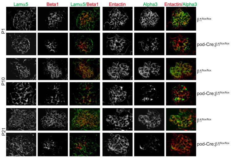
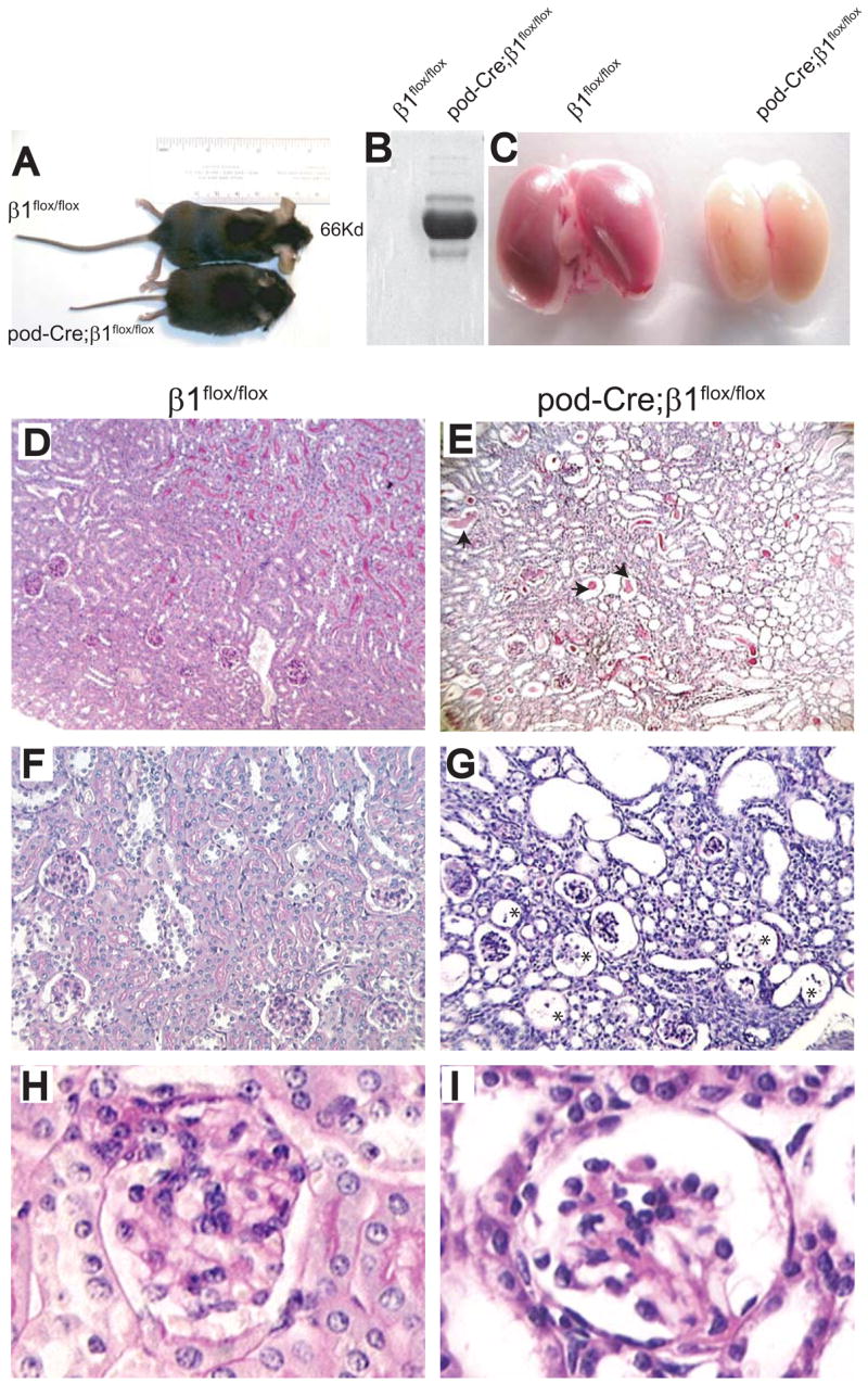
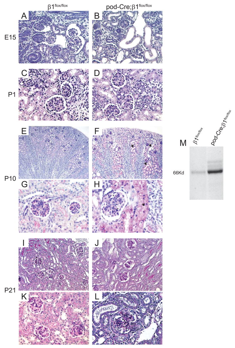
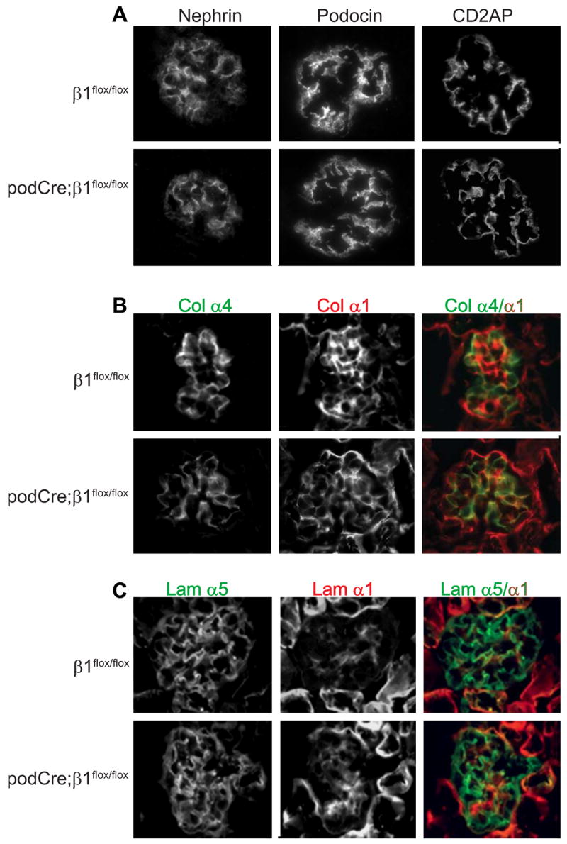
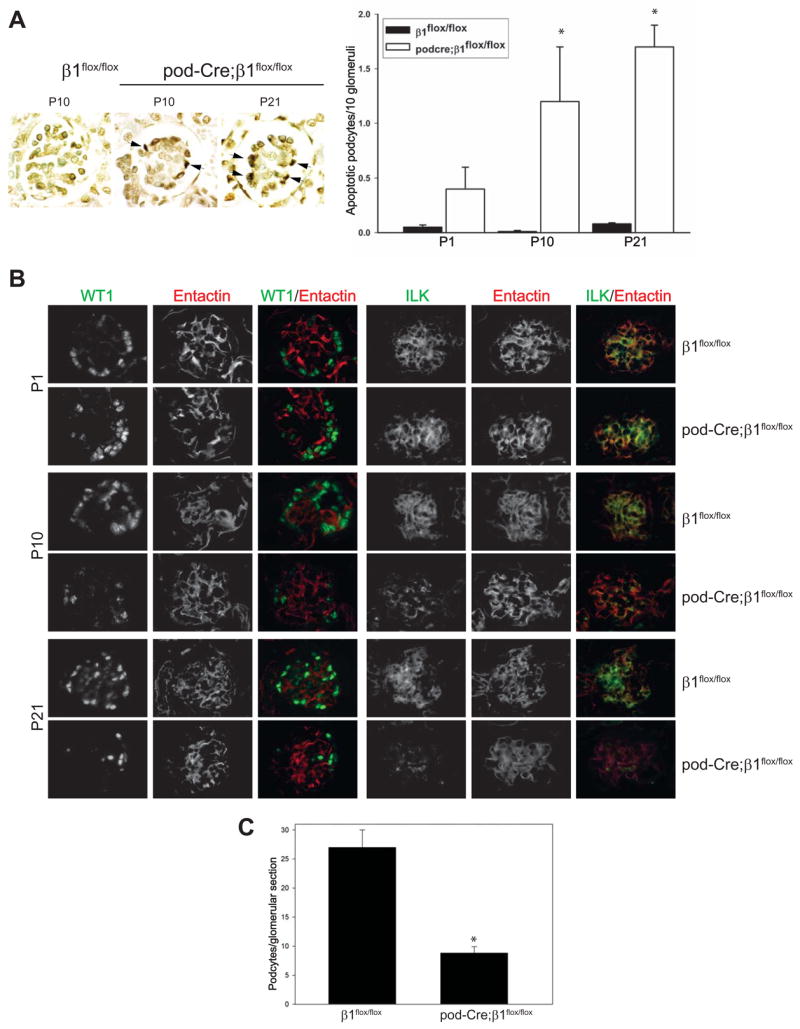
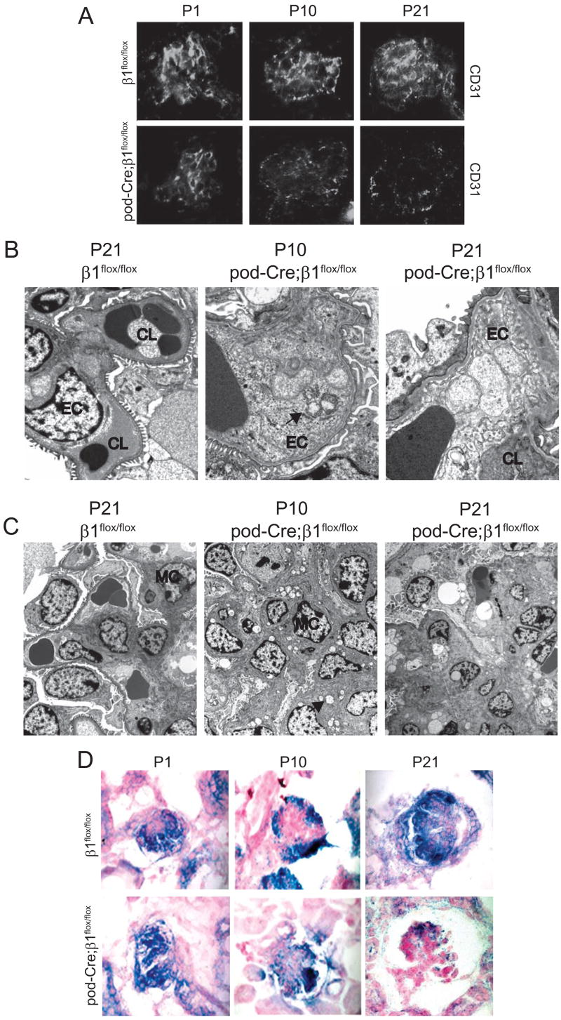
Integrins are transmembrane heteromeric receptors that mediate interactions between cells and extracellular matrix (ECM). beta1, the most abundantly expressed integrin subunit, binds at least 12 alpha subunits. beta1 containing integrins are highly expressed in the glomerulus of the kidney; however their role in glomerular morphogenesis and maintenance of glomerular filtration barrier integrity is poorly understood. To study these questions we selectively deleted beta1 integrin in the podocyte by crossing beta1(flox/flox) mice with podocyte specific podocin-cre mice (pod-Cre), which express cre at the time of glomerular capillary formation. We demonstrate that podocyte abnormalities are visualized during glomerulogenesis of the pod-Cre;beta1(flox/flox) mice and proteinuria is present at birth, despite a grossly normal glomerular basement membrane. Following the advent of glomerular filtration there is progressive podocyte loss and the mice develop capillary loop and mesangium degeneration with little evidence of glomerulosclerosis. By 3 weeks of age the mice develop severe end stage renal failure characterized by both tubulointerstitial and glomerular pathology. Thus, expression of beta1 containing integrins by the podocyte is critical for maintaining the structural integrity of the glomerulus.


Figures

Figure 1
Figure 1. β1 integrin subunit is deleted in pod-Cre;β1flox/flox mice
Frozen sections of kidneys from P1, P10 and P21 β1flox/flox and pod-Cre; β1flox/flox mice were co-stained with anti-mouse integrinβ1 (red) and anti laminin-α5 chain (green) or anti-mouse integrinα3 (green) and anti-entactin (red), respectively.
Figure 2
Figure 2. Six week old pod-Cre;β1flox/flox mice develop severe proteinuria and end stage renal disease
(A–C) Six week old pod-Cre; β1flox/flox mice are smaller with evidence of severe edema (A), albuminuria (2 μl urine/lane) (B) and end stage kidneys (C) compared to aged matched β1flox/flox mice. (D–I) PAS staining of kidneys derived from the mice described above showing glomerular and tubular interstitial abnormalities in the mutant group. The arrows in panel E show dilated tubules filled with hyaline material and the asterisks in panel G show the remnants of glomeruli in the pod-Cre; β1flox/flox mice. (D, E = 100 ×; F, G = 200 ×; H, I =630 ×).
Figure 3
Figure 3. Kidneys from pod-Cre; β1flox/flox mice exhibit severe abnormalities in the glomerulus and tubulointerstitium
(A, B) PAS staining of kidneys derived from E15.5 β1flox/flox and pod-Cre; β1flox/flox mice showing no significant differences between the two genotypes (200 ×). (C, D) PAS staining of kidneys derived from newborn β1flox/flox and pod-Cre; β1flox/flox mice showing no overall significant differences between the two genotypes (200 ×). (E, F) PAS staining of kidneys derived from P10 β1flox/flox and pod-Cre; β1flox/flox mice revealed tubulodilatation (arrow) in the mutant mice (100 ×). (G, H) In P10 mutant mice there was evidence of “ballooned” glomerular capillary loops (asterisk) and protein containing vacuoles (arrow) in the tubules of the pod-Cre; β1flox/flox mice (400 ×). (I–L) PAS staining of kidneys derived from 3 week old β1flox/flox and pod-Cre; β1flox/flox mice revealed dilated tubules, evidence of “ballooned” capillary loops and mesangial hypercellularity (I, J = 200 ×; K, L= 400 ×). (M) Coomassie staining of urine (2 μl/lane) from newborn β1flox/flox and pod-Cre; β1flox/flox mice showing albuminuria in the latter group.
Figure 4
Figure 4. Glomeruli from pod-Cre; β1flox/flox mice demonstrate podocyte foot process effacement
EM analysis was performed on kidneys of mice at various ages. In the E15 mutant mice there is evidence of foot process effacement of the podocytes but the GBM (arrow) was normal. Similar findings are present in P1 kidneys. In P10 mice, in addition to the foot process effacement, there was evidence of very mild segmental splitting of the GBM (arrow) which progressed by day P21. Abbreviations: FP=Foot Process; Po=Podocyte
Figure 5
Figure 5. Glomeruli from newborn pod-Cre; β1flox/flox mice demonstrate normal expression of podocyte-specific structural proteins glomerular basement membrane components
Frozen sections of kidneys derived from newborn mice were stained with antibodies to (A) Nephrin, podocin or CD2AP, (B) the α4 (green) or α1 (red) chains of collagen IV and (C) the α5 (green) orα1 (red) laminin chains.
Figure 6
Figure 6. Podocytes of pod-Cre; β1flox/flox mice undergo apoptosis
(A) Examples of apoptosis, as determined by TUNEL assay, detected in the glomeruli of β1flox/flox or pod-Cre; β1flox/flox mice at the time points indicated. The number of apoptotic podocytes expressed per 10 glomeruli is demonstrated graphically. The (*) indicates significant differences (p < 0.01) between the two genotypes. (B) Frozen sections of glomeruli were co-stained for WT-1 (green) and entactin (red) or ILK (green) and entactin (red) as described in the Methods. (C) The number of podocytes in EM sections were evaluated as described in the Methods and expressed as mean +/− SD. (*) indicates significant differences (p < 0.05) between P21 β1flox/flox and P21 pod-Cre; β1flox/flox mice.
Figure 7
Figure 7. Glomerular capillary and mesangium injury in pod-Cre; β1flox/flox mice
(A) Frozen sections of kidneys derived from newborn, P10 and P21 mice were stained with CD31 antibodies to visualize the glomerular vasculature. (B) EM of kidneys from P21 β1flox/flox (3,000x) as well as P10 (11,000x) and P21 (11,000x) pod-Cre; β1flox/flox mice revealed normal morphology of endothelial cells in the β1flox/flox mice. Vacuoles (arrow) in the endothelial cells (arrow) were evident in the P10 and P21 mutant mice. Abbreviations: EC = endothelial cells; CL = capillary loops; (C) EM of kidneys emphasizing the mesangium. Note the presence of vesicles (arrow) in the mesangial cells of mutant mice by P10 (8,900 ×) and the increased mesangial matrix in the P21 pod-Cre; β1flox/flox mice (4th panel) (2,200 ×). Abbreviations: MC= mesangial cell. (D) In situ hybridizations on kidneys of newborn, P10 and P21 day old mice showing VEGF mRNA expression.

References

    1. Abrahamson DR. Origin of the glomerular basement membrane visualized after in vivo labeling of laminin in newborn rat kidneys. J Cell Biol. 1985;100:1988–2000. - PMC - PubMed
    1. Aumailley M, et al. A simplified laminin nomenclature. Matrix Biol. 2005;24:326–332. - PubMed
    1. Bjarnegard M, et al. Endothelium-specific ablation of PDGFB leads to pericyte loss and glomerular, cardiac and placental abnormalities. Development. 2004;131:1847–57. - PubMed
    1. Borza CM, et al. Integrin alpha3beta1, a novel receptor for alpha3(IV) noncollagenous domain and a trans-dominant Inhibitor for integrin alphavbeta3. J Biol Chem. 2006;281:20932–9. - PubMed
    1. Brakebusch C, et al. Skin and hair follicle integrity is crucially dependent on beta 1 integrin expression on keratinocytes. Embo J. 2000;19:3990–4003. - PMC - PubMed

Publication types