. 2008 Mar 11;18(5):311-21.
doi: 10.1016/j.cub.2008.02.006.

MOBKL1A/MOBKL1B phosphorylation by MST1 and MST2 inhibits cell proliferation

Affiliations

MOBKL1A/MOBKL1B phosphorylation by MST1 and MST2 inhibits cell proliferation

Maria Praskova et al. Curr Biol. .

Abstract

Background: MST1 and MST2 are the mammalian Ste20-related protein kinases most closely related to Drosophila Hippo, a major regulator of cell proliferation and survival during development. Overexpression of MST1 or MST2 in mammalian cells is proapototic; however, little is known concerning the physiologic regulation of the endogenous MST1/MST2 kinases, their role in mammalian cell proliferation, or the identity of the MST1/MST2 substrates critical to proliferative regulation.

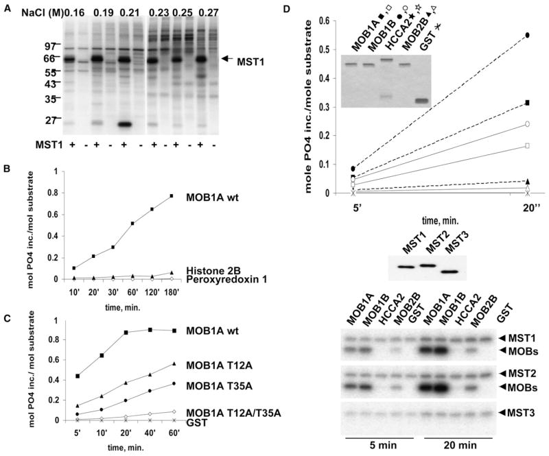
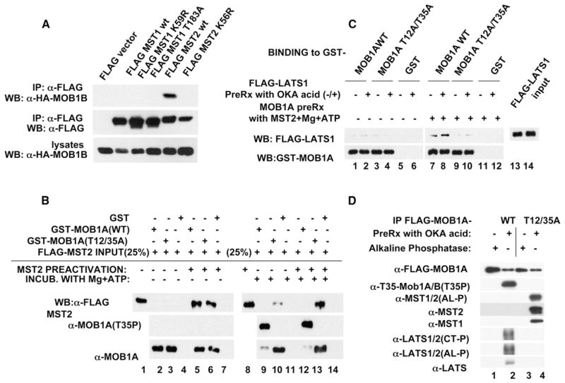
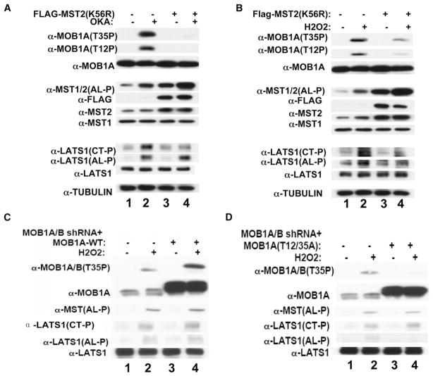
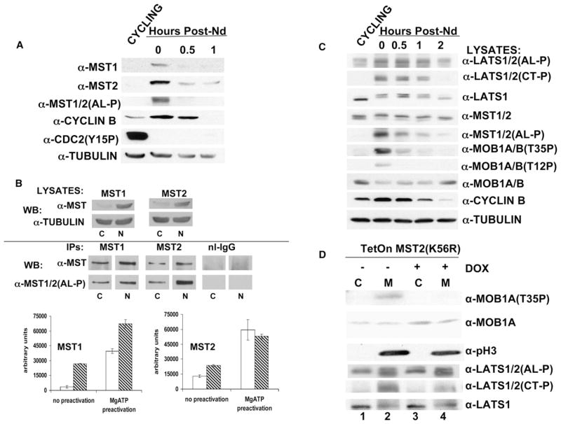
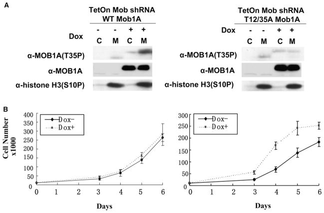
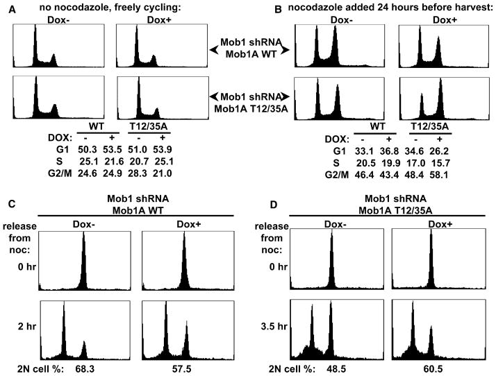
Results: We show that MST1 and MST2 activity increases during mitosis, especially in nocodazole-arrested mitotic cells, where these kinases exhibit both an increase in both abundance and activation. MST1 and MST2 also can be activated nonphysiologically by okadaic acid or H2O2. The MOBKL1A and MOBKL1B polypeptides, homologs of the Drosophila MATS polypeptide, are identified as preferred MST1/MST2 substrates in vitro and are phosphorylated in cells in an MST1/MST2-dependent manner in mitosis and in response to okadaic acid or H2O2. MST1/MST2-catalyzed MOBKL1A/MOBKL1B phosphorylation alters the ability of MOBKL1A/MOBKL1B to bind and regulate downstream targets such as the NDR-family protein kinases. Thus, MOBKL1A/MOBKL1B phosphorylation in cells promotes MOBKL1A/MOBKL1B binding to the LATS1 kinase and enables H2O2-stimulated LATS1 activation loop phosphorylation. Most importantly, replacement of endogenous MOBKL1A/MOBKL1B by a nonphosphorylatable mutant is sufficient to accelerate cell proliferation substantially by speeding progression through G1/S as well as mitotic exit.

Conclusions: These results establish that MST1 and MST2 are activated in mitosis and catalyze the mitotic phosphorylation of MOBKL1A/MOBKL1B. MOBKL1A/MOBKL1B phosphorylation, in turn, is sufficient to inhibit proliferation through actions at several points in the cell cycle.


Figures

Figure 1
Figure 1. Identification and Characterization of a Substrate for the MST1 and MST2 Kinases
(A) Shown is the detection of an MST1 substrate in vitro. L1210 cell extract was fractionated by NaCl gradient elution. Fractions were subjected to phosphorylation ± MST1 as described in the Supplemental Experimental Procedures. (B) Time course of MST1-catalyzed 32P incorporation into MOBKL1A (■), histone H2B (▲), and Peroxiredoxin-1 (Prx-1) (◇). (C) The effect of mutation of MOBKL1A Thr12 and Thr35 on MOBKL1A phosphorylation by MST1. (D) Different MOB family members as substrates for MST1/MST2/MST3. Substrates were phosphorylated by preactivated MST1 (solid lines), MST2 (dashed lines), and MST3 as in (B) for the time points indicated. The insert shows a Coomassie blue stain reflecting the relative amount of the substrates in the reaction. The open symbols are MST1, and the closed symbols are MST2. The panel below the graph is an anti-FLAG blot reflecting the relative amount of each kinase in the reaction. The autoradiograph shows the relative amount of 32P incorporation into the kinases and the substrates.
Figure 2
Figure 2. Characterization of MOBKL1A/MOBKL1B Binding to MST1/MST2 and LATS1
(A) MOBKL1B binds native MST2 in preference to kinase-dead MST2 or native MST1. HA-MOBKL1B was cotrasfected in HEK293T cells with the Flag-tagged MST1 and MST2 variants indicated. (B) MST2 autophosphorylation enhances, whereas MST2-catalyzed MOBKL1A phosphorylation inhibits, the MST-MOBKL1A interaction in vitro. Immobilized GST (lanes 4, 7, 11, and 14), GST-MOBKL1A wild-type (lanes 3, 6, 10, and 13), and the GST-MOBKL1A (Thr12/35Ala) (lanes 2, 5, 9, and 12) were incubated with purified MST2, nonpreactivated (lanes 2–4 and 9–11), or preactivated (lanes 5–7 and 12–14), with (lanes 9–14) or without (lanes 2–7) ATP. Note that phosphorylation of MOBKL1A (lanes 9 and 12) reduces its reactivity with the anti-MOBKL1A/MOBKL1B antibody (lowest panel). (C) Characterization of LATS1 binding to unphosphorylated and phosphorylated MOBKL1A in vitro is shown. Immobilized GST, GST-MOB1A wild-type, or GST-MOBKL1A (Thr12/35Ala) were preincubated with preactivated MST2, with (lanes 7–12) or without (lanes 1–6) MgCl2 and ATP. After washing, okadaic-acid-treated (lanes 2, 4, 6, 8, 10, and 12) or untreated (1, 3, 5, 7, 9, and 11) HEK293T cell lysates containing Flag-LATS1 were added for pull-down assay. (D) Characterization of the effect of okadaic acid on the ability of recombinant MOBKL1A wild-type or MOBKL1A (Thr12/35Ala) to bind endogenous MST1/MST2 and LATS1 during transient expression is shown. HEK293T cells expressing FLAG-tagged MOBKL1A wild-type (lanes 1 and 2) or MOBKL1A (Thr12/T35Ala) (lanes 3 and 4) were treated with okadaic acid (OA) (lanes 2 and 4) or not (lanes 1 and 3). Aliquots of the cell extracts containing 2 mg protein were incubated with either okadaic acid (lanes 2 and 4) or alkaline phosphatase (lanes 1 and 3) at room temperature for 30 min. and then subjected to anti-Flag immunoprecipitation.
Figure 3
Figure 3. The Role of MST1/MST2 in MOBKL1A/MOBKL1B and LATS1 Phosphorylation in Response to Okadaic Acid and H2O2
(A) Overexpression of a kinase-dead MST2 inhibits okadaic-acid-induced MOBKL1A/MOBKL1B and LATS1/LATS2 (Thr1079), but not LATS1/LATS2 (Ser909) phosphorylation. U2OS cells stably expressing a tetracycline (Tet) inducible Flag-MST2 (K56R) were treated with doxycycline (lanes 3 and 4) or carrier (lanes 1 and 2) and 12 hr later with okadaic acid (1 μM) (lanes 2 and 4) or carrier (lanes 1 and 3) for 30 min prior to harvest. (B) Overexpression of a kinase-dead MST2 inhibits H2O2-induced MOBKL1A/MOBKL1B and phosphorylation of both LATS1/LATS2 (Thr1079) and LATS1 (Ser909). The experiment was carried out exactly as in (A), except the stimulation was with H2O2 (2.5 mM), which was added for 20 min. (C) Replacement of endogenous MOBKL1A/MOBKL1B with recombinant MOBKL1A wild-type does not alter H2O2 stimulated LATS1/LATS2 phosphorylation. U2OS cells carry cassettes of shRNA against 3′ UTR of MOBKL1A, shRNA directed against the coding region of MOBKL1B, and cDNA encoding Flag-MOBKL1A. The first cassette is constitutively expressed, whereas the latter two are tetracycline inducible. Five days prior to stimulation, doxycycline (5 μg/ml) (lanes 3 and 4) or carrier (lanes 1 and 2) was added. Cells were treated with H2O2 as in (B) (lanes 2 and 4). Note that the recombinant Flag-MOBKL1A runs slightly slower than the endogenous MOBKLIA/MOBKL1B. (D) Elimination of endogenous MOBKL1A/MOBKL1B phosphorylation selectively eliminates H2O2-stimulated phosphorylation of LATS1/LATS2 (Ser909), but not of LATS1/LATS2 (Thr1079). The experiment was carried out exactly as in (C), except that the cDNA sequences employed encodes MOBKL1A (Thr12/35Ala).
Figure 4
Figure 4. MST1/MST2 Activity, MOBKL1A/MOBKL1B, and LATS1 Phosphorylation in Nocodazole-Arrested and Unimpeded Mitotic U2OS Cells
(A) The MST1 and MST2 kinases exhibit increased abundance in nocodazole-arrested mitotic cells. U2OS cells either were exponentially growing (cycling) or were arrested in metaphase by nocodazole (0 hr) and then harvested 0.5 and 1 hr after washing. (B) The MST1 and MST2 kinases show increased fractional activation in nocodazole-arrested cells. Top: Extracts from cycling (marked as “C”) or nocodazole (“N”)-arrested mitotic U2OS were analyzed by immunoblot for MST1 and MST2. Bottom: Immunoprecipitates of MST1 and MST2 were prepared from the extracts of cycling and nocodazole-arrested U2OS cells shown in the top panels. The bar graphs show the MST activities from three independent MBP kinase assays. The fractional activation (% maximal) in the cycling and nocodazole-arrested cells (n = 3) were as follows. MST1: cycling, 10.3 ± 1.2; mitotic, 40.8 ± 6.4 (p < 0.01); MST2, cycling 26.4 ± 6.7; mitotic, 45.6 ± 1.3 (p < 0.03). (C) Characterization of the effect of nocodazole-induced mitotic arrest on the phosphorylation of MOBKL1A/MOBKL1B and LATS1/LATS2 is shown. U2OS cells were treated as in (A). (D) Overexpression of kinase-dead MST2 in unimpeded mitotic cells eliminates mitotic MOBKL1A/MOBKL1B phosphorylation, modestly reduces LATS1 (Thr1079P), and does not alter LATS (Ser909P). U2OS cells carrying a tetracycline-inducible kinase-dead MST2 (K56R) were treated with doxycycline (1 μg/ml) (lanes 3 and 4) or carrier (lanes 1 and 2) 12 hr prior to a mitotic shake-off in the absence of nocodazole. Histone H3 (Ser10) phosphorylation marks mitosis.
Figure 5
Figure 5. Elimination of MST1/MST2-Catalyzed MOBKL1A/MOBKL1B Phosphorylation Accelerates U2OS Cell Proliferation
(A) Doxycycline-induced replacement of endogenous MOBKL1A/MOBKL1B with recombinant MOBKL1A wild-type or MOBKL1A (Thr12/35Ala) is shown. U2OS cells described in Figures 3C and 3D were treated with doxycycline for 5 days and then replated at an appropriate density. Unimpeded mitotic cells were collected as in Figure 4D. Note in the experiment illustrated on the left that the mitotic phosphorylation of the endogenous MOBKL1A/MOBKL1B polypeptides is greatly reduced after doxycycline induction, whereas the doxycycline-induced recombinant, overexpressed wild-type MOBKL1A is heavily phosphorylated in mitosis. In the experiment illustrated on the right, endogenous MOBKL1A/MOBKL1B polypeptides and phosphorylation also are abolished. The Thr35P immunoreactivity of the MOBKL1A (Thr12/35Ala) is indistinguishable in the cycling and shaken-off cells; this signal probably reflects the very minor nonphosphospecific immunoreactivity of this antibody, uncovered by the marked overexpression of recombinant MOBKL1A (Thr12/35Ala). (B) Elimination of MST1/MST2-catalyzed MOBKL1A/MOBKL1B phosphorylation accelerates U2OS cell proliferation. The doxycycline induction and the replating was carried out exactly as described in (A). The cell counts at each day after replating are shown. The error bars represent the SEM. On the right, the differences between Dox and Dox+ on days 3–6 are all significant at p < 0.01.
Figure 6
Figure 6. Elimination of MOBKL1A Phosphorylation Accelerates Progression through G1/S and Mitotic Exit
(A and B) Elimination of MOBKL1A phosphorylation accelerates progression through G1/S. The U2OS cells were treated with doxycycline and replated exactly as described in Figure 5A. (A) Three days after replating, aliquots of the cells were subjected to DNA content analysis by FACS. Note that when growing exponentially in the absence of nocodazole, neither cell line shows significant effects of doxycycline on cell-cycle distribution. (B) A parallel set of cells were treated with nocodazole, harvested 24 hr later, and analyzed for DNA content by FACS. Note that replacement with MOBKL1A (Thr12/35Ala) is accompanied by fewer cells in G1 and more in G2/M. (C and D) Elimination of MOBKL1A phosphorylation accelerates exit from mitosis into G1. Doxycycline, replating, and nocodazole treatments were performed as in (A) and (B). Cells arrested in mitosis (4N) were then collected by shake-off. A portion of mitotic cells were washed free of nocodazole and replated. The cells expressing MOBKL1A wild-type (C) were harvested 2 hr after replating, whereas the cells expressing MOBKL1A (Thr12/35Ala) (D) were harvested 3.5 hr after replating. Cells are analyzed by FACS (lower FACS scans) The fraction of replated cells exhibiting 2N DNA content is shown below the FACS scans and reflects the fraction of cells exiting mitosis into G1. Note that replacement of endogenous MOBKL1A/MOBKL1B with recombinant MOBKL1A wild-type diminishes the fraction of cells exiting M, whereas replacement with MOBKL1A (Thr12/35Ala) increases the fraction of cells exiting M into G1.

References

    1. Dan I, Watanabe NM, Kusumi A. The Ste20 group kinases as regulators of MAP kinase cascades. Trends Cell Biol. 2001;11:220–230. - PubMed
    1. Creasy CL, Chernoff J. Cloning and characterization of a human protein kinase with homology to Ste20. J Biol Chem. 1995;270:21695–21700. - PubMed
    1. Taylor LK, Wang WCR, Erikson RL. Newly identified stress-responsive protein kinases, Krs-1 and Krs-2. Proc Natl Acad Sci USA. 1996;93:10099–10104. - PMC - PubMed
    1. Graves JD, Gotoh Y, Draves KE, Ambrose D, Han DK, Wright M, Chernoff J, Clark EA, Krebs EG. Caspase-mediated activation and induction of apoptosis by the mammalian Ste20-like MST1. EMBO J. 1998;17:2224–2234. - PMC - PubMed
    1. Lee KK, Murakawa M, Nishida E, Tsubuki S, Kawashima SI, Safkamaki K, Yonehara S. Proteolytic activation of MST/Krs, STE20-related protein kinase by caspase during apoptosis. Oncogene. 1998;16:3029–3037. - PubMed

Publication types

MeSH terms