

Synapses are lost during aging in the primate prefrontal cortex
- PMID: 18329176
- PMCID: PMC2441531
- DOI: 10.1016/j.neuroscience.2007.07.014
Synapses are lost during aging in the primate prefrontal cortex
Abstract
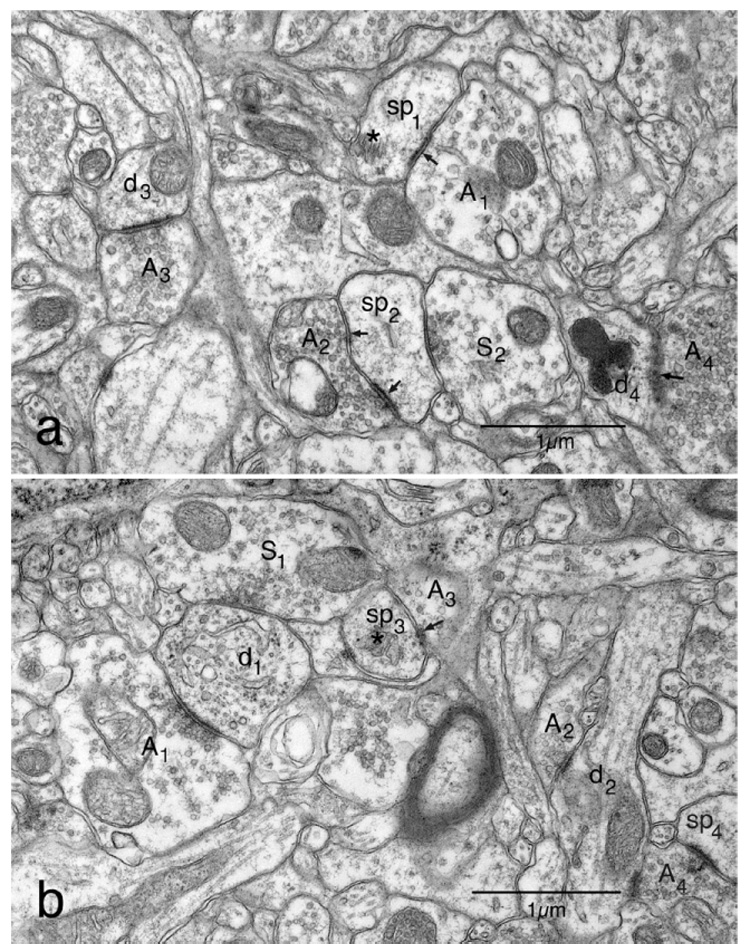
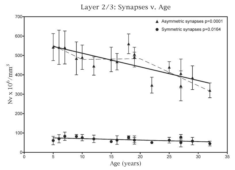
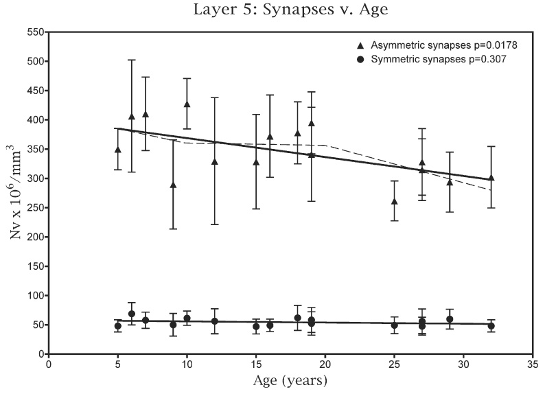
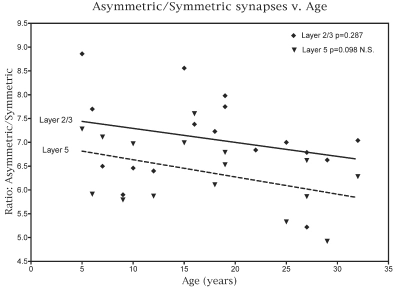
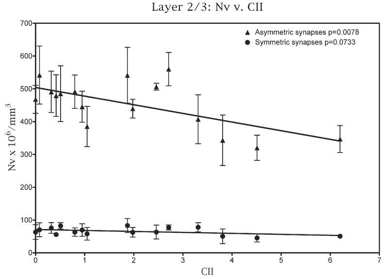
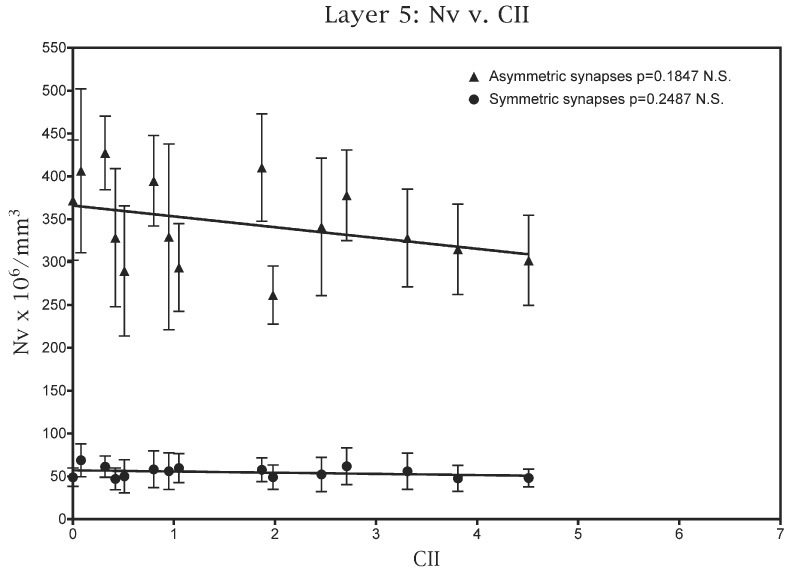
An electron microscopic analysis has been carried out on the effects of age on the numerical density of both excitatory (asymmetric) and inhibitory (symmetric) synapses in the neuropil of layers 2/3 and of layer 5 in area 46 from the frontal cortex of behaviorally tested rhesus monkeys. There is no change in the lengths of synaptic junctions with age or in the percentage distribution of synapses relative to the postsynaptic spines and dendritic shafts. However, in layers 2/3 there is an overall loss of about 30% of synapses from 5 to 30 years of age, and both asymmetric and symmetric synapses are lost at the same rate. In layer 5 the situation is different; the overall loss of synapses is only 20% and this is almost entirely due to a loss of asymmetric synapses, since there is no significant loss of symmetric synapses from this layer with age. When the synapse data are correlated with the overall cognitive impairment shown by the monkeys, it is found that there is a strong correlation between the numerical density of asymmetric synapses in layers 2/3 and cognitive impairment, with a weaker correlation between symmetric synapse loss and cognitive impairment. In layer 5 on the other hand there is no correlation between synapse loss and cognitive impairment. However synapse loss is not the only factor causing cognitive impairment, since in previous studies of area 46 we have found that age-related alteration in myelin in this frontal area also significantly contributes to cognitive decline. The synapse loss is also considered in light of earlier studies, which show that the frequency of spontaneous excitatory synaptic responses is reduced with age in layers 2/3 neurons.
Figures
References
-
- Adams I. Comparison of synaptic changes in the precentral and postcentral cerebral cortex of aging humans: a quantitative ultra-structural study. Neurobiol Aging. 1987;8:203–212. - PubMed
-
- Calverley RKS, Lewis DA, Jones DG. Estimation of the numerical density of synapses in rat neocortex: comparisons of the ‘disector’ with an ‘unfolding’ method. J Neurosci Methods. 1988;23:195–205. - PubMed
-
- Chang Y-M, Rosene DL, Killiany RJ, Mangiamele LA, Luebke JI. Increased action potential firing rates of layers 2/3 pyramidal cells in the prefrontal cortex are significantly related to cognitive performance in aged monkeys. Cereb Cortex. 2005;15:409–418. - PubMed
-
- Chen KS, Masliah E, Mallory M, Gage FH. Synaptic loss in cognitively impaired aged rats is ameliorated by chronic human growth factor infusion. Neuroscience. 1995;68:19–27. - PubMed
-
- Colonnier M. Synaptic patterns on different cell types in the different laminae of the cat visual cortex. Brain Res. 1968;9:268–287. - PubMed
Publication types
MeSH terms
Grants and funding
LinkOut - more resources
Full Text Sources
Other Literature Sources
Medical
