. 2008 Mar 7;132(5):807-17.
doi: 10.1016/j.cell.2007.12.041.

Structural basis of membrane invagination by F-BAR domains

Affiliations

Structural basis of membrane invagination by F-BAR domains

Adam Frost et al. Cell. .

Abstract

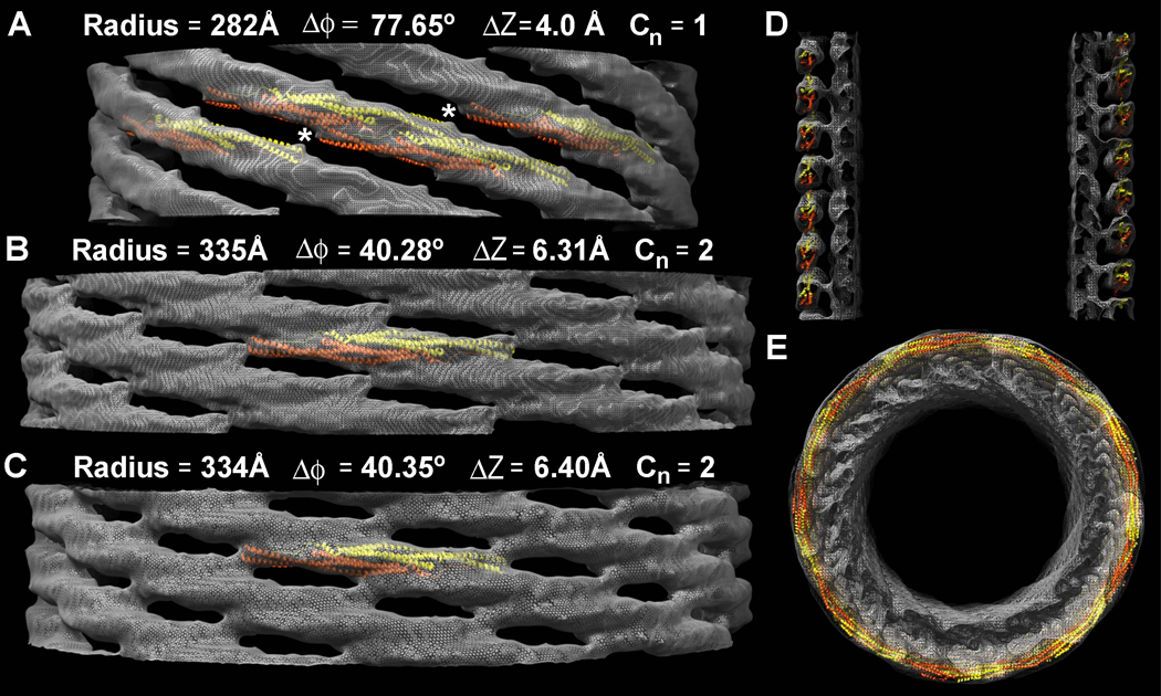
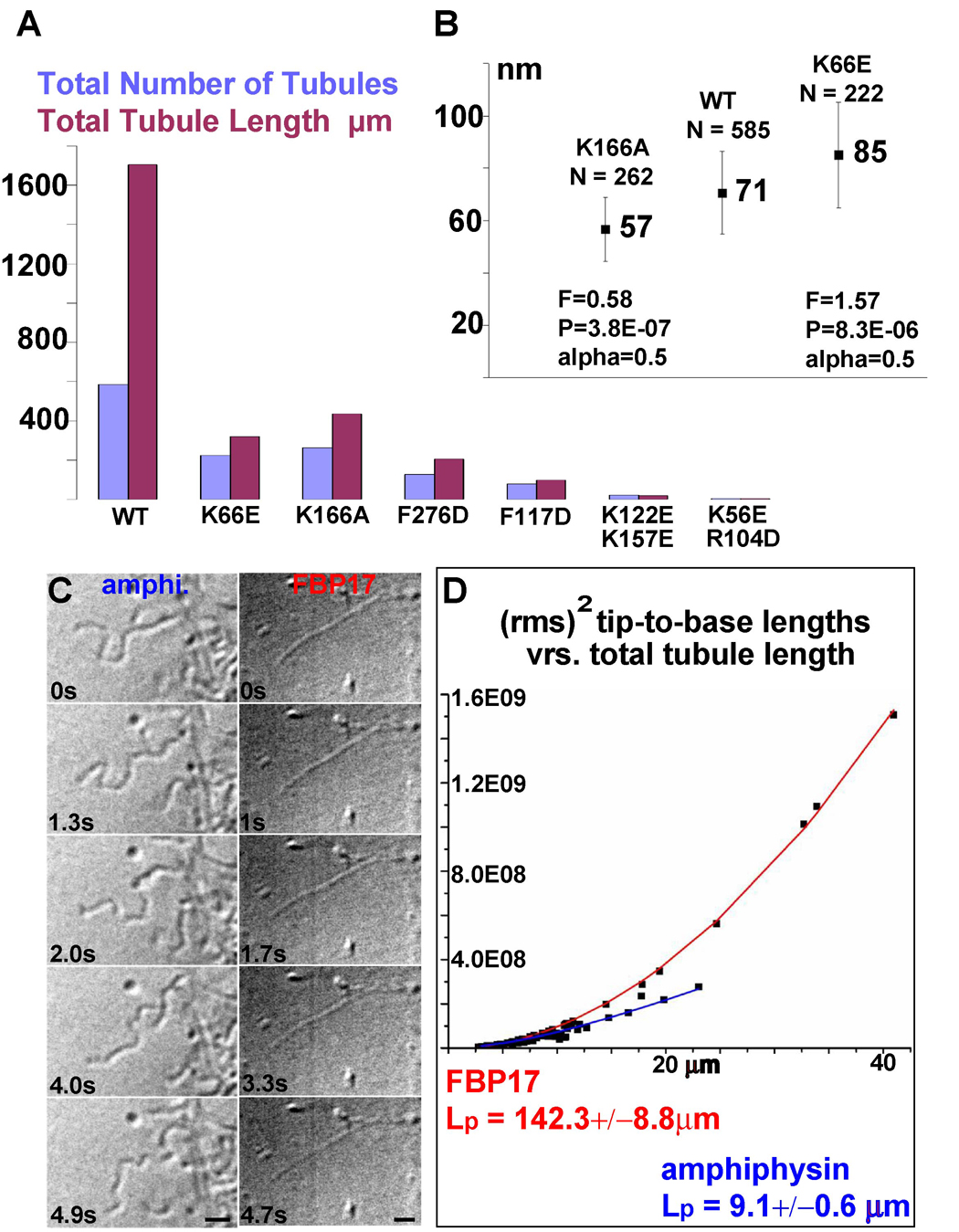
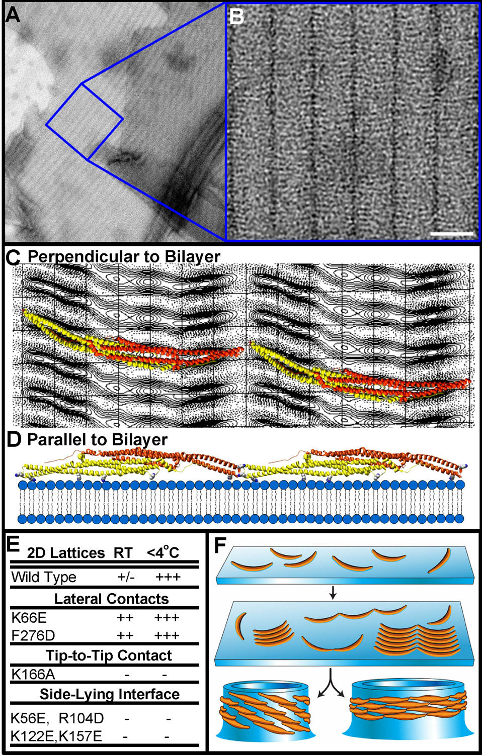
BAR superfamily domains shape membranes through poorly understood mechanisms. We solved structures of F-BAR modules bound to flat and curved bilayers using electron (cryo)microscopy. We show that membrane tubules form when F-BARs polymerize into helical coats that are held together by lateral and tip-to-tip interactions. On gel-state membranes or after mutation of residues along the lateral interaction surface, F-BARs adsorb onto bilayers via surfaces other than their concave face. We conclude that membrane binding is separable from membrane bending, and that imposition of the module's concave surface forces fluid-phase bilayers to bend locally. Furthermore, exposure of the domain's lateral interaction surface through a change in orientation serves as the crucial trigger for assembly of the helical coat and propagation of bilayer bending. The geometric constraints and sequential assembly of the helical lattice explain how F-BAR and classical BAR domains segregate into distinct microdomains, and provide insight into the spatial regulation of membrane invagination.


Figures

Figure 1
Figure 1. F-BAR versus N-BAR Tubulation in Living Cells: Spontaneous Segregation, Differences in Diameter and Rigidity
A) COS7 cell simultaneously transfected with amphiphysin2-GFP (green) and RFP-FBP17 (red) produces tubular networks in which the two proteins segregate from each other. Insets show the GFP, RFP, and merged channels. B) High magnification image of a cell transfected with GFP-CIP4 (left) and mRFP-FBP17 (middle) demonstrating the absence of segregation between the two proteins (merge; right). C) large invaginations of the plasma membrane observed by electron microscopy of thin-sections from COS7 cells transfected with full-length human GFP-FBP17 and D) GFP-CIP4; in comparison with the smaller tubules formed by amphiphysin2-GFP (E). Bars (A) 1 µm, (B) 0.5 µm, (C–E) 70 nm.
Figure 2
Figure 2. Reconstitution of CIP4 F-BAR Induced Tubulation and Segregation from Endophilin N-BAR Domains in vitro
A) electron (cryo)micrograph of a nascent tubule generated in vitro by F-BAR domains (human CIP4, residues 1–284). The yellow arrow points to the demarcation between the membrane surface with and without F-BAR domains, revealing a smooth bilayer to the right and adsorbed protein to the left, as seen in the 2x enlarged inset surrounded by the yellow box. To the left of the yellow arrow, the curvature of the membrane has changed little, if at all, despite the presence of bound proteins. Induction of tubule formation accompanies self-organization of F-BAR domains into a helical coat (cyan arrow and enlarged inset). B) Histogram of tubule widths generated in vitro measured from electron (cryo)micrographs. C) electron (cryo)micrograph of a tubule following temperature annealing and its corresponding Fourier Transform (D), which displays high signal-to-noise ratio layerlines beyond ~27 Å. E,F) Liposomes co-incubated with F-BAR (CIP4) and N-BAR (endophilin-1) proteins in vitro observed after negative staining with uranyl formate (E) or uranyl acetate (F), displaying contiguous membrane tubules whose change in diameter corresponds with the change in the radius of curvature for F-BAR versus N-BAR domains. Bars (A) 300 Å; (C) 25 nm; (E,F) 40 nm.
Figure 3
Figure 3. Single Particle Helical Reconstruction of a CIP4 F-BAR Domain-Induced Membrane Tubule
A) Surface of a ~67 nm diameter membrane tubule at ~17 Å resolution. The protein coat is colored blue-gray and the underlying membrane is green. B) Zoom in on the lattice seen orthogonal to the cylindrical axis, highlighting the tip-to-tip interactions and the broad contacts between laterally-adjacent dimers. C) Cross-sectional slab through one dimer parallel with the plane of the tip-to-tip interaction. There are four clearly resolved points of membrane binding. The hydrophobic core of the phospholipid bilayer is ~26 Å thick and the headgroup regions are ~12Å thick.
Figure 4
Figure 4. Fitting F-BAR Crystal Structures into the CryoEM Map Reveals Membrane-Binding Residues and Possible Lattice Contacts
A) Surface representation of a membrane tubule perpendicular to the cylindrical axis, focused on the interactions between four neighboring F-BAR molecules. The underlying membrane is colored in grey and the protein coat in grey mesh. One monomer of each F-BAR module is in yellow, the other in orange-red. Conserved residues hypothesized to contribute to the tip-to-tip and lateral interactions are annotated and shown with space-filling atoms. B) Cross-sectional slab through one dimer parallel with the plane of the tip-to-tip interactions. The four resolved points of membrane binding correspond with clusters of conserved, cationic residues found along the concave faces of both dimers, where R/K indicates the amino acid found in CIP4 or FBP17, respectively (Shimada et al., 2007). C) Representative images of COS7 cells with high levels of expression of wild type or mutated constructs of GFP-FBP17 or D) GFP-CIP4. Some mutations completely abolish membrane localization, while others only compromise tubule formation. Bars 10 µm.
Figure 5
Figure 5. Independent Reconstructions of Tubules with Different Diameters and Symmetries
A) The narrowest tubule reconstructed is ~56 nm in diameter, with ~8 tip-to-tip dimers around its circumference. Tilting the long axis of the dimer relative to the cylindrical axis produces a narrower tubule. In this case, the dimers are so steeply tilted that the tip-to-tip contacts appear to be broken (white asterisks). The tubule has no rotational symmetry; the fundamental (J+1) helical symmetry does not describe an inter-molecular contact. Only the near side of the lattice is shown and the underlying membrane has been masked out to emphasize differences in the protein coat. Atomic models of F-BAR domains were fit into the map as rigid bodies. B&C) Two tubules with the same apparent diameter and ~9.5 tip-to-tip dimers around their circumference have resolvable differences in their helical symmetry. D) Central section along the longitudinal axis of the thinnest tubule shown in ‘A’, demonstrating that the density of the protein coat accommodates rigid atomic models of the F-BAR module that are tilted relative to the cylindrical axis, but whose radius of curvature is unchanged. E) View along the cylindrical axis of the thinner reconstruction shown in ‘A’ and ‘D’.
Figure 6
Figure 6. Mutant Phenotypes & Tubule Persistence Lengths
A) Quantification of total tubule number, total tubule length (sum of all tubule lengths measured) as determined from 50 low magnification images evenly sampling one EM grid. B) Quantification of mean tubule diameter from low-magnification images like those in Figure S5A–B. Error bars are the standard deviation for the population of tubules. C) Still images of video-DIC recordings of amphiphysin and FBP17. D) Each data point is the rms2 (root mean square to the square) of 100 tip-to-base measurements of a tubule as a function of total tubule length. The persistence length (Lp) was determined by fitting the data with a theoretical curve according to the equation: δR2(t) = 2(Lp)2[x/Lp − 1 + e(−x Lp)]; where δR2(t) is the rms2, x the total length and Lp the persistence length (Derenyi et al., 2002; Le Goff et al., 2002a; Le Goff et al., 2002b). Bars (A,B) 10 µm (C) 5 µm.
Figure 7
Figure 7. F-BAR Domains Bind to Flat Membranes via a Surface Other Than Their Concave Face
A) Electron micrograph of negatively-stained membranes that were pre-cooled before incubation with human FBP17 F-BAR domains (see Figure S6). B) Higher magnification of the 2D F-BAR lattice; unit cell a = 33Å b = 214Å γ = 91.0° C) Projection view of the F-BAR domain, calculated from a 3D data set composed of images from a single-axis tilt series over ±40° (representative lattice lines in Figure S7, crystal statistics in Table S1). Ribbon diagrams of the domain are superimposed over the projection image, as seen from the orientation with the highest correlation perpendicular to the membrane surface. D) Two dimers interacting tip-to-tip viewed parallel with the membrane surface, or rotated by 90° with respect to the view in ‘C’. Residues likely to mediate membrane-binding in this side-lying state are shown as space-filing atoms from left-to-right: K122, R104, K56, and K157 (see also Figures S4–S5). E) Table of mutant propensity for forming flat lattices at different temperatures. F) Proposed model in which tubule formation proceeds through observable intermediate steps. F-BARs can bind to flat or curved bilayers, clustering in arrays by forming intermolecular interactions. Following the transition to high-affinity binding of the dimer’s concave surface, formation of the lateral contacts triggers the vectorial assembly of the helical coat and drives membrane invagination.

Comment in

References

    1. Ayton GS, Blood PD, Voth GA. Membrane remodeling from N-BAR domain interactions: insights from multi-scale simulation. Biophys J. 2007;92:3595–3602. - PMC - PubMed
    1. Blood PD, Voth GA. Direct observation of Bin/amphiphysin/Rvs (BAR) domain-induced membrane curvature by means of molecular dynamics simulations. Proc Natl Acad Sci U S A. 2006;103:15068–15072. - PMC - PubMed
    1. Brett TJ, Traub LM. Molecular structures of coat and coat-associated proteins: function follows form. Curr Opin Cell Biol. 2006;18:395–406. - PubMed
    1. Bruinsma R, Pincus P. Protein aggregation in membranes. Current Opinion in Solid State & Materials Science. 1996;1:401–406.
    1. Casal E, Federici L, Zhang W, Fernandez-Recio J, Priego EM, Miguel RN, DuHadaway JB, Prendergast GC, Luisi BF, Laue ED. The crystal structure of the BAR domain from human Bin1/amphiphysin II and its implications for molecular recognition. Biochemistry. 2006;45:12917–12928. - PMC - PubMed

Publication types

MeSH terms