. 2008 Mar 7;132(5):875-86.
doi: 10.1016/j.cell.2008.02.019.

Targeted deletion reveals essential and overlapping functions of the miR-17 through 92 family of miRNA clusters

Affiliations

Targeted deletion reveals essential and overlapping functions of the miR-17 through 92 family of miRNA clusters

Andrea Ventura et al. Cell. .

Abstract

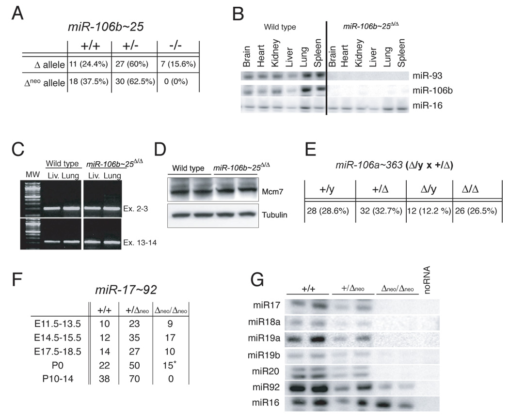
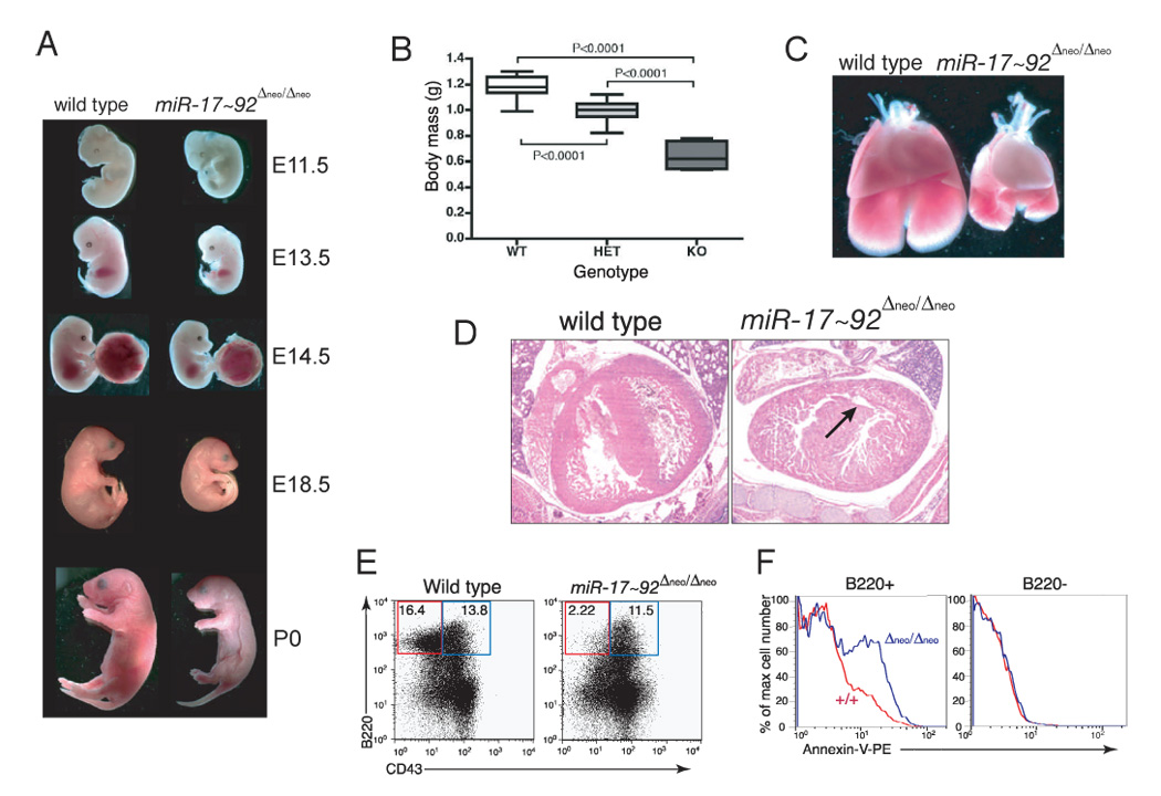
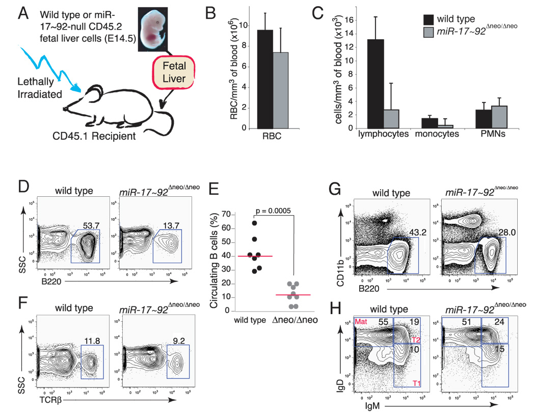
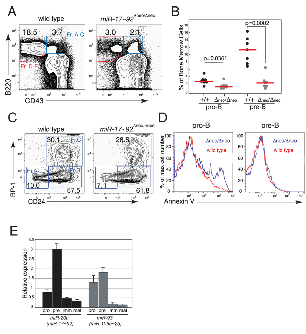
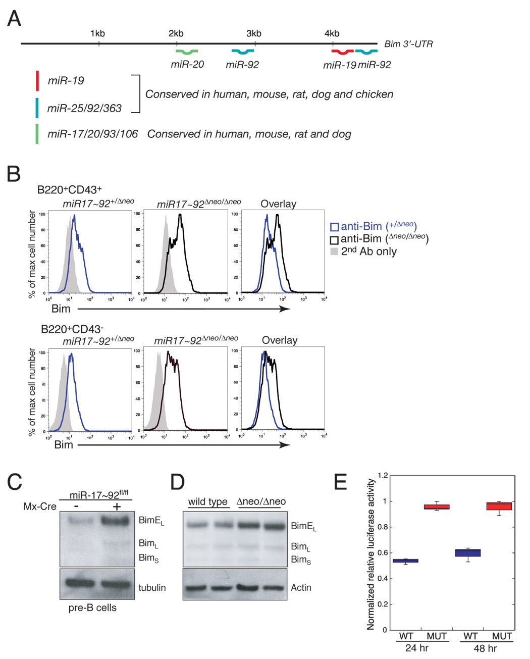
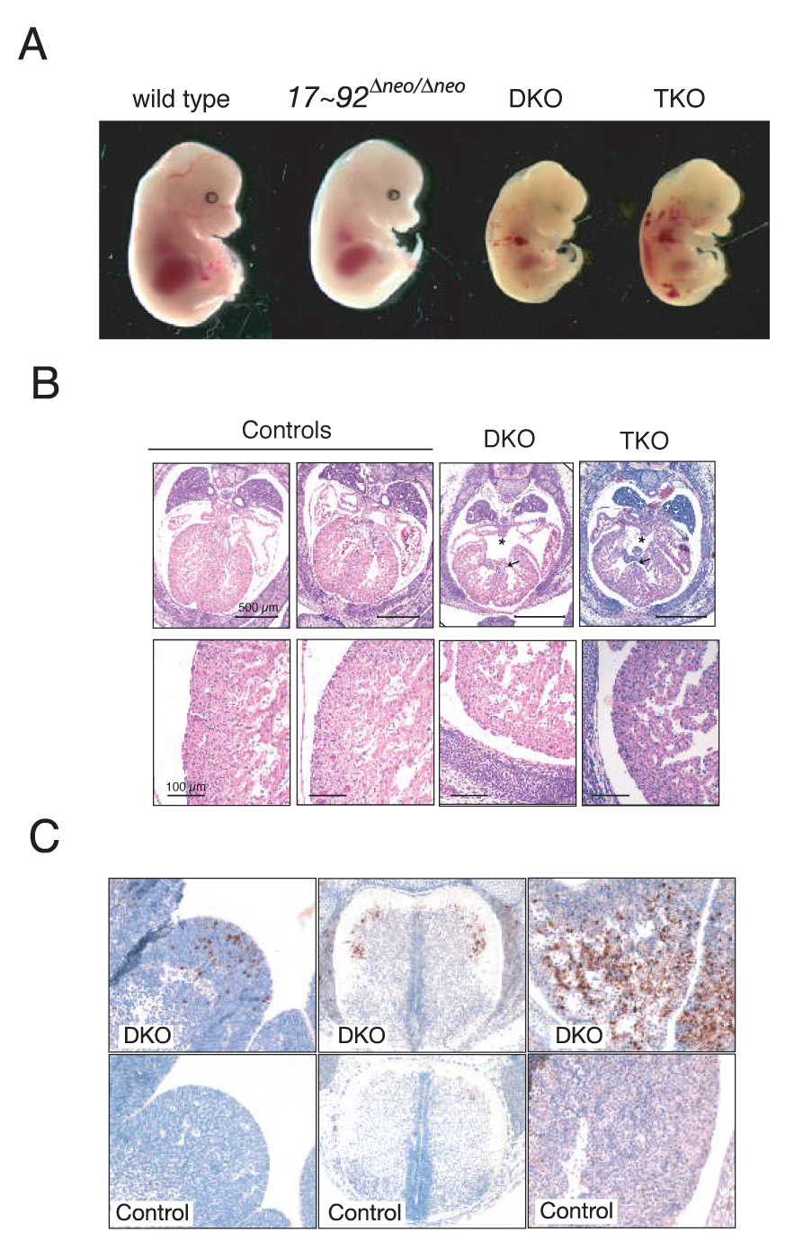
miR-17 approximately 92, miR-106b approximately 25, and miR-106a approximately 363 belong to a family of highly conserved miRNA clusters. Amplification and overexpression of miR-1792 is observed in human cancers, and its oncogenic properties have been confirmed in a mouse model of B cell lymphoma. Here we show that mice deficient for miR-17 approximately 92 die shortly after birth with lung hypoplasia and a ventricular septal defect. The miR-17 approximately 92 cluster is also essential for B cell development. Absence of miR-17 approximately 92 leads to increased levels of the proapoptotic protein Bim and inhibits B cell development at the pro-B to pre-B transition. Furthermore, while ablation of miR-106b approximately 25 or miR-106a approximately 363 has no obvious phenotypic consequences, compound mutant embryos lacking both miR-106b approximately 25 and miR-17 approximately 92 die at midgestation. These results provide key insights into the physiologic functions of this family of microRNAs and suggest a link between the oncogenic properties of miR-17 approximately 92 and its functions during B lymphopoiesis and lung development.


Figures

Figure 1
Figure 1. Generation of targeted deletions in miR-17~92, miR-106b~25 and miR-106a~363
(A) Schematic representation of the three miRNA clusters. Pre-miRNAs are indicated as color-coded boxes. Black boxes correspond to the mature miRNA. The color code identifies miRNAs with the same seed sequence. (B) Sequence comparison of miRNAs expressed from the three miRNA clusters. MiRNAs with the same seed sequence (bold) are grouped together and color-coded according to panel (A). (C) Targeting scheme for the deletion of miR-106b~25. Mcm-7 exons are represented by black boxes. The mature miRNAs are represented by colored boxes. (D) Genotyping PCR performed on tail DNA showing germline transmission of the targeted miR-106b~25 alleles. (E) Targeting scheme for the deletion of miR-106a~363. (F) Genotyping PCR performed on tail DNA showing germline transmission of the targeted miR-106a~363 allele. (G) Targeting scheme for the deletion of miR-17~92. (H) Genotyping PCR performed on DNA extracted from E13.5 embryos showing germline transmission of the miR-17~92-Δneo allele. (I) Genotyping PCR performed on DNA extracted from miR-17~92fl/fl;Cre-ERT2+ mouse embryo fibroblasts. Cre-mediated deletion of miR-17~92 was assessed 5 days after infection with Cre-expressing recombinant Adenoviruses (Ad-Cre) or after treatment with 4-Hydroxytamoxifen.
Figure 2
Figure 2. Deletion of miR-106a~363 or miR-106b~25 results in viable and fertile mice
(A) Genotypes of mice from miR-106b~25+/Δneo and miR-106b~25+/Δ intercrosses. (B) RPAs on total RNA extracted from wild-type and miR-106b~25Δ/Δ adult tissues. (C) RT-PCR on RNA extracted from miR-106b~25Δ/Δ and wild-type lungs and livers. Primers designed to amplify the junctions of exons 13–14 were used to determine whether the deletion of miR-106b~25 from intron 13 affects splicing between these two exons. Amplification of the junction between exons 2 and 3 was used as control. (D) Mcm-7 Western blot on whole cell lysates from wild type and miR-106b~25 Δneo/Δneo mouse embryo fibroblasts. (E) Genotypes of mice from miR-106a~363Δ/Y × miR-106a~363+/Δ crosses. (F) Genotypes of mice from miR-17~92+/Δneo intercrosses determined at the indicated gestation stages. The asterisk indicates that all miR-17~92Δneo/Δneo newborn mice died soon after birth. (G) RPAs on total RNA from E13.5 embryos with the indicated genotypes.
Figure 3
Figure 3. Characterization of miR-17~92-deficient mice
(A) Macroscopic appearance of wild-type and miR-17~92-deficient embryos at various developmental stages. (B) Body mass of wild-type (WT), heterozygous (HET) and miR-17~92-null (KO) E18.5 embryos. The boxes indicate the 25th–75th percentile range. The horizontal lines represent the median, and the error bars indicate the lowest and highest values. P values were calculated using the two-tailed t test. (C) Macroscopic appearance of wild-type and miR-17~92-deficient lungs and hearts from E17.5 embryos. Notice the markedly hypoplastic lungs in miR-17~92-null embryos. (D) Hematoxylin-eosin staining of comparable transverse sections through the hearts of E18.5 wild-type and miR-17~92-null embryos. The arrow indicates a ventricular septal defect. (E) Flow cytometry plots of fetal liver cells from E18.5 wild-type and mutant embryos showing a marked reduction of B220+;CD43−;IgM− pre-B cells in mice lacking miR-17~92. The results are representative of three independent experiments. (F) Annexin-V staining showing increased apoptosis in miR-17~92-deficient fetal liver B cell progenitors (blue) compared to wild-type control (red). The right panel shows the same analysis performed on B220-negative cells.
Figure 4
Figure 4. Deletion of miR-17~92 impairs adult B cell development
(A) Schematic of the reconstitution experiment. (B) Total number of circulating RBC per mm3 of blood in mice reconstituted with wild-type (black) or miR-17~92-deficient (gray) fetal livers 8 weeks post transplantation. Error bar indicates +1 S.D. The average of seven wild-type and eight miR-17~92-deficient mice is shown. (C) Differential white blood cells counts in mice reconstituted with wild-type (black) and miR-17~92-deficient (gray) fetal livers. PMNs: polymorphonucleates. Error bar is +1 S.D. n=7 wild-type and 8 miR-17~92 mutants. (D) Representative flow cytometry plot showing reduction of B220+ B cells in the blood of mice reconstituted with miR-17~92-deficient fetal liver cells. (E) Dot plot showing the number of circulating B cells in reconstituted mice. Horizontal bars in red indicate the median value for each genotype. (F) Flow cytometry plot of T cells in the periphery of reconstituted mice. On representative example per genotype is shown. (G) Flow cytometry plot of spleen cells stained for CD11b and B220, showing a reduction in splenic B cells in mice reconstituted with miR-17~92-deficient cells. (H) Flow cytometry plot on B-220-positive splenic B cells stained with IgM and IgD to determine the relative fraction of Transition (T1 and T2) and Mature B cells.
Figure 5
Figure 5. miR-17~92 regulates early B cell survival
(A) Representative flow cytometry plots of bone marrow cells from mice reconstituted with wild-type or miR-17~92-deficient fetal liver cells. (B) Percentage of pro-B and pre-B cells in reconstituted mice. The median (red bars) and the p-values are indicated. (C) Representative flow cytometry plots showing relative proportion of fraction A–C pro-B cells in the bone marrow of reconstituted mice. (D) Histogram overlays of Annexin-V staining of pro-B (left panel) and pre-B cells (right panel), showing increased apoptosis of pro-B cells in the bone marrow of mice reconstituted with miR-17~92-deficient fetal liver cells. The analysis was performed on the bone marrow of mice reconstituted with wild-type (red line) or miR-17~92-deficient (blue line) fetal livers. (E) qPCR analysis of miR-17~92 and miR-106b~25 expression during B cell development. Pro-, pre-, immature and mature B cells were sorted and their RNA assayed for miR-20a and miR-93 expression. Data are normalized to sno-142 expression.
Figure 6
Figure 6. Bim regulation by miR-17~92
(A) Schematic representation of the 3’UTR of Bim and the predicted binding sites for members of the miR-17~92 cluster and its paralogs. (B) Detection of Bim expression by flow cytometry in miR-17~92+/Δneo and miR-17~92Δneo/Δneo pro-B (upper panels) and pre-B cells (lower panel) from E18.5 fetal livers. (C) Detection of Bim expression by western blotting in pre-B cells from adult mice following conditional deletion of miR-17~92. miR-17~92fl/fl (left lane) and miR-17~92fl/fl;Mx-Cre mice (right lane) were treated with pI:pC to induce expression of the Cre transgene. Pre-B cells were sorted and analyzed by Western blotting four weeks after treatment. (D) Detection of Bim expression by western blotting in E18.5 lungs from wild-type and miR-17~92Δneo/Δneo embryos. (E) Box plot of normalized relative luciferase activity of wildtype and mutant Bim UTR constructs 24 and 48 hours after transfection in HeLa cells. Data represent three independent experiments performed in triplicate.
Figure 7
Figure 7. Functional cooperation between miR-17~92 and miR-106b~25
(A) Effects of compound mutations of miR-17~92, miR-106b~25 and miR-106a~363 on embryonic development. E14.5 embryos with the indicated genotypes are shown. DKO = miR-17~92Δ/Δ;miR-106b~25Δ/Δ; TKO = miR-17~92Δ/Δ;miR-106b~25Δ/Δ;miR-106A~363Δ/y (B) Representative transverse sections of E14.5 mice showing the presence of ventricular (arrow) and atrial (asterisk) septal defects in DKO and TKO embryos (upper panels). Lower panels show a higher magnification of the lateral wall of the left ventricle. Notice the thinner wall in double and triple mutant embryos. The black bars correspond to 500 µm (upper panels) and 100 µm (lower panels). The controls were a miR-17~92+/+;miR-106b~25+/Δ;miR-106a~363Δ/y (left) and a miR-17~92+/Δ;miR-106b~25+/Δ;miR-106a~363wt (right) mouse. (C) Cleaved caspase 3 immunostaining of sections from an E14.5 DKO miR-17~92Δ/Δ;miR-106b~25Δ/Δ (DKO) embryo, and a miR-17~92+/+; miR-106b~25Δ/Δ (control) embryo showing the presence of areas of apoptosis in the liver (right panels), the ventral horns of the spinal cord (middle), and the lateral ganglionic eminence (left) in the DKO.

Comment in

References

    1. Abbott AL, Alvarez-Saavedra E, Miska EA, Lau NC, Bartel DP, Horvitz HR, Ambros V. The let-7 MicroRNA family members mir-48, mir-84, and mir-241 function together to regulate developmental timing in Caenorhabditis elegant. Dev Cell. 2005;9:403–414. - PMC - PubMed
    1. Bagga S, Bracht J, Hunter S, Massirer K, Holtz J, Eachus R, Pasquinelli AE. Regulation by let-7 and lin-4 miRNAs results in target mRNA degradation. Cell. 2005;122:553–563. - PubMed
    1. Bartel DP. MicroRNAs: genomics, biogenesis, mechanism, and function. Cell. 2004;116:281–297. - PubMed
    1. Bartel DP, Chen CZ. Micromanagers of gene expression: the potentially widespread influence of metazoan microRNAs. Nat Rev Genet. 2004;5:396–400. - PubMed
    1. Bernstein E, Kim SY, Carmell MA, Murchison EP, Alcorn H, Li MZ, Mills AA, Elledge SJ, Anderson KV, Hannon GJ. Dicer is essential for mouse development. Nat Genet. 2003;35:215–217. - PubMed

Publication types