. 2008 Jun;1783(6):1140-9.
doi: 10.1016/j.bbamcr.2008.01.029. Epub 2008 Feb 19.

Localization of A20 to a lysosome-associated compartment and its role in NFkappaB signaling

Affiliations

Localization of A20 to a lysosome-associated compartment and its role in NFkappaB signaling

Lianyun Li et al. Biochim Biophys Acta. 2008 Jun.

Abstract

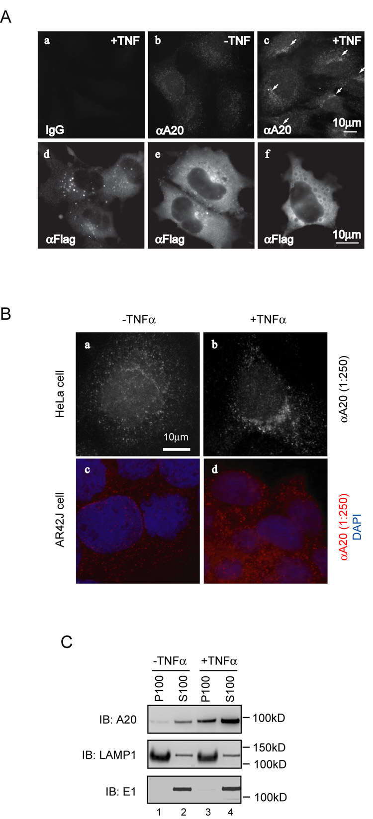
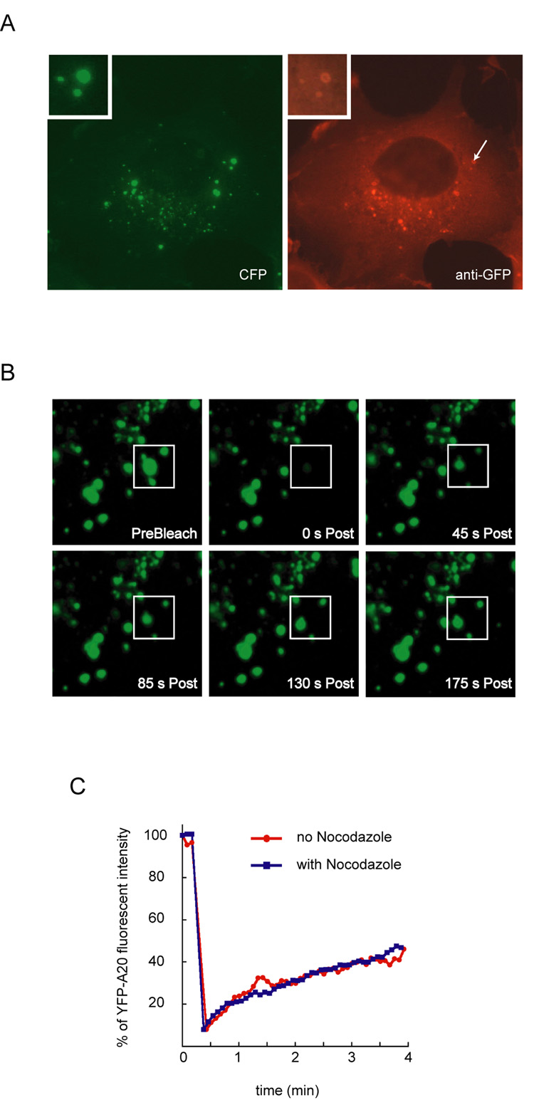
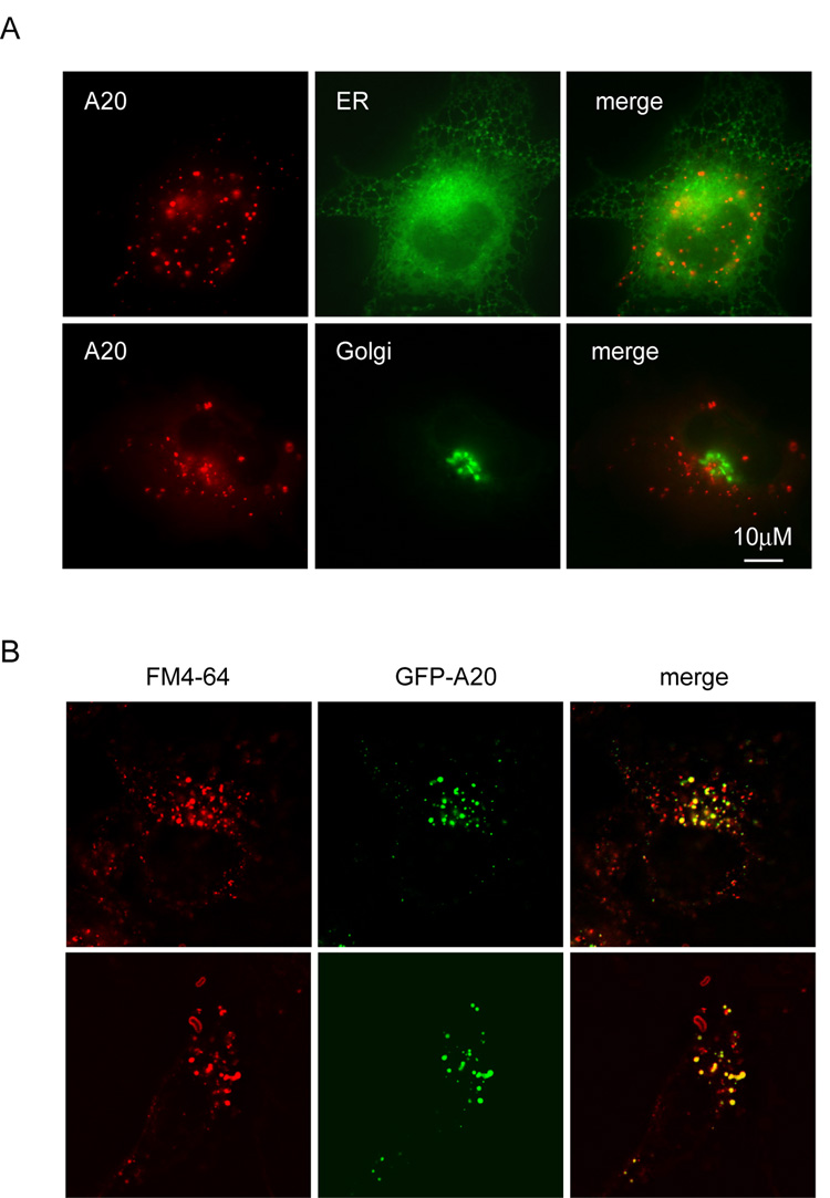
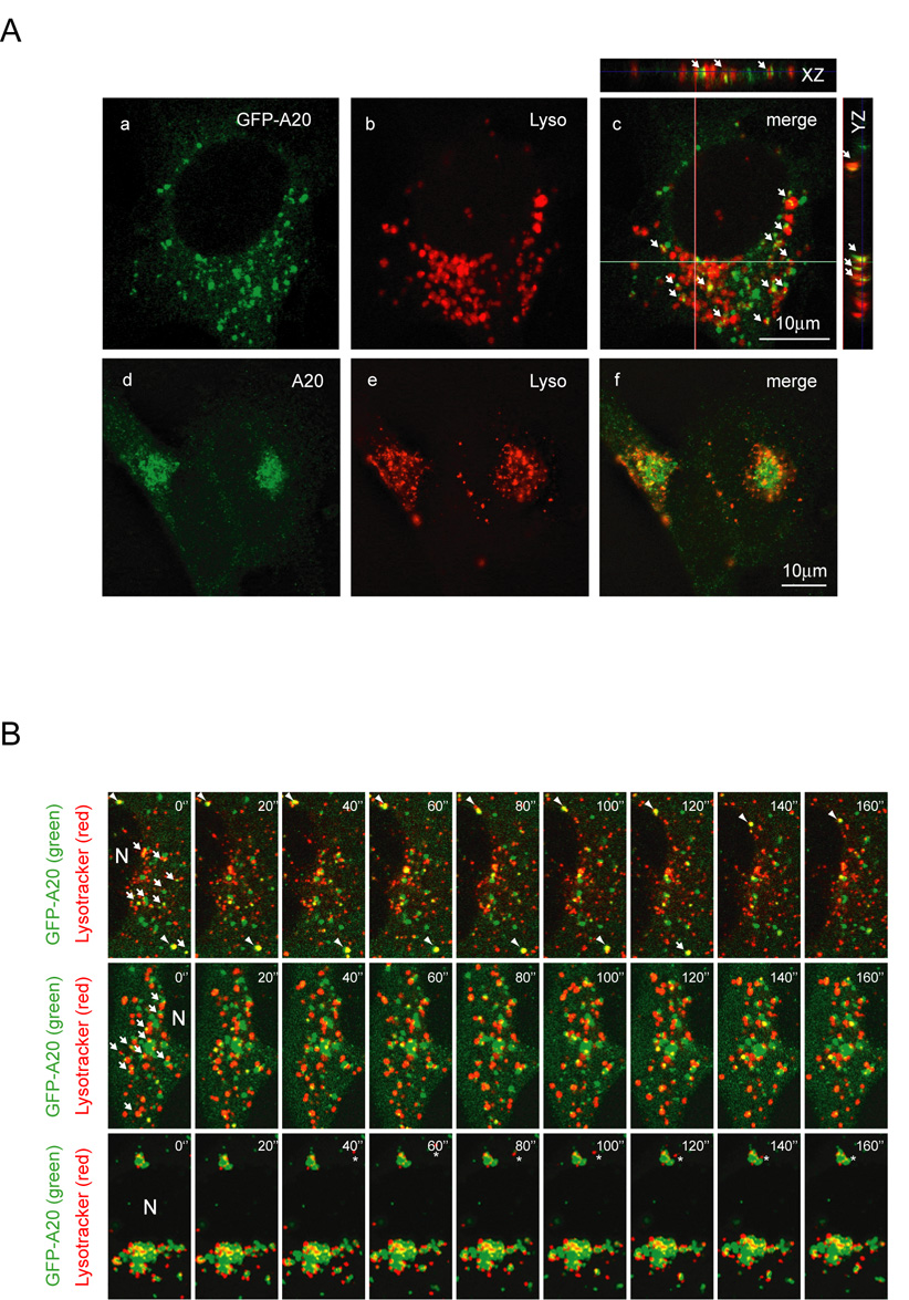
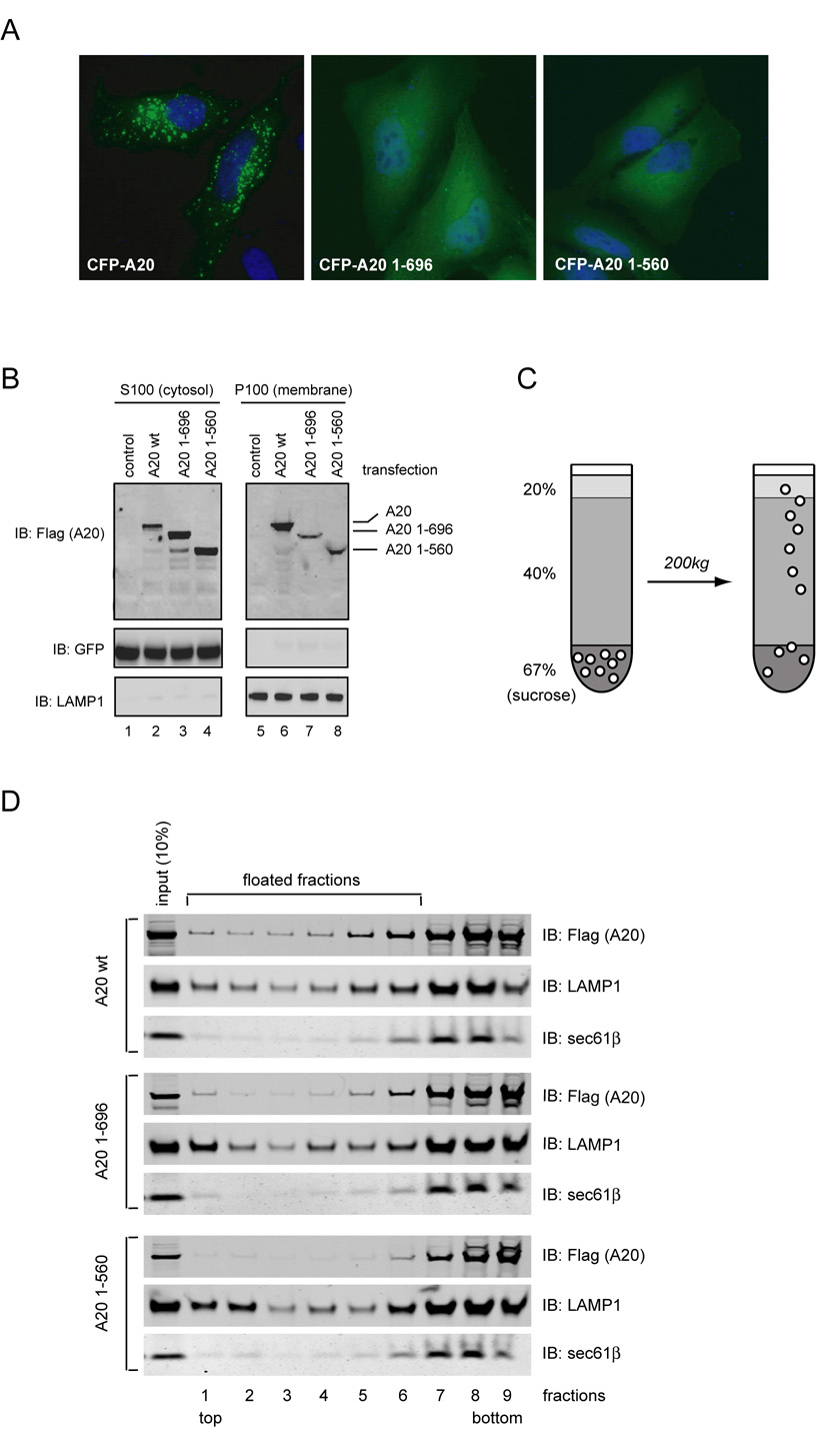
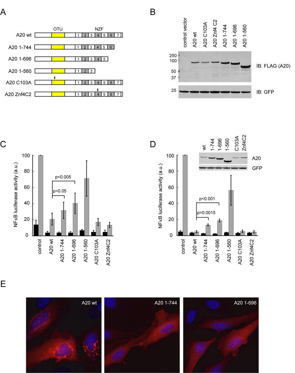
A20 is a tumor necrosis factor (TNF)-inducible zinc finger protein that contains both ubiquitinating and deubiquitinating activities. A20 negatively regulates NFkappaB (nuclear factor kappaB) signaling induced by TNF receptor family and Toll-like receptors, but the mechanism of A20 action is poorly defined. Here we show that a fraction of endogenous and ectopically expressed A20 is localized to an endocytic membrane compartment that is in association with the lysosome. The lysosomal association of A20 requires its carboxy terminal zinc finger domains, but is independent of its ubiquitin-modifying activities. Interestingly, A20 mutants defective in membrane association also contain reduced NFkappaB inhibitory activity. These findings suggest the involvement of a lysosome-associated mechanism in A20-dependent termination of NFkappaB signaling.


Figures

Figure 1
Figure 1. A20 is localized to an intracellular membrane compartment
(A) (a–c) Subcellular localization of endogenous A20 in HeLa cells. HeLa cells treated with TNFα (80ng/ml, 6h) or non-treated were stained with either an A20 specific monoclonal antibody (αA20) or control IgG. Arrows indicate the perinuclear enrichment of A20-containing vesicles. Same exposure time was used for these images. (d–f) COS7 cells expressing Flag-tagged wild type A20 were stained with an anti-Flag antibody (αFlag). Images represent cells with A20-containing vesicles of different size. (B) Membrane association of endogenous A20. HeLa cells (a, b) or AR42J cells (c, d) treated with TNFα or non-treated were stained with an anti-A20 monoclonal antibody at the indicated concentration. The bottom panels also show the nuclei stained by DAPI in blue. Images were obtained with a Zeiss Axiovert fluorescence microscope equipped with a 63X oil immersion Plan-Apochromat objective (N/A 1.4). (C) Biochemical fractionation experiment. HeLa cells untreated (−TNFα) or treated with TNFα (+TNFα) were lysed, fractionated, and analyzed by immunoblotting with antibodies to either A20, the lysosomal membrane protein LAMP1, or the cytosolic protein E1. Note that the loading of the supernatant fraction is equivalent to ~30% of the pellet fraction. The small amount of LAMP1 in S100 fractions was likely due to incomplete fractionation.
Figure 2
Figure 2. Dynamic association of A20 with membrane
(A) Membrane association of CFP-tagged A20. COS7 cells transfected with a CFP-A20 expressing constructs were stained with an anti-GFP antibody (in red), which also recognized the CFP tag. The cells were also imaged using a band pass filter specific for CFP (in green). The insets show an enlarged view of a vesicle indicated by the arrow. (B) FRAP experiment demonstrates the dynamic association of A20 with membranes. A small area (indicated by the boxes) in a COS7 cell expressing YFP-tagged A20 was repeatedly targeted with a high intensity laser light to photobleach the YFP-A20 signal. The recovery of the fluorescent signal was monitored by time-lapse confocal microscopy. (C) Quantification of the FRAP experiments as in B. Where indicated, cells were first treated with nocodazole (10µg/ml) for 10min before photobleaching.
Figure 3
Figure 3. Localization of A20 to an endocytic compartment
(A) A20 is not localized to either the ER or Golgi. COS7 cells expressing CFP-tagged A20 together with YFP-tagged markers for either the ER or Golgi were imaged. (B) Localization of A20 to an endocytic compartment. COS7 cells expressing GFP-A20 were stained with FM4-64 and imaged. Shown are horizontal sections of two A20 expressing cells stained with FM4-64.
Figure 4
Figure 4. Interaction of A20-containing vesicles with the lysosome
(A) (a–c) Colocalization of GFP-A20 with the lysosome. COS7 cells expressing GFP-tagged A20 (green) were stained with Lysotracker (red). Images were obtained with a laser scanning confocal microscope. The smaller panels show vertical sections along the XZ and YZ axes indicated by the lines. Arrows highlight some examples of A20-containing vesicles that are either colocalized or tethered with the lysosome. (d–f) Partial colocalization of endogenous A20 with the lysosome. HeLa cells treated with TNFα for 6 hours were stained with an anti-A20 antibody (green) and Lysotracker (Lyso, red). Shown is a deconvoluted horizontal section obtained with an Axiovert fluorescence microscope. (B) Co-migration of A20-containing vesicles with the lysosomal vesicles. COS7 cells expressing GFP-tagged A20 were stained with Lysotracker, and imaged in real time by a laser scanning confocal microscope in the interlace mode (see online supplemental material for the movies). Shown are three examples representing A20 vesicles of different size. Arrows highlight some examples of A20-containing structures (green) that interact with the lysosome (red). Arrowheads in the top panels indicate one example of an A20 vesicle that co-migrates with a lysosome. Asterisks in the bottom panels indicate a lysosomal vesicle that is being targeted to an A20-containing membrane compartment. N, nuclei.
Figure 5
Figure 5. Membrane association of A20 involves its carboxy terminal zinc finger domains
(A) The membrane localization of A20 requires its carboxy terminal zinc finger domains. Shown are HeLa cells expressing the indicated CFP-tagged A20 variants (green). Nuclei were counter-stained with DAPI (blue). (B) 293T cells transfected with a GFP expressing plasmid together with constructs expressing the indicated A20 variants were lysed in a hypotonic buffer and fractionated as described in the materials and methods. The proteins in the membrane pellet fraction (P100) and the cytosol (S100) were analyzed by immunoblotting (IB) with the indicated antibodies. Note that the loading of the supernatant fraction is equivalent to ~10% of the pellet fraction. (C) Scheme of the membrane floatation experiment shown in (D). (D) Membrane association of A20 variants examined by a membrane floatation assay. Fractions were analyzed by immunoblotting (IB) with antibodies to the indicated proteins.
Figure 6
Figure 6. Inhibition of TNFα-mediated NFκB activation by A20 variants
(A) Schematic representation of the A20 constructs. Arrows indicate the position of point mutations. (OTU, ovarian tumor; NZF, Npl4 zinc finger) (B) Expression of A20 variants in 293T cells. A GFP expressing construct was co-transfected as a control. (C) NFκB inhibiting activity of A20 variants in 293T cells. Luciferase activity in extracts of TNFα- stimulated 293T cells expressing a NFκB luciferase reporter together with the indicated A20 variants were measured and normalized by the level of co-expressed GFP. Error bars show s.d. (n=6). (D) NFκB inhibiting activity of the A20 variants in primary embryonic fibroblast cells. The inset shows the expression of the A20 variants. Error bars show s.d. (n=3). (E) Membrane localization of A20 requires the last zinc finger domain. HeLa cells expressing the indicated Flag-tagged A20 variants were stained with anti-Flag antibody (red). Nuclei were counter-stained with DAPI (blue).

References

    1. Chen G, Goeddel DV. TNF-R1 signaling: a beautiful pathway. Science. 2002;296:1634–1635. - PubMed
    1. Karin M, Greten FR. NF-kappaB: linking inflammation and immunity to cancer development and progression. Nat Rev Immunol. 2005;5:749–759. - PubMed
    1. Karin M, Lin A. NF-kappaB at the crossroads of life and death. Nat Immunol. 2002;3:221–227. - PubMed
    1. Dempsey PW, Doyle SE, He JQ, Cheng G. The signaling adaptors and pathways activated by TNF superfamily. Cytokine Growth Factor Rev. 2003;14:193–209. - PubMed
    1. Hsu H, Huang J, Shu HB, Baichwal V, Goeddel DV. TNF-dependent recruitment of the protein kinase RIP to the TNF receptor-1 signaling complex. Immunity. 1996;4:387–396. - PubMed

Publication types

MeSH terms