. 2008 Apr 14:1204:16-23.
doi: 10.1016/j.brainres.2008.02.018. Epub 2008 Feb 19.

Antioxidants prevent ethanol-associated apoptosis in fetal rhombencephalic neurons

Affiliations

Antioxidants prevent ethanol-associated apoptosis in fetal rhombencephalic neurons

Angeline M Antonio et al. Brain Res. .

Abstract

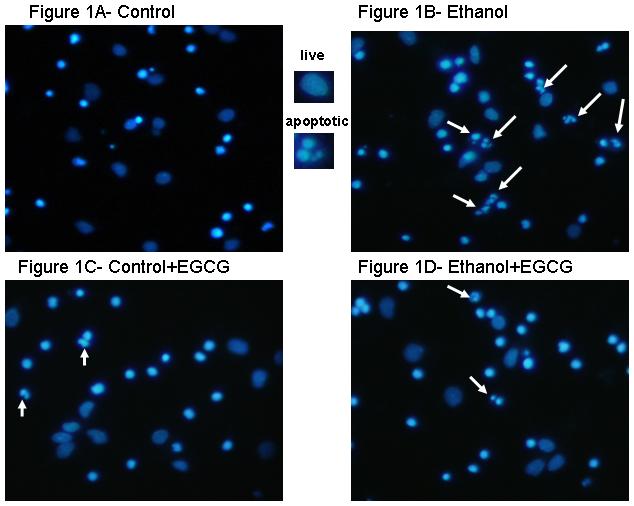
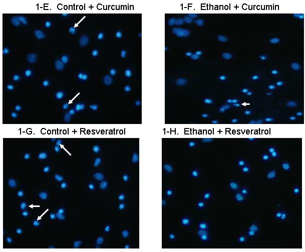
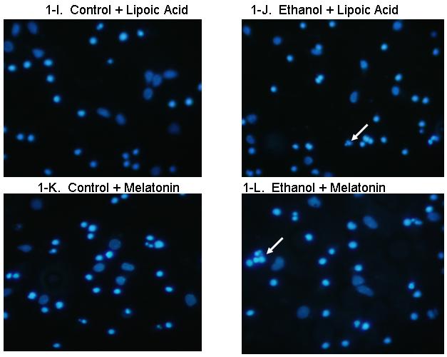
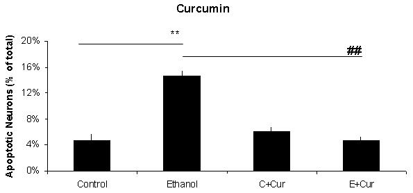
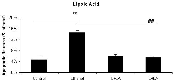
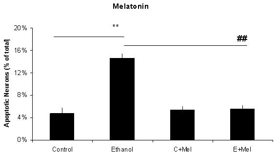
It is well known that ethanol damages the developing nervous system by augmenting apoptosis. Previously, this laboratory reported that ethanol augments apoptosis in fetal rhombencephalic neurons, and that the increased apoptosis is associated with reduced activity of the phosphatidylinositol 3-kinase pathway and downstream expression of pro-survival genes. Other laboratories have shown that another mechanism by which ethanol induces apoptosis in developing neurons is through the generation of reactive oxygen species (ROS) and the associated oxidative stress. The present study used an in vitro model to investigate the potential neuroprotective effects of several antioxidants against ethanol-associated apoptosis in fetal rhombencephalic neurons. The investigated antioxidants included three phenolics: (-)-epigallocatechin-3-gallate (EGCG), a flavanoid polyphenol found in green tea; curcumin, found in tumeric; and resveratrol (3,5,4'-trihydroxystilbene), a component of red wine. Additional antioxidants, including melatonin, a naturally occurring indole, and alpha-lipoic acid, a naturally occurring dithiol, were also investigated. These studies demonstrated that a 24-hour treatment of fetal rhombencephalic neurons with 75 mM ethanol caused a 3-fold increase in the percentage of apoptotic neurons. However, co-treatment of these cultures with any of the five different antioxidants prevented ethanol-associated apoptosis. Antioxidant treatment did not alter the extent of apoptosis in control neurons, i.e., those cultured in the absence of ethanol. These studies showed that several classes of antioxidants can exert neuroprotection against ethanol-associated apoptosis in fetal rhombencephalic neurons.


Figures

Figure 1
Figure 1. Ethanol augments apoptosis in fetal rhombencephalic neurons in vitro, and antioxidants prevent the pro-apoptotic effects of 75 mM ethanol
Figures 1A and 1B show fetal rhombencephalic neurons stained with Hoechst 33342 from cultures that were maintained under control conditions (no ethanol) or that were treated for the last 24 hours (DIV5 to DIV6) with 75 mM ethanol (ethanol). Control and ethanol-treated neurons treated with EGCG (1C, 1D), curcumin (1E, 1F), resveratrol (1G, 1H), LA (1I, 1J), and melatonin (1K, 1L) are also depicted. Hoechst-stained living and apoptotic neurons were identified as described. A higher magnification (see inset) was used to identify neurons that exhibit the characteristics of apoptotic cells. Arrows point to one or a cluster of fragmented/apoptotic nuclei.
Figure 2
Figure 2. A graphic depiction of the percentage of apoptotic neurons in cultures of fetal rhombencephalic neurons that were maintained under control conditions (no ethanol) or in the presence of 75 mM ethanol and treated with EGCG (2A), curcumin (2B), resveratrol (2C), LA (2D) or melatonin (2E)
The ** indicates that ethanol treatment significantly augmented apoptosis (p < .01); this treatment tripled the number of apoptotic neurons. Each of these antioxidants exerted a significant main effect when cultures of fetal rhombencephalic neurons were co-treated with the antioxidant and ethanol [(1 μM EGCG (F(1,23) = 13.5, p < .01), 1 μM curcumin (F(1,23) = 20.2, p < .01), 10 μM resveratrol (F(1,23) = 22.3, p < .01), 10 μM α-lipoic acid (F(1,22) = 24.9, p < .01) , and 1 μM melatonin (F(1,23) = 19.4, p < .01)]. The ## indicates that the percentage of apoptotic neurons in ethanol-treated cultures was significantly decreased by co-treatment with antioxidant (p < .01). In fact, the percentage of apoptotic neurons in cultures co-treated with ethanol and each of the antioxidants was comparable to that in control cultures (p> .05). In addition, antioxidant treatment did not significantly alter the percentage of apoptotic neurons (p > .05) in control (no ethanol) cultures. In each of the 5-7 separate experiments, treatments with ethanol and each of five antioxidants were conducted simultaneously. Results are graphically depicted for each antioxidant for clarity of presentation.

References

    1. Antonio AM, Druse MJ. Protective effects of antioxidants on ethanol-treated rhombencephalic neurons. Alcohol. Clin. Exp. Res. 2006;30:116A.
    1. Barlow-Walden L, Reiter RJ, Abe M, Pablos M, Menendez-Pelaez A, Chen LD, Poeggeler B. Melatonin stimulates brain glutathione peroxidase activity. Neurochem. Int. 1995;26:497–502. - PubMed
    1. Bastianetto S, Zheng W-H, Quirion R. Neuroprotective abilities of resveratrol and other red wine constituents against nitric oxide-related toxicity in cultured hippocampal neurons. Br. J. Pharmacol. 2000;131:711–720. - PMC - PubMed
    1. Cheema ZF, West JR, Miranda RC. Ethanol induces Fas/Apo [apoptosis]-1 mRNA and cell suicide in the developing cerebral cortex. Alcohol. Clin. Exp. Res. 2000;24:535–543. - PubMed
    1. Chen WJ, Berryhill EC, West JR. Zinc supplementation does not attenuate alcohol-induced cerebellar Purkinje cell loss during the brain growth spurt period. Alcohol. Clin. Exp. Res. 2001;25:600–605. - PubMed

Publication types

MeSH terms