. 2008 Jun 5;375(2):442-51.
doi: 10.1016/j.virol.2008.01.044. Epub 2008 Mar 10.

Productive infection of human immunodeficiency virus type 1 in dendritic cells requires fusion-mediated viral entry

Affiliations

Productive infection of human immunodeficiency virus type 1 in dendritic cells requires fusion-mediated viral entry

Alicia M Janas et al. Virology. .

Abstract

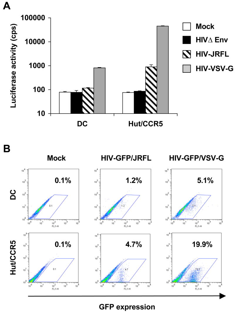
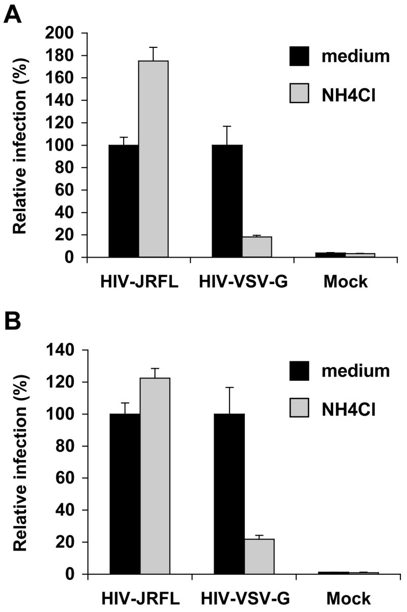
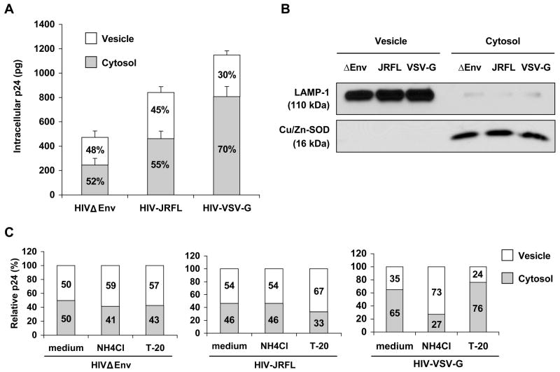
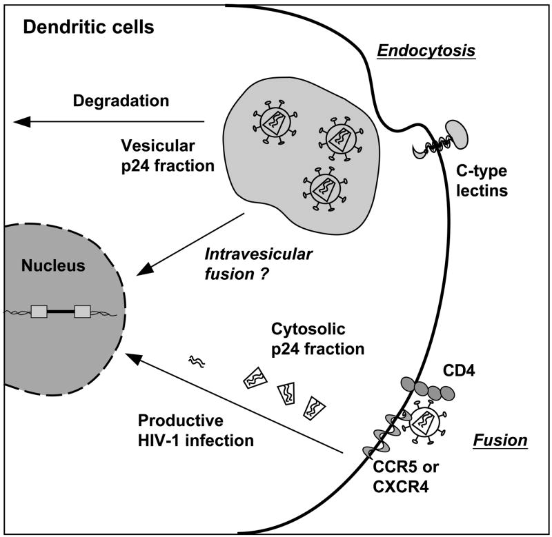
Human immunodeficiency virus type 1 (HIV-1) enters dendritic cells (DCs) through endocytosis and viral receptor-mediated fusion. Although endocytosis-mediated HIV-1 entry can generate productive infection in certain cell types, including human monocyte-derived macrophages, productive HIV-1 infection in DCs appears to be dependent on fusion-mediated viral entry. It remains to be defined whether endocytosed HIV-1 in DCs can initiate productive infection. Using HIV-1 infection and cellular fractionation assays to measure productive viral infection and entry, here we show that HIV-1 enters monocyte-derived DCs predominately through endocytosis; however, endocytosed HIV-1 cannot initiate productive HIV-1 infection in DCs. In contrast, productive HIV-1 infection in DCs requires fusion-mediated viral entry. Together, these results provide functional evidence in understanding HIV-1 cis-infection of DCs, suggesting that different pathways of HIV-1 entry into DCs determine the outcome of viral infection.


Figures

Fig. 1
Fig. 1
HIV-1 infection in immature DCs is blocked by AZT. (A) Surface expression of CD4 and CCR5 on DCs. Monocyte-derived immature DCs were stained with specific MAbs and analyzed by flow cytometry. DCs were stained with either a murine IgG isotypic control antibody (filled peaks) or CD4 MAb (thick line, left panel) or CCR5 MAb (thick line, right panel). (B) DCs were infected with R5-tropic HIV-1 Env (JRFL)-pesudotyped, single-cycle luciferase reporter HIV-1 (MOI, 0.8) in the presence or absence of AZT. DCs were lysed 3, 5, and 7 dpi to determine viral infection by measuring the luciferase activity of cell lysates. Mock infection was used for a background control for the detection. cps, counts per second. (C) DCs were infected with HIV-1NLAD8 in the presence or absence of AZT. HIV-1 infection was determined at 3, 5 and 7 dpi by measuring the p24 levels in supernatants. All data show the means ± standard deviations (S.D.) of triplicate samples. Data for one representative experiment out of three are shown.
Fig. 2
Fig. 2
Comparison of HIV-1 infection in DCs and CD4+ T cells with different HIV-1 pseudotypes. (A) Direct infection of DCs or Hut/CCR5 cells with HIV-JRFL and HIV-VSV-G; the same amount of HIV Env was used as a negative control. DCs were pulsed with different HIV-1 pseudotypes separately for 2 h at 37°C, washed and cultured 4 dpi before measuring the luciferase activity of cell lysates. Mock infection was used for a background control for the detection. cps, counts per second. All data show the means ± S.D. of triplicate samples. (B) Direct infection of DCs or Hut/CCR5 cells with HIV-GFP/JRFL or HIV-GFP/VSV-G. Infected cells were analyzed by flow cytometry for GFP expression 4 dpi. Percentages of GFP-positive cells are shown in the plots. Data for one representative experiment out of three are shown.
Fig. 3
Fig. 3
Productive HIV-1 infection in DCs requires fusion-mediated viral entry. (A) Blockade of fusion-mediated HIV-1 entry into DCs diminishes viral replication. DCs were infected with HIV-1NLAD8 for 2 h at 37°C in the presence or absence of T-20. Supernatants of infected DCs were measured for p24 levels at 3, 5 and 7 dpi. (B) T-20 does not impair HIV-1NLAD8 endocytosis into DCs. DC-associated p24 was measured after incubation with HIV-1NLAD8 for 2 h at 37°C in the presence or absence of T-20. HIV-1-pulsed DCs were extensively washed, trypsinized and lysed for p24 detection. (C) Endocytosed HIV-1 in DCs does not generate significant amounts of late reverse transcription (RT) products. DCs were infected with HIV-1NLAD8, washed, trypsinized, and cultured for 12 h before the cells were lysed for real-time PCR detection (40 ng of cellular DNA per sample was used). T-20 was present during the viral incubation and the 12-h culture. All data show the means ± S.D. of triplicate samples. Data for one representative experiment out of four are shown.
Fig. 4
Fig. 4
HIV-1 infection of DCs is pH-independent. HIV-JRFL and HIV-VSV-G infection of (A) DCs and (B) CD4+ Hut/CCR5 T cell line. DCs or Hut/CCR5 cells were incubated with NH4Cl at 37°C for 30 min and then pulsed separately with HIV-VSV-G and HIV-JRFL (MOI, 0.5) at 37°C for 2 h in the presence of NH4Cl. NH4Cl was washed away after the 2.5-h incubation with DCs. HIV-1 infection was detected 3 dpi by measuring the luciferase activity of cell lysates and relative infection is shown. Values for medium controls were set at 100%. Data for one representative experiment out of three are shown.
Fig. 5
Fig. 5
Analysis of HIV-1 entry into DCs by cellular fractionation. (A) Cellular fractionation of HIV-pulsed DCs. DCs were separately exposed to HIV Env, HIV-JRFL, and HIV-VSV at 37°C for 2 h. After extensive washes, DCs were treated with pronase to remove HIV-1 bound on cell surface. DCs were then washed, permeabilized, and fractionated. Intracellular p24 levels were measured in the cytosolic and vesicular fractions. The percentages of cytosolic and vesicular p24 are shown inside the bars. The average results of four independent experiments using DCs from different donors are shown. (B) Western blot analysis of the cytosolic and vesicular fractionations of HIV-infected DCs. HIV-pulsed DCs were fractionated as described above in (A) and analyzed by Western blotting with antibodies specific for Cu/Zn-SOD (cytosol marker) and LAMP-1 (vesicle marker). Data for one representative experiment out of three are shown. (C) Relative p24 of the cytosolic and vesicular fractionations of DCs treated with fusion inhibitors. DCs were pretreated separately with NHCl4 and T-20, pulsed with different HIV-1 pseudotypes in the presence of the appropriate inhibitors. DCs were then washed, fractionated, and the p24 levels were measured as described above in (A). The relative percentages of cytosolic and vesicular p24 are shown inside the bars. Data for one representative experiment out of three are shown.
Fig. 6
Fig. 6
A proposed model for HIV-1 entry and productive infection in DCs. HIV-1 enters DCs through the endocytosis and plasma membrane fusion pathways. The endocytic pathway appears to be the predominate route of HIV-1 entry into DCs; however, only fusion-mediated HIV-1 entry can lead to productive HIV-1 infection. In the cellular fractionation assay, the vesicular p24 fraction represents the HIV-1 confined in the endocytic compartments; whereas the cytosolic p24 fraction represents the HIV-1 entered through the fusion pathway. Endocytosed HIV-1 could be eventually degraded. However, it is currently uncertain whether a small proportion of internalized authentic HIV-1 in DCs can escape the vesicular compartment via potential intravisecular fusion.

References

    1. Aiken C. Pseudotyping human immunodeficiency virus type 1 (HIV-1) by the glycoprotein of vesicular stomatitis virus targets HIV-1 entry to an endocytic pathway and suppresses both the requirement for Nef and the sensitivity to cyclosporin A. J Virol. 1997;71(8):5871–5877. - PMC - PubMed
    1. Arrighi JF, Pion M, Garcia E, Escola JM, van Kooyk Y, Geijtenbeek TB, Piguet V. DC-SIGN-mediated infectious synapse formation enhances X4 HIV-1 transmission from dendritic cells to T cells. J Exp Med. 2004;200(10):1279–1288. - PMC - PubMed
    1. Burleigh L, Lozach PY, Schiffer C, Staropoli I, Pezo V, Porrot F, Canque B, Virelizier JL, Arenzana-Seisdedos F, Amara A. Infection of dendritic cells (DCs), not DC-SIGN-mediated internalization of human immunodeficiency virus, is required for long-term transfer of virus to T cells. J Virol. 2006;80(6):2949–2957. - PMC - PubMed
    1. Cavrois M, De Noronha C, Greene WC. A sensitive and specific enzyme-based assay detecting HIV-1 virion fusion in primary T lymphocytes. Nat Biotechnol. 2002;20(11):1151–1154. - PubMed
    1. Cavrois M, Neidleman J, Kreisberg JF, Fenard D, Callebaut C, Greene WC. Human immunodeficiency virus fusion to dendritic cells declines as cells mature. J Virol. 2006;80(4):1992–1999. - PMC - PubMed

Publication types