Review
. 2007;18(6):417-38.
doi: 10.1515/revneuro.2007.18.6.417.

Circuits formultisensory integration and attentional modulation through the prefrontal cortex and the thalamic reticular nucleus in primates

Affiliations
Review

Circuits formultisensory integration and attentional modulation through the prefrontal cortex and the thalamic reticular nucleus in primates

Basilis Zikopoulos et al. Rev Neurosci. 2007.

Abstract

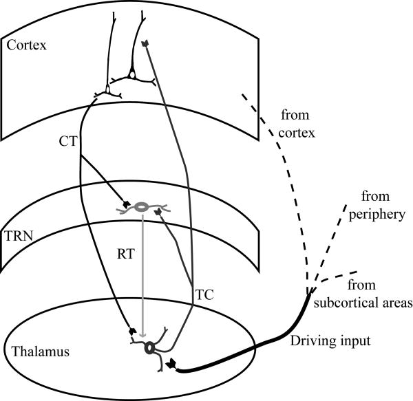
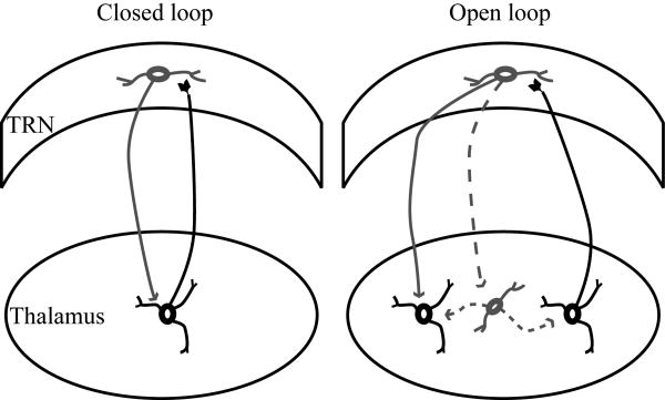
Converging evidence from anatomic and physiological studies suggests that the interaction of high-order association cortices with the thalamus is necessary to focus attention on a task in a complex environment with multiple distractions. Interposed between the thalamus and cortex, the inhibitory thalamic reticular nucleus intercepts and regulates communication between the two structures. Recent findings demonstrate that a unique circuitry links the prefrontal cortex with the reticular nucleus and may underlie the process of selective attention to enhance salient stimuli and suppress irrelevant stimuli in behavior. Unlike other cortices, some prefrontal areas issue widespread projections to the reticular nucleus, extending beyond the frontal sector to the sensory sectors of the nucleus, and may influence the flow of sensory information from the thalamus to the cortex. Unlike other thalamic nuclei, the mediodorsal nucleus, which is the principal thalamic nucleus for the prefrontal cortex, has similarly widespread connections with the reticular nucleus. Unlike sensory association cortices, some terminations from prefrontal areas to the reticular nucleus are large, suggesting efficient transfer of information. We propose a model showing that the specialized features of prefrontal pathways in the reticular nucleus may allow selection of relevant information and override distractors, in processes that are deranged in schizophrenia.


Figures

Figure 1
Figure 1
Position of TRN in the rhesus monkey brain. Reconstructed hemisphere shown from the lateral surface, which was rendered transparent to show the position of the TRN (gray) surrounding the thalamus (black).
Figure 2
Figure 2
The TRN nucleus and some of its neurochemical features. Brightfield photomicrographs of coronal sections from rostral (top) to caudal (bottom) levels of the rhesus monkey thalamus showing the TRN in sections treated for: Column A, acetylcholinesterase (AChE) histochemistry, showing TRN as it enveils the dorsal thalamus. Column B, High magnification of sections at the same rostrocaudal levels as in column A, immunohistochemically labeled with PV, showing that the majority of TRN neurons are PV+. Black and white arrows indicate the borders of TRN. Scale bars A, 5 mm; B, 500 μm.
Figure 3
Figure 3
Features of TRN neurons. Brightfield photomicrographs showing differences in the morphology of TRN neurons from a rostral to a caudal direction (top to bottom panels), with larger multipolar neurons with round perikarya appearing rostrally (top panels), and smaller, fusiform, bipolar neurons appearing caudally (bottom panels). Column A, Coronal sections of a rhesus monkey thalamus stained for Nissl. Column B, Coronal sections of a rhesus monkey thalamus stained for PV. Column C, Coronal sections of a rhesus monkey thalamus stained for nonphosphorylated neurofilament H (SMI-32). Scale bars A, 500 μm; B, C, 200 μm.
Figure 4
Figure 4
Basic circuits linking TRN with the cortex and the thalamus. Driving input reaching the thalamus from the sensory periphery, subcortical regions or the cortex, activates thalamocortical projection neurons, which then transmit that information to the cortex. Thalamocortical axons, on their way to the cortex, give off collateral branches that terminate in TRN. In the feedback loop of this circuit, corticothalamic axons give off collateral branches that innervate the same TRN regions. The TRN sends inhibitory projections only to the thalamus.
Figure 5
Figure 5
Reticulothalamic connections. TRN neurons send inhibitory projections (gray line) to thalamic relay neurons that excite them (black line), forming closed loops (left panel). In some cases, a TRN neuron can receive excitatory projections from one thalamic neuron (black line) and send inhibitory projections to another thalamic relay neuron (gray line) or a local inhibitory neuron (dotted gray line), forming open loops (right panel).
Figure 6
Figure 6
Sensory sectors of the rhesus monkey TRN. A, Three-dimensional reconstruction of TRN (light gray) showing the approximate sectors receiving projections from auditory (green), visual (blue) and somatosensory (yellow) cortices in non-human primates based on available evidence. The colors between the different sectors change gradually to illustrate the blurred topography of the sectors and their overlaps. B, Three-dimensional reconstruction of TRN (light gray), which was rendered transparent to show the position of axonal terminations from temporal sensory association cortices, including auditory areas Ts1 (light green) and Ts2 (dark green), visual area TE1 (dark blue), and polymodal area 36 (light green-blue gradient).
Figure 7
Figure 7
Widespread prefrontal terminations in TRN and projection neurons to MD overlap with projections from sensory association cortices. A, Three-dimensional reconstruction of TRN (light gray), which was rendered transparent to show the position of axonal terminations from prefrontal areas (red) and their overlaps with somatosensory (yellow), visual (blue) and auditory (green) sectors. Black asterisks indicate some projections from dorsal area 46 and white asterisks indicate some projections from orbital area 13. B, TRN neurons projecting to MD (purple) superimposed on a 3D model of TRN with color coded sensory sectors (as in A).
Figure 8
Figure 8
Large and small bouton populations in TRN. 3D-reconstructions of large (Lb) and small (Sb) boutons (blue), synapsing (red) on PV+ dendrites of TRN neurons (gray).
Figure 9
Figure 9
Schematic diagram summarizing the involvement of prefrontal and sensory systems in attentional mechanisms through TRN. A-D depict all possible combinations of salient (black) and distracting (brown) inputs interacting with open or closed reticulothalamic loops. Reticulo-MD loops can be either closed or open. A, Salient and distracting inputs interact with open reticulothalamic loops. Salient and distracting input is relayed from the thalamus to the cortex (green dots and lines) and back (gray triangles and lines); Activated TRN neurons inhibit other thalamic neurons (dotted red lines) or thalamic GABAergic neurons (red squares), allowing prolonged access of the stimulus to the cortex. The sensory system can suppress distracting stimuli if reticular neurons activated by the salient signal inhibit neighboring thalamic neurons that transmit distracting signals (1a). Dimorphic prefrontal input (blue), through small and large terminals, and input from MD (cyan) can also activate neighboring reticular neurons and inhibit the thalamic neurons that relay distractors (2) and disinhibit thalamic neurons relaying relevant information (1b). B, Salient and distracting inputs interact with closed reticulothalamic loops, reaching the cortex briefly before they are inhibited by TRN neurons. Input from prefrontal cortex and MD can reverse this outcome by activating neighboring TRN neurons that inhibit the TRN neurons, that prevent transmission of the salient input (3a), or by inhibiting thalamic GABAergic neurons and disinhibiting the relay of salient input (3b). Prefrontal-MD input could also lead to increased inhibition of distractors (3c). C, Salient input has prolonged access to the cortex, interacting with open reticulothalamic loops, and distracting input reaches the cortex briefly, interacting with closed reticulothalamic loops. This is the best case scenario for selection of relevant information and suppression of distractors, and can occur even at early processing stages. Input from the prefrontal cortex and MD to TRN results in enhancement of selection of relevant stimuli by activating TRN neurons that suppress distractors (4a, 5) or disinhibit neurons relaying salient input (4b). D, Salient input reaches the cortex briefly, before TRN feedback inhibition through closed loops, and distracting input has prolonged access to the cortex, interacting with open reticulothalamic loops. Projections from prefrontal cortex and MD to TRN provide a mechanism to allow prolonged passage of relevant input, through lateral inhibition in TRN (6a) or disinhibition of the thalamus (6b). The same projections have a concomitant consequence of suppressing distractors (6c).

Similar articles

Cited by

References

    1. Abramson BP, Chalupa LM. The laminar distribution of cortical connections with the tecto- and cortico-recipient zones in the cat's lateral posterior nucleus. Neuroscience. 1985;15:81–95. - PubMed
    1. Alexander GE, Fuster JM. Effects of cooling prefrontal cortex on cell firing in the nucleus medialis dorsalis. Brain Res. 1973;61:93–105. - PubMed
    1. Alvarez P, Squire LR. Memory consolidation and the medial temporal lobe: a simple network model. Proc Natl Acad Sci U S A. 1994;91:7041–7045. - PMC - PubMed
    1. Arnsten AF, Li BM. Neurobiology of executive functions: catecholamine influences on prefrontal cortical functions. Biol Psychiatry. 2005;57:1377–1384. - PubMed
    1. Asanuma C. GABAergic and pallidal terminals in the thalamic reticular nucleus of squirrel monkeys. Exp Brain Res. 1994;101:439–451. - PubMed

Publication types

LinkOut - more resources