. 2007 Sep;17(5):285-302.
doi: 10.1055/s-2007-985193.

Microanatomy and surgical approaches to the infratemporal fossa: an anaglyphic three-dimensional stereoscopic printing study

Affiliations

Microanatomy and surgical approaches to the infratemporal fossa: an anaglyphic three-dimensional stereoscopic printing study

Gustavo Rassier Isolan et al. Skull Base. 2007 Sep.

Abstract

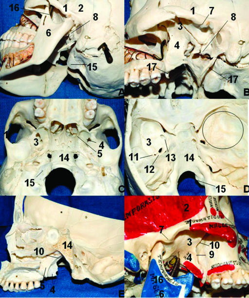
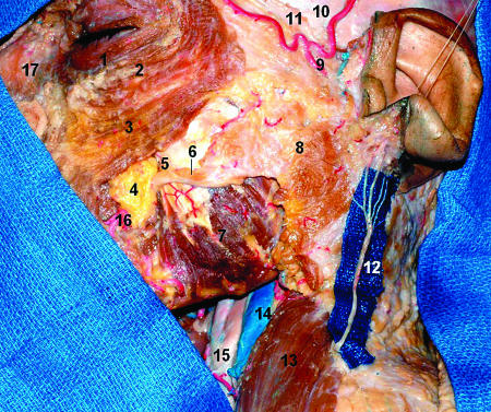
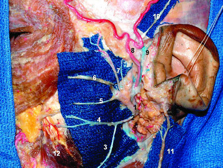
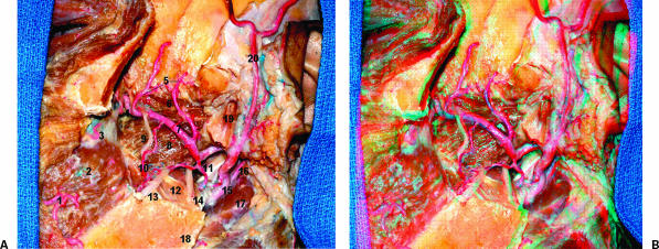
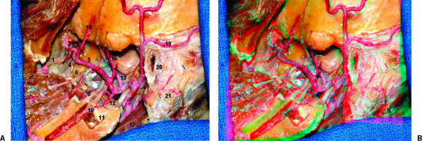
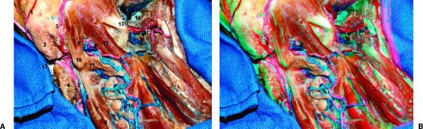
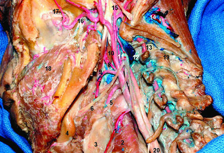
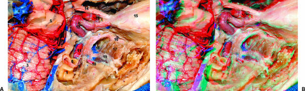
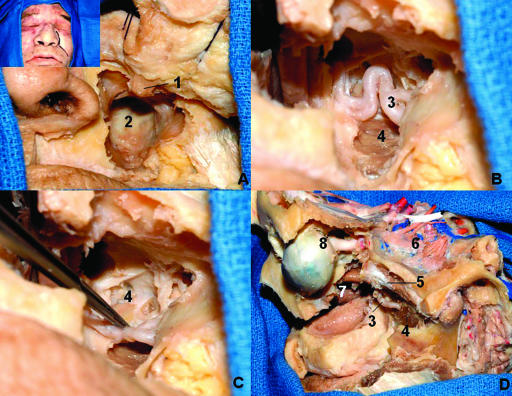
Objective: The infratemporal fossa (ITF) is a continuation of the temporal fossa between the internal surface of the zygoma and the external surface of the temporal bone and greater wing of the sphenoid bone that is sitting deep to the ramus of the mandible. The principal structure to understanding its relationships is the lateral pterygoid muscle. Other important structures are the medial pterygoid muscle, the maxillary artery, the pterygoid venous plexus, the otic ganglion, the chorda tympani nerve and the mandibular nerve. In this study, we describe the microsurgical anatomy of the ITF, as viewed by step-by-step anatomical dissection and also through the perspective of three lateral approaches and one anterior surgical approach.

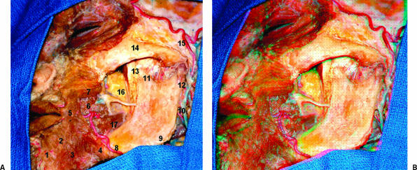
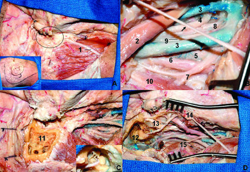
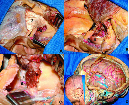
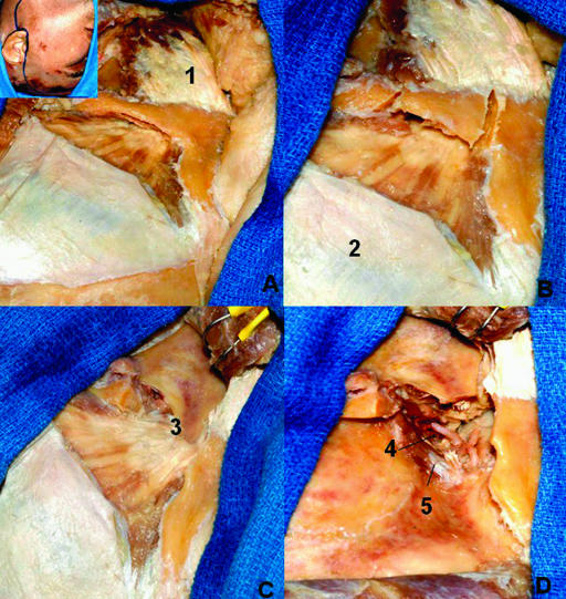
Methods: Eight cadaver specimens were dissected. In one side of all specimens, an anatomical dissection was done in which a wide preauricular incision from the neck on the anterior border of the sternoclidomastoid muscle at the level of the cricoid cartilage to the superior temporal line was made. The flap was displaced anteriorly and the structures of the neck were dissected followed by a zygomatic osteotomy and dissection of the ITF structures. On the other side were the surgical approaches to the ITF. The combined infratemporal and posterior fossa approach was done in two specimens, the subtemporal preauricular infratemporal fossa approach in two, the zygomatic approach in two, and the lateral transantral maxillotomy in two. The anatomical dissections were documented on the three-dimensional (3D) anaglyphic method to produce stereoscopic prints.

Results: The lateral pterygoid muscle is one of the principal structures to enable understanding of the relationships into the ITF. The tendon of the temporal muscle inserts in the coronoid process at the ITF. The maxillary artery is the terminal branch of the external carotid artery that originates at the neck of the mandible and runs into the parotid gland. In our dissections the maxillary artery was lateral to the buccal, lingual, and inferior alveolar nerves. We found the second part of the maxillary artery superficial to the lateral pterygoid muscle in all specimens The anterior and posterior branches of the deep temporal artery supply the temporal muscle. In two cases we found a middle deep temporal artery. The different approaches that we used provided different views of the same anatomical landmarks and this provides not only safer surgery but also the best choice to approach the ITF according with the pathology extension.

Conclusions: The ITF is a complex region on the skull base that is affected by benign and malignant tumors. The study through different routes is helpful to disclose the relationship among the anatomical structures. Although the authors have shown four approaches, there are a variety of approaches and even a combination of these can be used. This type of anatomical knowledge is essential to choosing the best approach to treat lesions in this area.

Keywords: Infratemporal fossa; approaches; mandibular nerve; paraganglioma; surgical anatomy.


Figures

Figure 1
Figure 1
Limits of the ITF and bone relationships. (A) Oblique lateral view of the ITF. (B) Same view after removal of the mandible. (C) Inferior aspect of the cranium. (D) Interior view of the cranial base. The circle on the right middle fossa represents approximately the correspondence of the ITF in the middle fossa. (E) Lateral view of the ITF after sagittal paramedian section. (F) Note that the depression of the mandible (open mouth) gives more access to the ITF laterally. 1, zygomatic process of the temporal bone; 2, temporal fossa; 3, greater wing of the sphenoid; 4, lateral pterygoid plate; 5, medial pterygoid plate; 6, mandibular ramus; 7, articular tubercle of the temporal bone; 8, spine of the sphenoid bone; 9, pterygomaxillary fissure; 10, pterygopalatine fossa; 11, foramen spinosum; 12, foramen ovale; 13, foramen rotundum; 14, clivus; 15, occipital condyle; 16, coronoid process; 17, styloid process. ITF, infratemporal fossa.
Figure 2
Figure 2
Lateral view of the superficial dissection of the left face. 1, orbicularis oculi (palpebral part); 2, orbicularis oculi (orbital part); 3, zygomaticus minor and major; 4, buccal fat pad; 5, parotid duct; 6, accessory parotid gland; 7, masseter muscle; 8, parotid fascia over the parotid gland; 9, superficial temporal artery; 10, temporal fascia (deep temporal fascia); 11, temporoparietal fascia (superficial temporal fascia); 12, great auricular nerve; 13, sternocleidomastoid muscle; 14, internal jugular vein; 15, common carotid artery; 16, facial artery; 17, nasalis (more medially) and levator labii superioris alaeque nasi.
Figure 3
Figure 3
Lateral view of the superficial dissection of the left face. The majority of the parotid tissue was removed to expose the facial nerve branches. A blue field was put under the nerves to enhance them. The distal ramification as well as the parotid plexus are not shown. 1, facial nerve (superior trunk); 2, facial nerve (inferior trunk); 3, cervical branch; 4, mandibular branch; 5, buccal branch; 6, zygomatic branch; 7, temporal branches; 8, superficial temporal artery; 9, superficial temporal vein; 10, auriculotemporal nerve; 11, great auricular nerve; 12, masseter muscle.
Figure 4
Figure 4
(A) Lateral view of the deep dissection of the left face. The masseter muscle was removed. Note the insertion of the temporal tendon into the coronoid process and anterior aspect of the mandible. 1, mentalis muscle; 2, depressor labii inferioris; 3, depressor anguli oris; 4, fibers of the platisma muscle; 5, orbicularis oris muscle; 6, risorius muscle; 7, zygomatic major muscle; 8, facial artery; 9, angle of the mandible; 10, retromandibular vein; 11, coronoid process; 12, condylar process; 13, coronoid process; 14, zygomatic bone; 15, superficial temporal artery; 16, buccal fat pad; 17, medial pterygoid muscle. (B) The figure shows the stereoscopic view.
Figure 5
Figure 5
(A) Lateral view of the ITF. The zygoma and part of the mandible were removed. The condylar process was left to show the insertion of the lateral pterygoid muscle in the pterygoid fovea in the neck of the mandible. 1, facial artery; 2, buccinator muscle; 3, posterior superior alveolar artery; 4, sphenopalatine artery; 5, anterior and posterior deep temporal arteries; 6, lateral pterygoid muscle (upper head); 7, maxillary artery; 8, lateral pterygoid muscle (lower head); 9, buccal nerve; 10, buccal artery; 11, styloid process; 12, medial pterygoid muscle; 13, lingual nerve; 14, inferior alveolar nerve; 15, external carotid artery; 16, posterior auricular artery; 17, digastric muscle (posterior belly); 18, angle of the mandible; 19, condylar process; 20, superficial temporal artery.(B) The figure shows the stereoscopic view. ITF, infratemporal fossa.
Figure 6
Figure 6
(A) Lateral view of the ITF. The lateral pterygoid process and the condylar process were removed. 1, posterior superior alveolar artery; 2, infraorbital artery; 3, sphenopalatine artery; 4, descending palatine artery (not injected); 5, maxillary artery; 6, lateral pterygoid plate; 7, buccal nerve; 8, buccal artery; 9, lingual nerve; 10, inferior alveolar nerve; 11, medial pterygoid muscle; 12, inferior alveolar artery; 13, middle meningeal artery; 14, deep temporal nerve; 15, anterior deep temporal artery; 16, posterior deep temporal artery; 17, digastric muscle (posterior belly); 18, posterior auricular artery; 19, posterior auricular artery; 20, external acoustic meatus; 21, mastoid. (B) The figure shows the stereoscopic view. ITF, infratemporal fossa.
Figure 7
Figure 7
Posterior oblique view of the neck and part of the left ITF. 1, posterior auricular artery; 2, mastoid; 3, occipital artery; 4, middle pterygoid muscle; 5, external carotid artery; 6, internal jugular vein; 7, accessory nerve; 8, levator scapula muscle; 9, superior oblique muscle; 10, transverse process of atlas; 11, inferior oblique; 12, rectus capitis posterior major muscle; 13, rectus capitis posterior minor; 14, right jugular bulb; 15, right vertebral artery; 16, deep cervical vein and artery. (B) The figure shows the stereoscopic view. ITF, infratemporal fossa.
Figure 8
Figure 8
Lateral view of the ITF and neck, emphasizing the branches of the external carotid artery. 1, superior thyroid artery; 2, inferior pharyngeal constrictor muscle; 3, thyroid cartilage; 4, submandibular gland; 5, lingual artery; 6, hypoglossal nerve; 7, facial artery; 8, ascending palatine artery; 9, stylohyoid muscle; 10, styloglossus muscle; 11, ascending pharyngeal artery; 12, internal jugular vein sectioned; 13, vertebral artery; 14, suboccipital triangle with venous plexus and muscular branches from the vertebral artery; 15, styloid process and posterior auricular artery; 16, maxillary artery; 17, infraorbital nerve; 18, buccinator muscle. ITF, infratemporal fossa.
Figure 9
Figure 9
(A) Lateral superior view of the ITF. Part of the middle fossa floor was removed to expose the ITF. The gasserion ganglion was resected to expose the intrapetrous portion of the ICA. The floor of the anterior fossa was resected also. 1, semicircular canals; 2, superior petrosal sinus; 3, superior surface of the cerebellum; 4, great cerebral vein (Galen); 5, substantia nigra; 6, cranial nerves VII and VIII; 7, cranial nerve V; 8, cochlea; 9, geniculate ganglion; 10, middle meningeal artery; 11, ICA (intrapetrous portion); 12, ICA (intracavernous portions); 13, ICA (subclinoidal portion); 14, cranial nerve II; 15, orbit; 16, maxillary nerve; 17, lateral pterygoid muscle (upper head); 18, part of the pterygoid plexus; 19, V3. (B) The figure shows the stereoscopic view. ITF, infratemporal fossa; ICA, internal carotid artery.
Figure 10
Figure 10
Combined infratemporal and posterior fossa approach. (A) After the incision (inset) the flap is reflected anteriorly and the external meatus is sectioned and closed in a blind sac as described by Ugo Fisch (oval). (B) The neurovascular structures are exposed in the neck to proximal control. The arrow indicates the greater auricular nerve. (C) The petrosectomy is performed. The inset shows the facial nerve, the ossicles, and semicircular canals. (D) Final anatomical view of the neurovascular structures in the neck and the presigmoid and temporal fossa dura after petrosectomy. The semicircular canals are preserved and the facial nerve is transposed anteriorly. 1, great auricular nerve; 2, digastric muscle (posterior belly); 3, duplicated internal jugular vein; 4, glossopharyngeal nerve; 5, internal carotid artery; 6, sympathetic trunk; 7, accessory nerve; 8, hypoglossal nerve; 9, vagus nerve; 10, inferior oblique nerve; 11, mastoid drilled; 12, facial nerve; 13, semicircular canals; 14, superficial temporal artery; 15, transverse process of C1.
Figure 11
Figure 11
Subtemporal preauricular ITF approach. (A) The preauricular incision on the right side was performed, the flap is displaced anteriorly, and the zygomatic process was removed. (B) The temporal muscle is displaced all the way down. (C) Exposition of the middle meningeal artery and V3 in the ITF. (D) Relationship among ITF, mastoid, and temporal lobe; the inset shows the incision. 1, temporal superficial fascia; 2, tendon of the temporal muscle; 3, mandibular incisura and maxillary artery; 4, superficial temporal artery; 5, mandibular condyle; 6, middle meningeal artery entering in the foramen spinosum; 7, V3; 8, sigmoid sinus exposed after mastoidectomy; 9, mastoid portion of the facial nerve.
Figure 12
Figure 12
Zygomatic approach. Part of the deep temporal fascia was removed to show the muscular fibers. (A) Preauricular incision and anterior displacement of the flap. (B) Section of the zygomatic arch. (C) The masseter and the zygomatic arch are displaced inferiorly. (D) The coronoid process is sectioned and displaced upward with the temporal muscle. 1, masseter muscle; 2, deep temporal fascia; 3, coronoid process; 4, maxillary artery; 5, lateral pterygoid muscle (upper head).
Figure 13
Figure 13
Lateral transantral maxillotomy. (A) The anterior and lateral walls of the maxilla are resected. (B) After removal of the sinus mucosa, the posterior wall is drilled out, exposing the ITF. (C) The maxillary is displaced to show the lateral pterygoid plate. (D) This dissection exposes the ITF via an anterior and lateral view. The maxilla was totally removed. 1, infraorbital nerve; 2, posterior wall of the maxilla; 3, maxillary artery; 4, lateral pterygoid plate; 5, lateral pterygoid muscle (upper head); 6, lateral pterygoid muscle (lower head); 7, nasal cavity; 8, eyeball and optic nerve. ITF, inferior temporal fossa.

Similar articles

Cited by

References

    1. Gray H. Anatomy of the Human Body. New York: Bartley.com; 2000.
    1. Vrionis F D, Cano W G, Heilman C B. Microsurgical anatomy of the infratemporal fossa as viewed laterally and superiorly. Neurosurgery. 1996;39:777–786. - PubMed
    1. Bejjani G K, Sullivan B, Salas-Lopez E, et al. Surgical anatomy of the infratemporal fossa: the styloid diaphragm revisited. Neurosurgery. 1998;43:842–852. - PubMed
    1. Schramm V L., Jr In: Sekhar LN, Schramm VL, editor. Tumors of the Cranial Base: Diagnosis and Treatment. Mount Kisco, NY: Futura Publishing Co; 1987. Infratemporal fossa surgery. pp. 421–437.
    1. Ribas G C, Bento R F, Rodrigues A J. Anaglyphic three-dimensional stereoscopic printing: revival of an old method for anatomical and surgical teaching and reporting. J Neurosurg. 2001;95:1057–1066. - PubMed