. 2008 Jul;216(1):29-37.
doi: 10.1002/jcp.21426.

Topical administration of a multi-targeted kinase inhibitor suppresses choroidal neovascularization and retinal edema

Affiliations

Topical administration of a multi-targeted kinase inhibitor suppresses choroidal neovascularization and retinal edema

John Doukas et al. J Cell Physiol. 2008 Jul.

Abstract

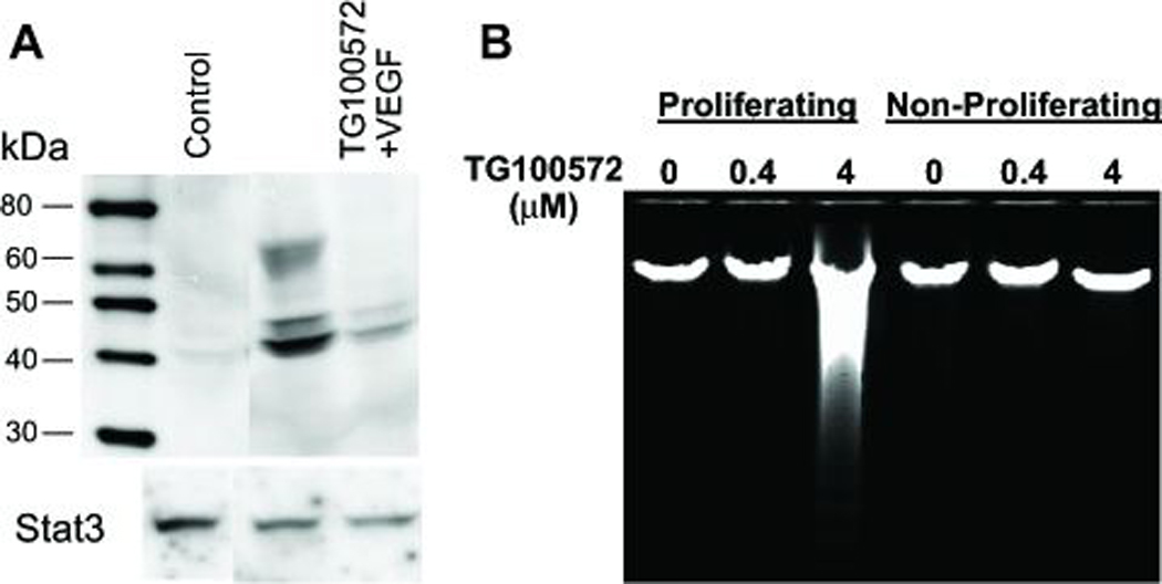
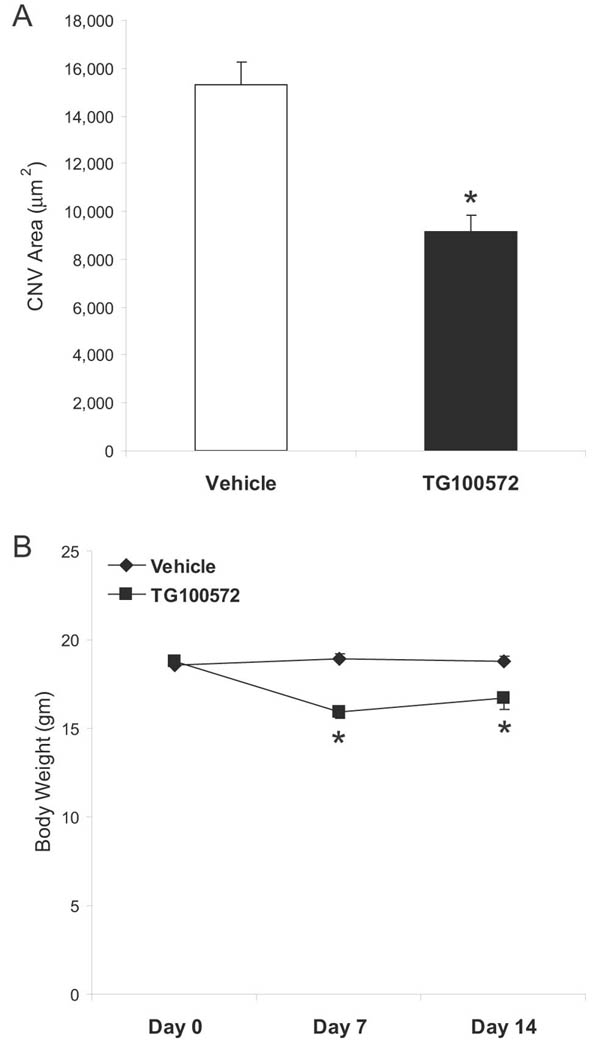
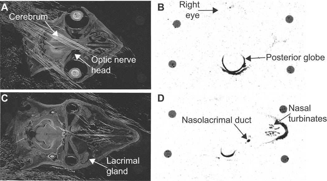
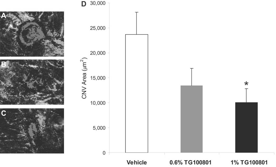
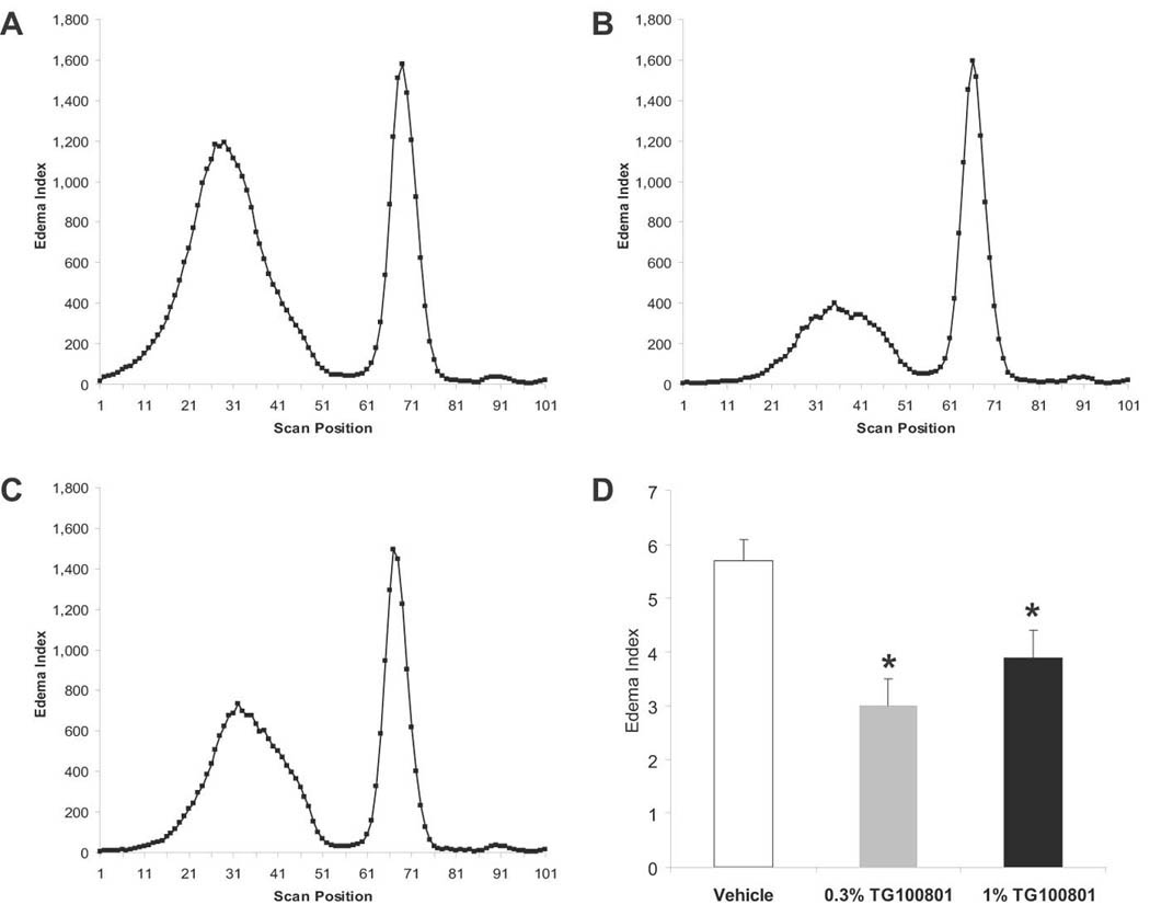
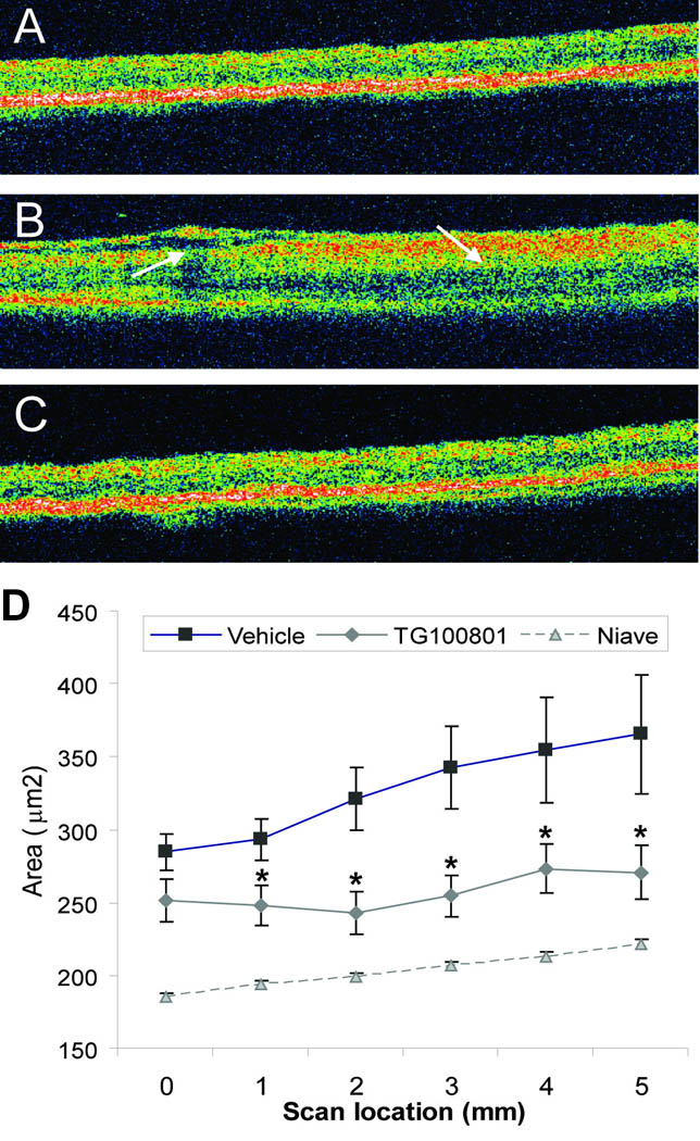
Age-related macular degeneration, diabetic retinopathy, and retinal vein occlusions are complicated by neovascularization and macular edema. Multi-targeted kinase inhibitors that inhibit select growth factor receptor tyrosine kinases and/or components of their down-stream signaling cascades (such as Src kinases) are rationale treatment strategies for these disease processes. We describe the discovery and characterization of two such agents. TG100572, which inhibits Src kinases and selected receptor tyrosine kinases, induced apoptosis of proliferating endothelial cells in vitro. Systemic delivery of TG100572 in a murine model of laser-induced choroidal neovascularization (CNV) caused significant suppression of CNV, but with an associated weight loss suggestive of systemic toxicity. To minimize systemic exposure, topical delivery of TG100572 to the cornea was explored, and while substantial levels of TG100572 were achieved in the retina and choroid, superior exposure levels were achieved using TG100801, an inactive prodrug that generates TG100572 by de-esterification. Neither TG100801 nor TG100572 were detectable in plasma following topical delivery of TG100801, and adverse safety signals (such as weight loss) were not observed even with prolonged dosing schedules. Topical TG100801 significantly suppressed laser-induced CNV in mice, and reduced fluorescein leakage from the vasculature and retinal thickening measured by optical coherence tomography in a rat model of retinal vein occlusion. These data suggest that TG100801 may provide a new topically applied treatment approach for ocular neovascularization and retinal edema.


Figures

Figure 1
Figure 1. TG100572 inhibits VEGF signaling and induces apoptosis in proliferating vascular endothelial cells
(A) Endothelial cells cultured in the absence of VEGF (Control), in the presence of VEGF, or in the presence of both VEGF and TG100572 were processed for Western blot detection of phosphorylated Erk1/2 (or total STAT3 as a loading control). (B) Endothelial cells cultured under either proliferating (low density/high serum) or non-proliferating (high density/low serum) conditions were exposed to TG100572 at the indicated concentrations for 24 hr, after which genomic DNA was extracted and resolved on agarose gels in order to visualize DNA laddering indicative of apoptosis.
Figure 2
Figure 2. Systemic delivery of TG100572 in a murine model of choroidal neovascularization (CNV)
(A) Laser-induced rupture of Bruch’s membrane was performed in C57BL/6 mice, after which animals were dosed i.p daily with either TG100572 (5 mg/kg) or vehicle. After 14 days, animals were perfused with FITC-dextran and choroidal whole mounts were photographed for measurement of CNV area by computerized image analysis (data shown as means ± SEM, n= 55–60 lesion sites; * vehicle and TG100572 groups differ with P< 0.001). (B) Body weights taken prior to study initiation, Day 7, and at study end are shown (means ± SEM, n= 11–12 animals; *Vehicle and TG100572 groups differ with P< 0.005).
Figure 3
Figure 3. Ocular distribution of 14C-TG100801 following topical instillation in the rabbit
Transillumination (A & C) and corresponding autoradiograph (B & D) images of cross sections through a rabbit’s head taken 30 minutes after a single unilateral (left eye) topical instillation of 1% TG100801 containing radiotracer levels of 14C-TG100801. A 40 µm section at the horizontal midline (A & B) or inferior to the midline capturing the nasolacrimal duct (C & D). The four circles visible at the perimeter of each tissue section are remnants of mounting posts.
Figure 4
Figure 4. Topical delivery of TG100801 in a murine model of choroidal neovascularlization (CNV)
Laser-induced rupture of Bruch’s membrane was performed in C57BL/6 mice, after which animals were dosed topically twice a day with either TG100801 (1% or 0.6% formulations) or vehicle. After 14 days, animals were perfused with FITC-dextran and choroidal whole mounts were photographed for measurement of CNV area by computerized image analysis. (A–C) Representative fluorescent photomicrographs taken from the three treatment groups (A= vehicle, B= 0.6% TG100801, C= 1% TG100801), with fluorescence of the CNV highlighted in red. (D) CNV area graph (data shown as means ± SEM, n= 24 lesion sites; * 1% TG100801 and vehicle groups differ with P= 0.036).
Figure 5
Figure 5. Fluorescein leakage after topical delivery of TG100801 in a rat model of retinal vein occlusion
Retinal vein thrombosis was induced in the right eye of Long Evans rats, and both eyes were dosed topically twice a day with either vehicle or TG100801 (1% or 0.3% formulations). Following 5 doses, animals were injected i.p. with fluorescein and leakage into the retina and vitreous was quantified by ocular fluorophotometry. (A–C) Representative scans taken along the vertical axis from the three treatment groups; the peaks on the left represent fluorescein within the retina and vitreous compartments (A= vehicle, B= 0.3% TG100801, C= 1% TG100801) (D) Retina/vitreous peak area shown as an Edema Index corrected for basal vascular permeability in the non-thrombosed left eye (means ± SEM, n= 9–11; *, both TG100801 groups differ from vehicle group with P< 0.04).
Figure 6
Figure 6. Retinal thickness after topical delivery of TG100801 in a rat model of retinal vein occlusion
A retinal vein occlusion study was performed as described in Fig. 5, except that only one dose of TG100801 (1%) was investigated, and at study end retinal thickness was measured by optical coherence tomography (OCT; A–C). Representative OCT scans taken from naïve (A), thrombosed, vehicle-treated (B), or thrombosed, 1% TG100801-treated (C) animals. (D) Retinal area (µm2) determined from six 5 mm OCT scans (means ± SEM, n= 17–21; * TG100801 group differs from vehicle group with P< 0.05).

Similar articles

Cited by

References

    1. Armulik A, Abramsson A, Betsholtz C. Endothelial/pericyte interactions. Circ Res. 2005;97:512–523. - PubMed
    1. Bergers G, Song S, Meyer-Morse N, Bersland E, Hanahan D. Benefits of targeting both pericytes and endothelial cells in the tumor vasculature with kinase inhibitors. J Clin Invest. 2003;111:1287–1295. - PMC - PubMed
    1. Brown DM, Kaiser PK, Michels M, Soubrane G, Heier JS, Kim RY, Sy JP, Schneider S, Group AS. Ranibizumab versus verteporfin for neovascular age-related macular degeneration. N Eng J Med. 2006;355:1432–1444. - PubMed
    1. Campochiaro PA. Retinal and choroidal neovascularization. J Cell Physiol. 2000;184:301–310. - PubMed
    1. Campochiaro PA. Ocular neovascularisation and excessive vascular permeability. Expert Opin Biol Ther. 2004;4:1395–1402. - PubMed

MeSH terms

Substances

LinkOut - more resources