Review
. 2008 Jul;10(7):1185-98.
doi: 10.1089/ars.2007.1959.

Nitric oxide in the vasculature: where does it come from and where does it go? A quantitative perspective

Affiliations
Review

Nitric oxide in the vasculature: where does it come from and where does it go? A quantitative perspective

Kejing Chen et al. Antioxid Redox Signal. 2008 Jul.

Abstract

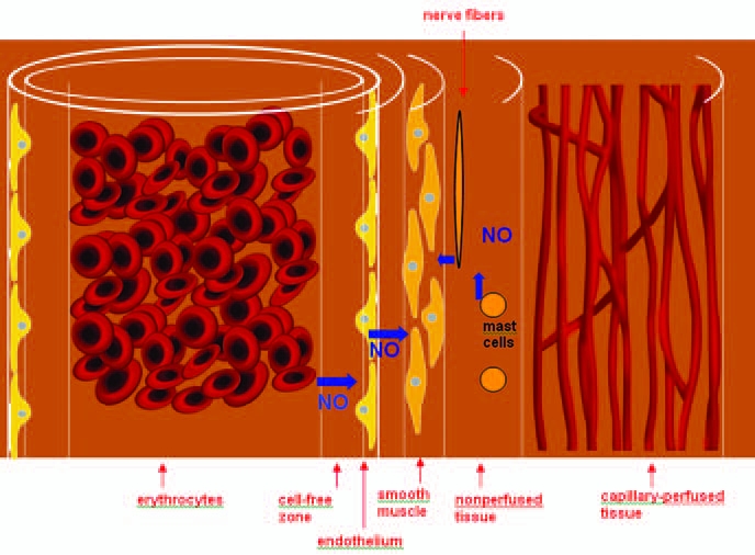
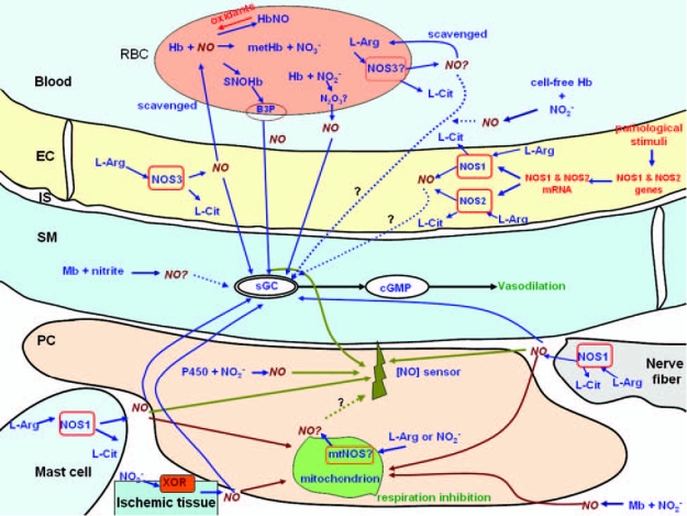
Nitric oxide (NO) affects two key aspects of O2 supply and demand: It regulates vascular tone and blood flow by activating soluble guanylate cyclase (sGC) in the vascular smooth muscle, and it controls mitochondrial O2 consumption by inhibiting cytochrome c oxidase. However, significant gaps exist in our quantitative understanding of the regulation of NO production in the vascular region. Large apparent discrepancies exist among the published reports that have analyzed the various pathways in terms of the perivascular NO concentration, the efficacy of NO in causing vasodilation (EC50), its efficacy in tissue respiration (IC50), and the paracrine and endocrine NO release. In this study, we review the NO literature, analyzing NO levels on various scales, identifying and analyzing the discrepancies in the reported data, and proposing hypotheses that can potentially reconcile these discrepancies. Resolving these issues is highly relevant to improving our understanding of vascular biology and to developing pharmaceutical agents that target NO pathways, such as vasodilating drugs.


Figures

FIG. 1.
FIG. 1.
The distribution of NO in the vasculature. The vasculature consists of the lumen containing erythrocytes, the erythrocyte-free zone resulting from blood flow, the endothelium, smooth muscle, the nonperfused region, and the capillary perfused region. (Adapted from ref. .) (For interpretation of the references to color in this figure legend, the reader is referred to the web version of this article at www.liebertonline.com/ars).
FIG. 2.
FIG. 2.
Major pathways of NO release that can regulate the activities of sGC and cytochrome c oxidase in the immediate vicinity of the vascular wall. A variety of enzymatic and nonenzymatic sources of NO can contribute to the NO concentration measured in the perivascular region. [NO] sensor represents a device that can be placed in the perivascular region and measures NO concentration. NO sources by nitric oxide synthase include NOS3 in the endothelium and erythrocytes, NOS1 in the nerve fibers, mast cells, and other tissues, mtNOS in the mitochondria, and NOS2 in a variety of tissues under pathologic conditions. Xanthine oxidoreductase and cytochrome P450 reductase can reduce nitrite to NO, respectively. Nonenzymatic NO release can potentially come from S-nitrosylated blood proteins, nitrite reduction by heme-containing proteins, and iron-nitrosylhemoglobin, among other sources. See the text for the details of these pathways. EC, endothelial cell; IS, interstitial space between the endothelium and smooth muscle; PC, parenchymal cell; RBC, red blood cell; Hb, hemoglobin; Mb, myoglobin; B3P, band 3 protein; metHb, methemoglobin; NO2, nitrite; NO3, nitrate; L-arg, l-arginine; L-cit, l-citrulline; cAMP, adenosine 3′,5′-cyclic monophosphate; cGMP, 3′,5′-cyclic guanosine monophosphate; Mt, mitochondrion; XOR, xanthine oxidoreductase; P450, P450 reductase.
FIG. 3.
FIG. 3.
Mechanism for the formation of NO via eNOS catalysis after the binding of the coenzyme tetrahydrobiopterin (H4B). The heme iron (Fe) is the major catalytic site and represents NOS3 here. NOS3 undergoes a series of redox reactions. Arg represents l-arginine, and NOHA represents Nω-hydroxyl-l-arginine. Parentheses around H4B, H4B+•, or H3B mean that that species is bound to the enzyme. NO is released from Fe3+NO. All three NOS isoforms share a similar catalytic mechanism. [Adapted from (66, 85).]

References

    1. Alderton WK. Cooper CE. Knowles RG. Nitric oxide synthases: structure, function and inhibition. Biochem J. 2001;357:593–615. - PMC - PubMed
    1. Andersen MR. Walker LR. Stender S. Reduced endothelial nitric oxide synthase activity and concentration in fetal umbilical veins from maternal cigarette smokers. Am J Obstet Gynecol. 2004;191:346–351. - PubMed
    1. Angelo M. Singel DJ. Stamler JS. An S-nitrosothiol (SNO) synthase function of hemoglobin that utilizes nitrite as a substrate. Proc Natl Acad Sci U S A. 2006;103:8366–8371. - PMC - PubMed
    1. Arnal JF. Clamens S. Pechet C. Negre-Salvayre A. Allera C. Girolami JP. Salvayre R. Bayard F. Ethinylestradiol does not enhance the expression of nitric oxide synthase in bovine endothelial cells but increases the release of bioactive nitric oxide by inhibiting superoxide anion production. Proc Natl Acad Sci U S A. 1996;93:4108–4113. - PMC - PubMed
    1. Artz JD. Toader V. Zavorin SI. Bennett BM. Thatcher GR. In vitro activation of soluble guanylyl cyclase and nitric oxide release: a comparison of NO donors and NO mimetics. Biochemistry. 2001;40:9256–9264. - PubMed

Publication types

LinkOut - more resources