

D2 dopamine modulation of corticoaccumbens synaptic responses changes during adolescence
- PMID: 18331340
- PMCID: PMC2613327
- DOI: 10.1111/j.1460-9568.2008.06107.x
D2 dopamine modulation of corticoaccumbens synaptic responses changes during adolescence
Abstract
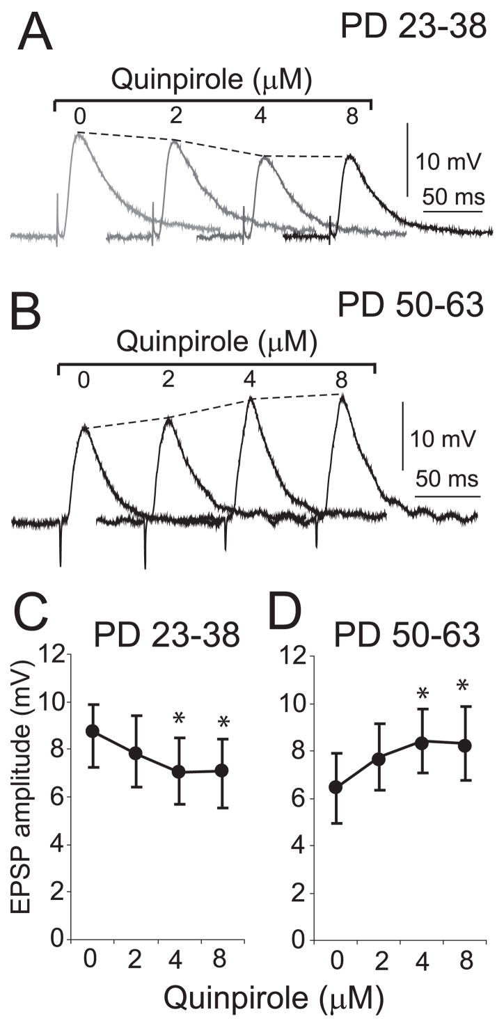
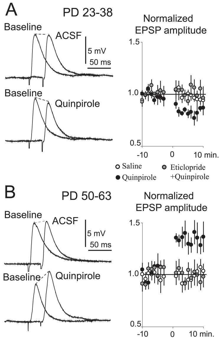
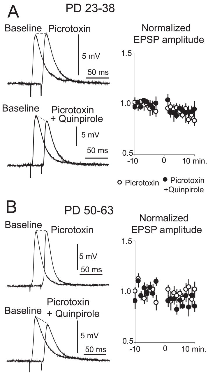
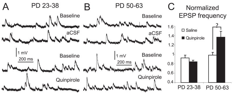
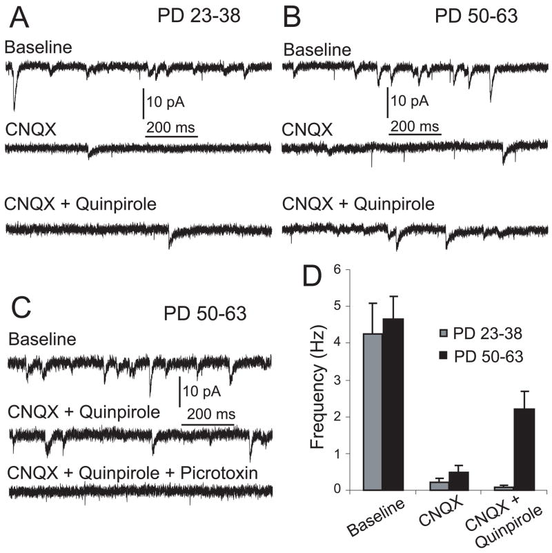
Dopaminergic afferents from the ventral tegmental area (VTA) modulate information processing in the nucleus accumbens (NA), a brain region critical for motivation and reward mechanisms. In NA medium spiny neurons (MSNs) from young rats, D(2) agonists have been shown to decrease the amplitude of corticoaccumbens synaptic responses. As several dopamine-related functions change during adolescence, we assessed the D(2) modulation of cortical inputs with whole-cell recordings in slices obtained from adult and preadolescent rats. The D(2) agonist quinpirole (5 microM) decreased synaptic response of NA MSNs to electrical cortical stimulation in slices from preadolescent rats. In slices from adult rats, however, quinpirole increased both the amplitude of evoked synaptic responses and the frequency of spontaneous synaptic events. These effects were blocked by the GABA-A antagonist picrotoxin (50 microM), revealing a D(2)-mediated decrease These results suggest that D(2) receptors modulate NA neurons differently in young and adult rats, due to the emergence of a D(2)-facilitated GABA component in corticoaccumbens responses during adolescence.
Figures
Similar articles
-
Bidirectional dopaminergic modulation of excitatory synaptic transmission in orexin neurons.J Neurosci. 2006 Sep 27;26(39):10043-50. doi: 10.1523/JNEUROSCI.1819-06.2006. J Neurosci. 2006. Retraction in: J Neurosci. 2012 Jun 27;32(26):9116. doi: 10.1523/JNEUROSCI.1889-12.2012. PMID: 17005867 Free PMC article. Retracted.
-
D2 dopamine receptors recruit a GABA component for their attenuation of excitatory synaptic transmission in the adult rat prefrontal cortex.Synapse. 2007 Oct;61(10):843-50. doi: 10.1002/syn.20432. Synapse. 2007. PMID: 17603809 Free PMC article.
-
Electrophysiological characteristic of corticoaccumbens synapses in rat mesolimbic system reconstructed using organotypic slice cultures.Brain Res. 2004 Jul 23;1015(1-2):34-40. doi: 10.1016/j.brainres.2004.04.033. Brain Res. 2004. PMID: 15223364
-
Dual synaptic sites of D(1)-dopaminergic regulation of ethanol sensitivity of NMDA receptors in nucleus accumbens.Synapse. 2005 Oct;58(1):30-44. doi: 10.1002/syn.20181. Synapse. 2005. PMID: 16037948
-
The modulation of corticoaccumbens transmission by limbic afferents and dopamine: a model for the pathophysiology of schizophrenia.Adv Pharmacol. 1998;42:721-4. doi: 10.1016/s1054-3589(08)60849-2. Adv Pharmacol. 1998. PMID: 9328000 No abstract available.
Cited by
-
Microglial activation increases cocaine self-administration following adolescent nicotine exposure.Nat Commun. 2020 Jan 16;11(1):306. doi: 10.1038/s41467-019-14173-3. Nat Commun. 2020. PMID: 31949158 Free PMC article.
-
Olanzapine antipsychotic treatment of adolescent rats causes long term changes in glutamate and GABA levels in the nucleus accumbens.Schizophr Res. 2015 Feb;161(2-3):452-7. doi: 10.1016/j.schres.2014.10.034. Epub 2014 Dec 5. Schizophr Res. 2015. PMID: 25487700 Free PMC article.
-
Age-dependent effects of low-dose nicotine treatment on cocaine-induced behavioral plasticity in rats.Psychopharmacology (Berl). 2009 Nov;207(1):143-52. doi: 10.1007/s00213-009-1642-0. Psychopharmacology (Berl). 2009. PMID: 19727678 Free PMC article.
-
Dopamine neuron glutamate cotransmission: frequency-dependent modulation in the mesoventromedial projection.Neuroscience. 2009 Dec 15;164(3):1068-83. doi: 10.1016/j.neuroscience.2009.08.057. Epub 2009 Sep 1. Neuroscience. 2009. PMID: 19729052 Free PMC article.
-
Nicotine and the adolescent brain.J Physiol. 2015 Aug 15;593(16):3397-412. doi: 10.1113/JP270492. Epub 2015 Jun 23. J Physiol. 2015. PMID: 26018031 Free PMC article. Review.
References
-
- Akaike A, Ohno Y, Sasa M, Takaori S. Excitatory and inhibitory effects of dopamine on neuronal activity of the caudate nucleus neurons in vitro. Brain Res. 1987;418:262–272. - PubMed
-
- Belleau ML, Warren RA. Postnatal development of electrophysiological properties of nucleus accumbens neurons. J Neurophysiol. 2000;84:2204–2216. - PubMed
-
- Brog JS, Salyapongse A, Deutch AY, Zahm DS. The patterns of afferent innervation of the core and shell in the ‘accumbens’ part of the rat ventral striatum: immunohistochemical detection of retrogradely transported fluoro-gold. J Comp Neurol. 1993;338:255–278. - PubMed
Publication types
MeSH terms
Substances
Grants and funding
LinkOut - more resources
Full Text Sources
Research Materials
