. 2008 May;9(5):786-97.
doi: 10.1111/j.1600-0854.2008.00711.x. Epub 2008 Mar 4.

The lysophospholipid acyltransferase antagonist CI-976 inhibits a late step in COPII vesicle budding

Affiliations

The lysophospholipid acyltransferase antagonist CI-976 inhibits a late step in COPII vesicle budding

William J Brown et al. Traffic. 2008 May.

Abstract

The mechanism of coat protein (COP)II vesicle fission from the endoplasmic reticulum (ER) remains unclear. Lysophospholipid acyltransferases (LPATs) catalyze the conversion of various lysophospholipids to phospholipids, a process that can promote spontaneous changes in membrane curvature. Here, we show that 2,2-methyl-N-(2,4,6,-trimethoxyphenyl)dodecanamide (CI-976), a potent LPAT inhibitor, reversibly inhibited export from the ER in vivo and the formation of COPII vesicles in vitro. Moreover, CI-976 caused the rapid and reversible accumulation of cargo at ER exit sites (ERESs) containing the COPII coat components Sec23/24 and Sec13/31 and a marked enhancement of Sar1p-mediated tubule formation from ERESs, suggesting that CI-976 inhibits the fission of assembled COPII budding elements. These results identify a small molecule inhibitor of a very late step in COPII vesicle formation, consistent with fission inhibition, and demonstrate that this step is likely facilitated by an ER-associated LPAT.


Figures
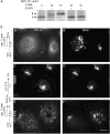
Figure 1
Figure 1. CI-976 inhibits the processing and transport of VSV-G from the ER
A) Processing of VSV-G from the endo H-sensitive (S) to the -resistant (R) form following shift to the permissive temperature is inhibited by CI-976. Cells were labeled with 35S-TransLabel, shifted to the permissive temperature in the absence or presence of 50 μm CI-976 for the times indicated and VSV-G was immunoprecipitated and subjected to digestion by endo H. B) Transport of VSV-G from the ER to the Golgi complex is inhibited by CI-976 as seen by immunofluorescence microscopy. When cells were fixed prior to shift to the permissive temperature, VSV-G was found diffused throughout the cytoplasm in the ER (A′) and separate from the Golgi complex as seen by ManII labeling (B′). When cells were shifted to the permissive temperature for 15 min in the absence of CI-976, VSV-G was found to colocalize with ManII in the Golgi complex (C′ and D′). In contrast, when CI-976 (50 μm) was added for the last 15 min at the restrictive temperature and included for 15 min following shift to the permissive temperature, VSV-G (E′) did not reach the ManII compartment (F′). Instead, VSV-G moved from a diffuse ER-staining pattern to numerous small, punctate foci located throughout the cytoplasm.
Figure 2
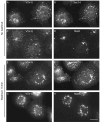
Figure 2. CI-976 inhibits export of VSV-G from the ER at sites enriched for the COPII components Sec24 and Sec31
Cells were infected with ts045 VSV for 3.5 h at the restrictive temperature to accumulate VSV-G in the ER, and CI-976 (50 μm) was then added for the last 15 min of this period. Cells were either fixed immediately (A and B) or shifted to the permissive temperature for an additional 15 min in the continuous presence of CI-976 (50 μm) (C–F). After each incubation period, cells were processed for double immunofluorescence localization of VSV-G (left side panels) and Sec24 or Sec31 (as indicated). Arrows show clusters of foci in which VSV-G and Sec24 or Sec31 colocalize. Bar = 5 μm.
Figure 3
Figure 3. VSV-G that accumulates in Sec24-enriched foci is rapidly transported to the Golgi complex following removal of CI-976
Cells were infected with ts045 VSV for 3.5 h at the restrictive temperature to accumulate VSV-G in the ER, and CI-976 (50 μm) was then added for the last 15 min of this period and then shifted to the permissive temperature in the presence of CI-976 for 15 min. Cells were either maintained under these conditions for an additional 15 min (A–D) or washed free of CI-976 and incubated for 15 min at the permissive temperature (E–H). After each incubation period, cells were processed for double immunofluorescence localization of VSV-G (left side panels) and Sec24 or ManII (as indicated). Arrows show foci containing both VSV-G and Sec24. Bar = 5 μm.
Figure 4
Figure 4. Recovery from CI-976 reduces the lag in ts045 VSV-G processing
A) VSV-G is converted from the endo H-sensitive (S) to the endo H-resistant (R) form following removal of CI-976. Cells were infected with ts045 VSV at the restrictive temperature to accumulate VSV-G in the ER, treated with CI-976 (50 μm) and then shifted to the permissive temperature in the continuous presence of CI-976 for 15 min as described in Figure 1. Cells were then washed free of CI-976 to permit export from the ER and then incubated for the times indicated above each lane. Cells were solubilized, and VSV-G was immunoprecipitated and treated with endo H prior to SDS–PAGE and fluorography. B) Rates of conversion of VSV-G to the endo H-resistant form following a simple shift to the permissive temperature as in Figure 1 (solid circles) or following removal of CI-976 from cells held at the permissive temperature (solid squares) as in (A). Bar = 5 μm.
Figure 5
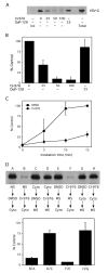
Figure 5. Formation of COPII vesicles in vitro is inhibited by CI-976
A) VSV-G served as cargo marker for Western blot analysis of COPII vesicle budding from ER membranes in an in vitro reconstitution assay. Samples were incubated with the indicated amounts of CI-976 or DuP-128 (μm) for 15 min at 32°C to produce vesicles. Alternatively, samples were kept on ice (as indicated) as a negative control. ‘Total’ lane is equivalent to one third of the total amount of VSV-G present in the incubation mix at the start of the experiment (volumes adjusted to prevent overexposing the ‘Total’ lane). B) Quantification of dose–response experiments similar to those shown in (A). C) Time–course of COPII vesicle budding in the absence (DMSO) or presence of CI-976 (50 μm). D) Inhibition of COPII budding in vitro is most effective when CI-976 is first added to microsomes, as determined by order-of-addition experiments. The order in which each component was added is shown below each lane (A–H). Cyto, cytosol; MS, microsomes. CI-976 was used at 50 μm, and DMSO served as a solvent control. Quantification of these results is shown in the lower graph, and the results are expressed as the % control for each pair of conditions, for example, B/A. For B–D, the results show the mean plus 1 standard deviation from three or more independent experiments.
Figure 6
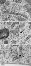
Figure 6. CI-976 induces the formation of chains of COPII vesicles at ERES
Electron microscopy of control (A) and CI-976-treated cells (B and C) reveals that CI-976 causes the formation of chains of vesicles (arrows) from ERESs that have apparently been unable to undergo a late fission step. RER, rough ER. Bar = 0.5 μm.
Figure 7
Figure 7. CI-976 enhances Sar1p-induced ER tubule formation
A) Sar1p-induced tubule formation from ERESs is enhanced by CI-976. Permeabilized NRK cells were incubated with varying amounts of Sar1p-GTP (as indicated on the left) in the absence (DMSO) or presence of CI-976 (50 μm) as indicated under conditions that promote Sar1p-induced tubule formation. Cells were then fixed and stained by immunofluorescence for Sar1p. B) Quantification of CI-976-enhanced Sar1p-mediated membrane tubule formation from ERESs from experiments shown in (A). The lengths of tubules were determined in permeabilized cells incubated with 37.5 or 62.5 μg/mL Sar1p-GTP in absence (−CI-976) or presence (+CI-976) of CI-976. Note that for lengths between 1.0–7.5 μm, tubules were placed into 0.5-μm bins. Number of tubules measured: (37.5 μg/mL Sar1p-GTP), −CI-976 (n = 495), +CI-976 (n = 621); (62.5 μg/mL Sar1p-GTP), −CI-976 (n = 670), +CI-976 (n = 974). C) CI-976-enhanced Sar1p membrane tubules contain VSV-G. Cells were incubated as in (A) and stained by double immunofluorescence for Sar1p and VSV-G. Bar = 5 μm.

Similar articles

Cited by

References

    1. Kooijman EE, Chupin V, de Kruijff B, Burger KN. Modulation of membrane curvature by phosphatidic acid and lysophosphatidic acid. Traffic. 2003;4:162–174. - PubMed
    1. Bonifacino JS, Glick BS. The mechanisms of vesicle budding and fusion. Cell. 2004;116:153–166. - PubMed
    1. Lee MC, Miller EA, Goldberg J, Orci L, Schekman R. Bi-directional protein transport between the ER and Golgi. Annu Rev Cell Dev Biol. 2004;20:87–123. - PubMed
    1. Aridor M, Weissman J, Bannykh S, Nuoffer C, Balch WE. Cargo selection by the COPII budding machinery during export from the ER. J Cell Biol. 1998;141:61–70. - PMC - PubMed
    1. Schekman R. Lasker Basic Medical Research Award. SEC mutants and the secretory apparatus. Nat Med. 2002;8:1055–1058. - PubMed

Publication types

MeSH terms

LinkOut - more resources