Review
. 2008 Jun;9(6):871-81.
doi: 10.1111/j.1600-0854.2008.00734.x. Epub 2008 Mar 6.

Exosome function: from tumor immunology to pathogen biology

Affiliations
Review

Exosome function: from tumor immunology to pathogen biology

Jeffrey S Schorey et al. Traffic. 2008 Jun.

Abstract

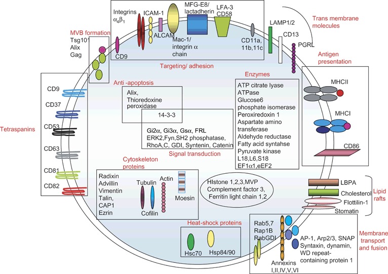
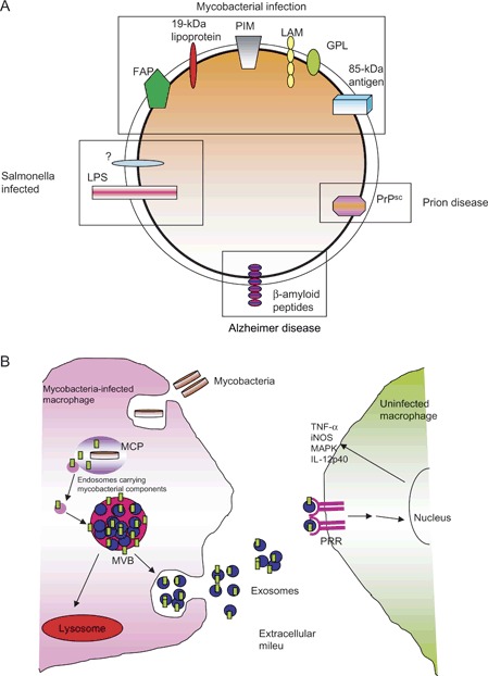
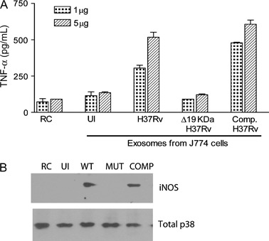
Exosomes are the newest family member of 'bioactive vesicles' that function to promote intercellular communication. Exosomes are derived from the fusion of multivesicular bodies with the plasma membrane and extracellular release of the intraluminal vesicles. Recent studies have focused on the biogenesis and composition of exosomes as well as regulation of exosome release. Exosomes have been shown to be released by cells of hematopoietic and non-hematopoietic origin, yet their function remains enigmatic. Much of the prior work has focused on exosomes as a source of tumor antigens and in presentation of tumor antigens to T cells. However, new studies have shown that exosomes might also promote cell-to-cell spread of infectious agents. Moreover, exosomes isolated from cells infected with various intracellular pathogens, including Mycobacterium tuberculosis and Toxoplasma gondii, have been shown to contain microbial components and can promote antigen presentation and macrophage activation, suggesting that exosomes may function in immune surveillance. In this review, we summarize our understanding of exosome biogenesis but focus primarily on new insights into exosome function. We also discuss their possible use as disease biomarkers and vaccine candidates.


Figures

Figure 1
Figure 1
MVB biogenesis and exosome release. Monoubiquitination or aggregation provides the signal for trafficking of proteins and lipids to MVBs. The machinery for sorting ubiquitinated proteins involves the multi‐domain Vps27/HRS protein that acts as a bridge between monoubiquitinated transmembrane proteins and clathrin on endosomes (94). ESCRT is also a key player in MVB biogenesis. ESCRT‐I, ‐II and ‐III recognize monoubiquitinated cargoes and promote their inclusion in MVBs (95). Once completed, the ESCRT complex dissociates from the MVB membrane. Interestingly, the proteins within the ILVs are enriched for ubiquitin, indicating that not all of the ubiquitin is removed from proteins upon targeting to the MVB. Fusion of the MVB and release of the ILVs as exosomes are regulated and are known to require PLD, calcium and Rab11. PLD, phospholipase D.
Figure 2
Figure 2
Protein composition of exosomes indicating their name, their location (i.e. membrane bound or soluble) and in some cases their function. GDI, GTP dissociation inhibitor; ICAM1, intercellular adhesion molecule‐1; CAP‐1, adenylyl cyclase‐associated protein; LAMP, lysosomal associated membrane protein‐1; PGRL, PG regulatory‐like protein.
Figure 3
Figure 3
Composition and trafficking of exosomes from infected macrophages with a focus on mycobacterial‐infected cells. A) A list of microbial or infectious components that have been shown to be on exosomes. These include ‘infectious’ proteins such as prions as well as HIV. Also shown are microbial molecules, which have been released from the pathogen and trafficked to MVBs and present on exosomes. B) General diagram of how mycobacterial PAMPs are released from the mycobacteria within a phagosome and transported to the MVB for release on exosomes. The exosomes containing the PAMPs bind to pattern recognition receptors (PRRs) on surrounding macrophages leading to macrophage activation. LPS, lipopolysaccharide; MCP, mycobacteria‐containing phagosome; FAP, fibronectin attachment protein; iNOS, inducible nitric oxide synthase; MAPK, mitogen activated protein kinase.
Figure 4
Figure 4
Exosomes isolated from macrophages infected with an LspA‐deficient H37Rv fail to stimulate macrophage activation. Exosomes were isolated from infected J774 cells as described (81) and used to treat bone marrow‐derived murine macrophages. After 24 h, supernatants were isolated and analyzed for TNF‐α by enzyme‐linked immunosorbent assay, and cells were harvested and analyzed for iNOS expression by Western blot as described (81). Total MAPK p38 was used as a loading control. COMP, the LspA mutant complemented with the wild‐type (WT) LspA gene; MUT, H37Rv mutant that lacks LspA and therefore fails to make the 19‐KDa lipoprotein; RC, untreated cells; UI, exosomes from uninfected cells.

References

    1. Johnstone RM, Adam M, Hammond JR, Orr L, Turbide C. Vesicle formation during reticulocyte maturation. Association of plasma membrane activities with released vesicles (exosomes). J Biol Chem 1987;262:9412–9420. - PubMed
    1. Couzin J. Cell biology: the ins and outs of exosomes. Science 2005;308:1862–1863. - PubMed
    1. Gould SJ, Booth AM, Hildreth JE. The Trojan exosome hypothesis. Proc Natl Acad Sci U S A 2003;100:10592–10597. - PMC - PubMed
    1. Fang Y, Wu N, Gan X, Yan W, Morrell JC, Gould SJ. Higher‐order oligomerization targets plasma membrane proteins and HIV gag to exosomes. PLoS Biol 2007;5:e158. - PMC - PubMed
    1. Chen BJ, Lamb RA. Mechanisms for enveloped virus budding: can some viruses do without an ESCRT? Virology 2007;372:221–232. - PMC - PubMed

Publication types

LinkOut - more resources