. 2008 Mar;14(3):331-41.
doi: 10.1016/j.devcel.2007.12.007.

sSgo1, a major splice variant of Sgo1, functions in centriole cohesion where it is regulated by Plk1

Affiliations

sSgo1, a major splice variant of Sgo1, functions in centriole cohesion where it is regulated by Plk1

Xiaoxing Wang et al. Dev Cell. 2008 Mar.

Abstract

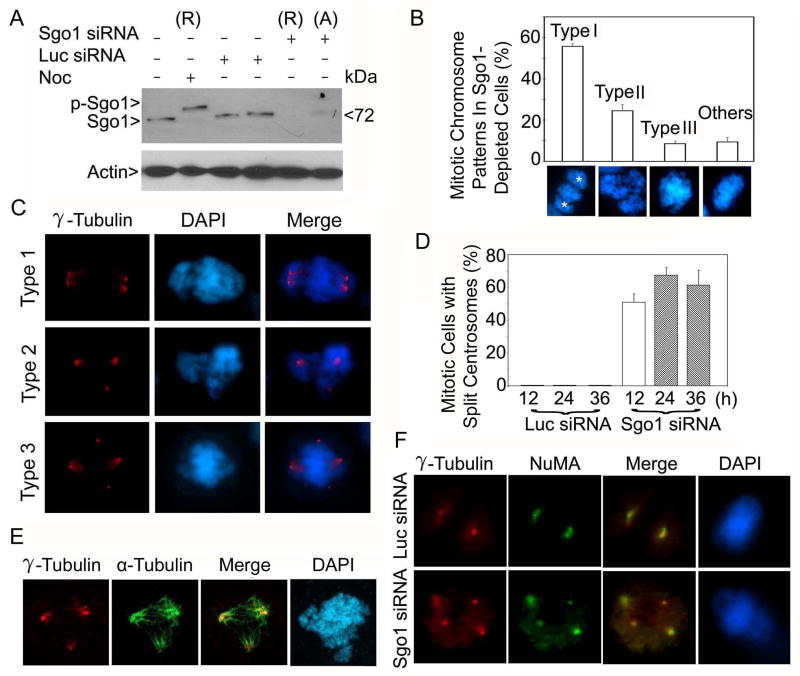
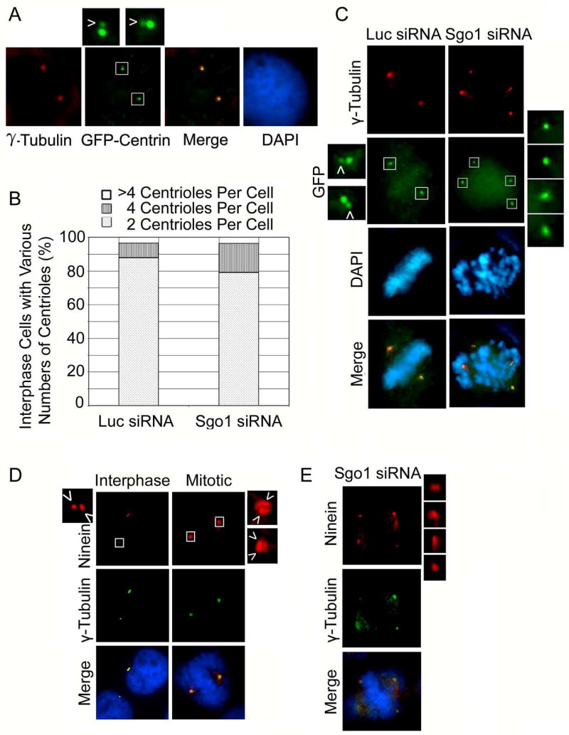
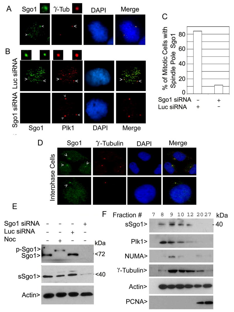
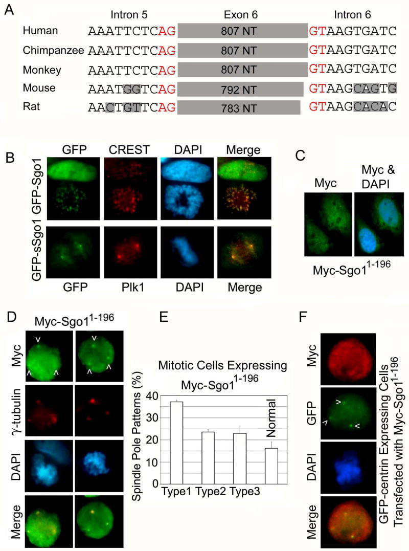
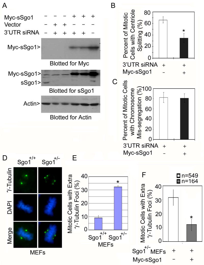
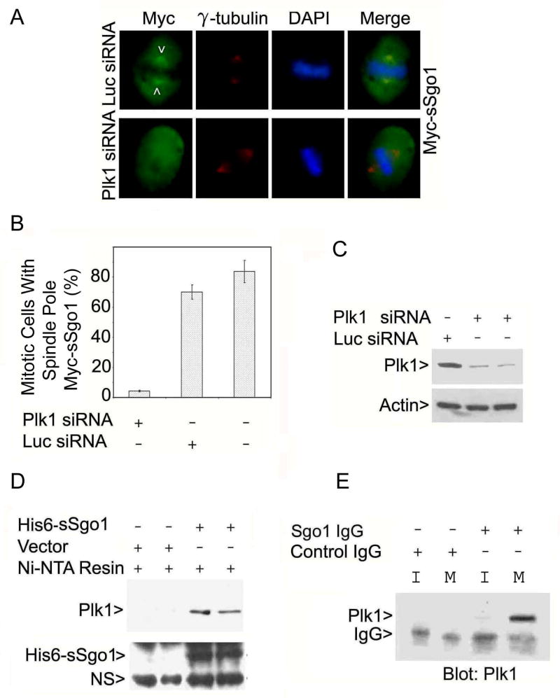
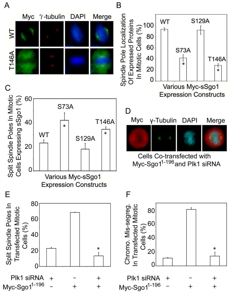
Shugoshin 1 (Sgo1) functions as a protector of centromeric cohesion of sister chromatids in higher eukaryotes. Here, we provide evidence for a previously unrecognized role for Sgo1 in centriole cohesion. Sgo1 depletion via RNA interference induces the formation of multiple centrosome-like structures in mitotic cells that result from the separation of paired centrioles. Sgo1+/- mitotic murine embryonic fibroblasts display split centrosomes. Localization study of two major endogenous splice variants of Sgo1 indicates that the smaller variant, sSgo1, is found at the centrosome in interphase and at spindle poles in mitosis. sSgo1 interacts with Plk1 and its spindle pole localization is Plk1 dependent. Centriole splitting induced by Sgo1 depletion or expression of a dominant negative mutant is suppressed by ectopic expression of sSgo1 or by Plk1 knockdown. Our studies strongly suggest that sSgo1 plays an essential role in protecting centriole cohesion, which is partly regulated by Plk1.


Figures

Figure 1
Figure 1
siRNA depletion of Sgo1 causes formation of extra γ-tubulin foci. (A) HeLa cells were transfected with Sgo1 or luciferase (Luc) siRNA for 24 h. Rounded-up (R) and adherent (A) cells in Sgo1 siRNA transfected dishes were collected separately. An equal amount of each cell lysate was blotted for Sgo1 and β-actin. Nocodazole (Noc)-treated cell lysates were loaded as control. Arrow p-Sgo1 indicates phospho-Sgo1. (B) Rounded-up cells induced as a result of Sgo1 depletion were examined for chromosome patterns after DAPI staining. The data were summarized from more than 300 mitotic cells depleted of Sgo1. The stars (*) denote spindle pole positions. (C) HeLa cells transfected with Sgo1 siRNA were stained with an antibody to γ-tubulin (red). DNA was stained with DAPI (blue). Representative images of cells with centrosome splitting are shown. (D) The percentage of siRNA-transfected cells with extra centrosomal foci was summarized from 200 mitotic cells at each time point. (E) A representative cell transfected with Sgo1 siRNA for 24 h and stained with antibodies to γ-tubulin (red) or α-tubulin (green) is shown. (F) HeLa cells transfected with Sgo1 or Luc siRNA were stained with antibodies to γ-tubulin (red) and NuMA (green). Representative images are shown.
Figure 2
Figure 2
Sgo1 depletion-induced formation of extra centrosomal foci results from centriole splitting. (A) HeLa cells transfected with a GFP-centrin expression plasmid for 24 h were stained with an antibody to GFP (green) or γ-tubulin (red). The daughter centrioles are indicated by arrows. (B) HeLa cells stably expressing GFP-centrin transfected with Sgo1 or Luc siRNA for 24 h were fixed and stained with the antibody to GFP. The number of centrioles in individual interphase cells (100 cells/experiment) was scored under the microscope. The data was summarized from three independent experiments. (C) GFP-centrin-expressing cells were transfected with Sgo1 or Luc siRNA for 24 h. Cells were fixed and stained with antibody to GFP or γ-tubulin. Representative cells are shown. The boxed areas were enlarged. Daughter centrioles are indicated by arrows. (D). HeLa cells were fixed and stained with the antibody to ninein (red) or γ-tubulin (green). Representative cells are shown. Paired centrioles are indicated by arrows. (E) HeLa cells transfected with Sgo1 siRNA for 24 h were stained with the antibody to ninein or γ-tubulin.. The ninein foci were enlarged.
Figure 3
Figure 3
Sgo1 is localized to the centrosomes/spindle poles. (A) HeLa cells were stained with antibody to Sgo1 (green) or γ-tubulin (red). Arrows indicate spindle pole signal. The area indicated by the upper right arrow was enlarged. (B) U2OS cells transfected with Luc or Sgo1 siRNA for 24 h were stained with the antibody to Sgo1 (green) or Plk1 (red). Arrows indicate spindle pole signal. The areas indicated by arrows were enlarged. (C) Data were summarized from over 100 mitotic cells shown in B after transfection of Sgo1 or Luc siRNA. (D) HeLa cells were stained with antibodies to Sgo1 γ-tubulin. Arrows indicate the centrosome positions. (E) An equal amount of lysates from HeLa cells transfected with Sgo1 or Luc siRNA for 24 h was blotted for Sgo1 and β-actin. Noc-treated cell lysates were loaded as controls. (F) Centrosomes were isolated from HeLa cells.. An equal volume of sample from selected centrosomal isolation fractions was blotted for sSgo1, Plk1, NuMA, γ-tubulin, β-actin, and PCNA.
Figure 4
Figure 4
sSgo1 functions in regulating spindle pole integrity. (A) The intronic sequences surrounding human exon 6 or its equivalent in other mammals were aligned. Red color marks the invariant splice donor and splice acceptor sequences. The shaded areas indicate non-conserved sequences in introns. (B) HeLa cells transfected with GFP-Sgo1 or GFP-sSgo1 expression constructs for 24 h were fixed and stained with antibodies to GFP (green), CREST (red, upper panel), or Plk1 (red, lower panel). (C) HeLa cells transfected with Myc-Sgo11-196 expression plasmid for 24 h were stained with antibody to Myc. (D&E) HeLa cells transfected with Myc-Sgo11-196 plasmid for 24 h were stained with antibodies to Myc (green) and γ-tubulin (red) (D). Various abnormal spindle pole patterns were summarized from three independent experiments (E) (150 mitotic cells/experiment). (F) HeLa cells expressing GFP-centrin transfected with Myc-Sgo11-196 plasmid for 24 h were stained with antibodies to Myc (red) and GFP (green). Arrows indicate the position of split centrioles.
Figure 5
Figure 5
Ectopic expression of sSgo1 suppresses centriole splitting induced by Sgo1 depletion. (A) HeLa cells co-transfected with the Myc-sSgo1 expression construct and Sgo1 3′UTR siRNA for 48 h were collected and an equal amount of cell lysate was blotted for Myc, endogenous sSgo1, and β-actin. (B & C) HeLa cells transfected with the Myc-sSgo1 expression construct and Sgo1 3′UTR siRNA for about 32 h were fixed and stained with antibodies to the Myc tag and γ-tubulin. The data were summarized from over 200 transfected mitotic cells. (D) Wild-type (Sgo1+/+) and Sgo1+/− MEF cells were stained with antibody to γ-tubulin (green). (E) The number of mitotic cells with extra γ-tubulin foci in Sgo1+/+ and Sgo1+/− MEF cells was scored. The data were summarized from three independent experiments (100 mitotic cells/experiment). (F) Sgo1+/− MEFs transfected with a Myc-sSgo1 expression construct for 24 h were stained with antibodies to γ-tubulin and the Myc tag. The number (n) of mitotic cells with extra γ-tubulin foci was scored (2 independent experiments). * denotes significant statistical difference (p<0.01).
Figure 6
Figure 6
Plk1 is physically associated with sSgo1. (A) HeLa cells co-transfected with Myc-sSgo1 plasmid and Plk1 or Luc siRNA for 24 h were stained with antibodies to the Myc tag (green) and γ-tubulin (red). (B) HeLa cells were cotransfected with Myc-sSgo1 expression plasmid and Plk1 or Luc siRNA. The data were summarized from three independent experiments (100 Plk1-depleted mitotic cells/experiment). (C) HeLa cells transfected with Plk1 or Luc siRNA for 24 h were blotted for Plk1 and β-actin. (D) Aqual amounts of HeLa cell lysates after transfection with a His6-sSgo1plasmid or the vector alone for 48 h were incubated with Ni-NTA resin. Proteins specifically bound to the resin were blotted for Plk1 or Sgo1. Arrow NS denotes a non-specific signal. (E) An equal amount of interphase (I) or mitotic (M) HeLa cell lysate was immunoprecipitated with the anti-Sgo1 antibody or with a control IgG. The immunoprecipitates were blotted for Plk1.
Figure 7
Figure 7
Plk1 regulates Sgo1 function. (A) HeLa cells transfected with Myc-sSgo1 or Myc-sSgo1T146A expression plasmid for 24 h were stained with antibodies to the Myc tag (green) and γ-tubulin (red). (B) HeLa cells transfected with Myc-sSgo1 expression plasmid (WT) or with various Myc-tagged sSgo1 mutant plasmids as indicated for 24 h were stained with antibody to the Myc tag. The data were summarized from three independent experiments (>100 transfected mitotic cells). * denotes significant statistical difference (p<0.01). (C) HeLa cells transfected with Myc-tagged sSgo1 (WT) or with mutant expression plasmids for 24 h were stained with antibodies to the Myc tag and γ-tubulin. The data were summarized from three independent experiments (>300 transfected mitotic cells). * denotes significant statistical difference (p<0.05). (D) HeLa cells co-transfected with Myc-Sgo11-196 expression plasmid and Plk1 siRNA for 24 h were stained with antibodies to the Myc tag (red) and γ-tubulin (green). (E and F) HeLa cells co-transfected with Myc-Sgo11-196 expression plasmid and Plk1 siRNA for 24 h were stained with antibodies to the Myc tag and γ-tubulin. The data were summarized from three independent experiments (>300 transfected mitotic cells). * denotes significant statistical difference (p<0.01).

Comment in

References

    1. Arnaud L, Pines J, Nigg EA. GFP tagging reveals human Polo-like kinase 1 at the kinetochore/centromere region of mitotic chromosomes. Chromosoma. 1998;107:424–429. - PubMed
    1. Blomberg-Wirschell M, Doxsey SJ. Rapid isolation of centrosomes. Methods Enzymol. 1998;298:228–238. - PubMed
    1. Clarke AS, Tang TT, Ooi DL, Orr-Weaver TL. POLO kinase regulates the Drosophila centromere cohesion protein MEI-S332. Dev Cell. 2005;8:53–64. - PubMed
    1. Dionne MA, Howard L, Compton DA. NuMA is a component of an insoluble matrix at mitotic spindle poles. Cell Motil Cytoskeleton. 1999;42:189–203. - PubMed
    1. Doxsey S, McCollum D, Theurkauf W. Centrosomes in cellular regulation. Annu Rev Cell Dev Biol. 2005;21:411–434. - PubMed

Publication types

MeSH terms