. 2008 Mar;14(3):424-36.
doi: 10.1016/j.devcel.2008.01.005.

Ajuba LIM proteins are snail/slug corepressors required for neural crest development in Xenopus

Affiliations

Ajuba LIM proteins are snail/slug corepressors required for neural crest development in Xenopus

Ellen M Langer et al. Dev Cell. 2008 Mar.

Abstract

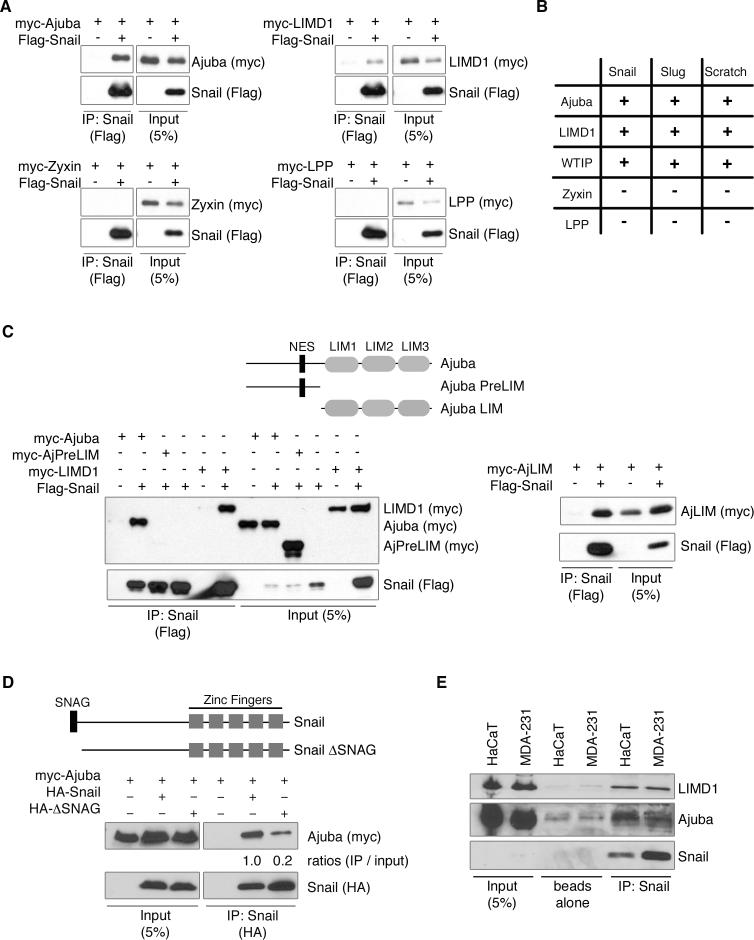
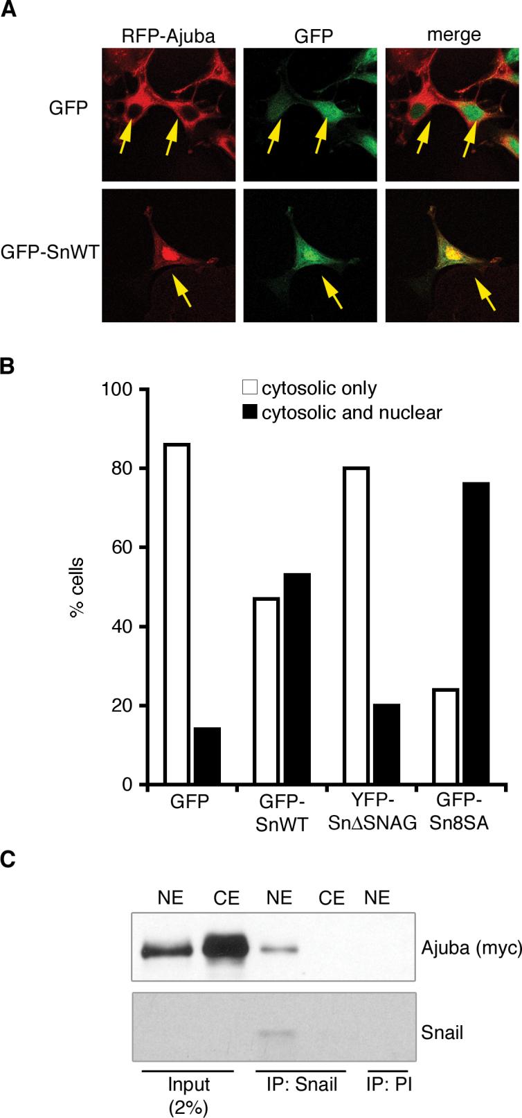
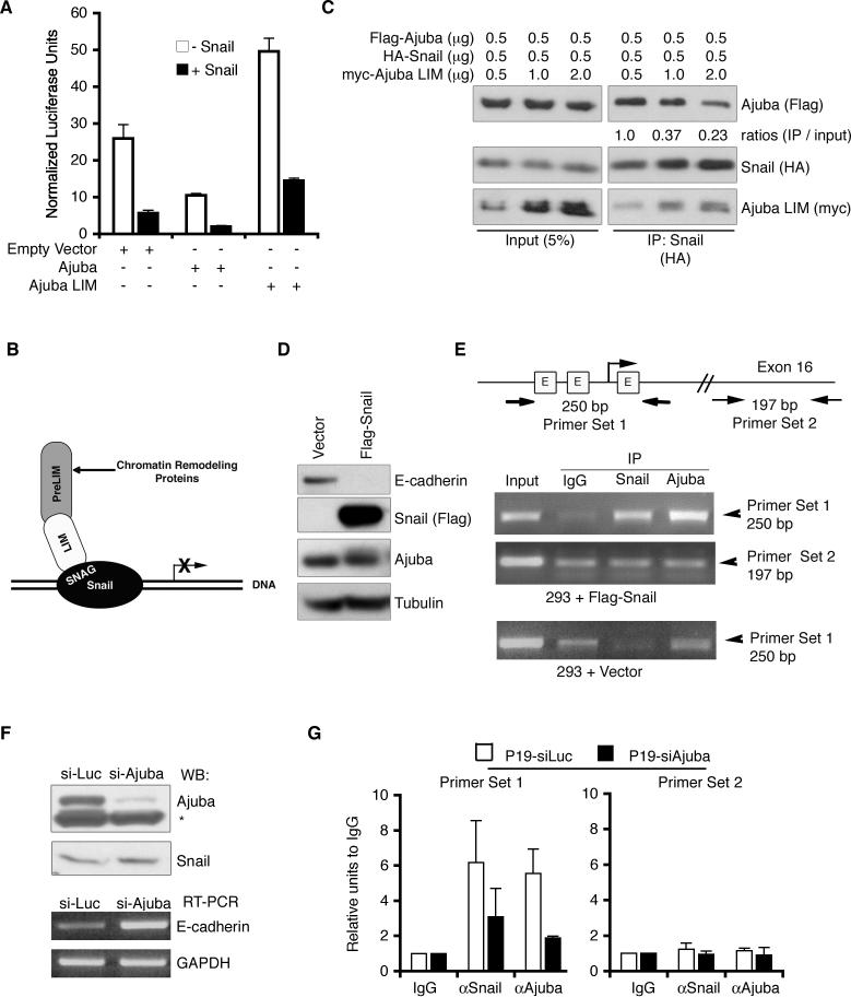
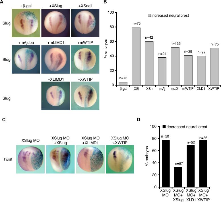
Snail family transcriptional repressors regulate epithelial mesenchymal transitions during physiological and pathological processes. A conserved SNAG repression domain present in all vertebrate Snail proteins is necessary for repressor complex assembly. Here, we identify the Ajuba family of LIM proteins as functional corepressors of the Snail family via an interaction with the SNAG domain. Ajuba LIM proteins interact with Snail in the nucleus on endogenous E-cadherin promoters and contribute to Snail-dependent repression of E-cadherin. Using Xenopus neural crest as a model of in vivo Snail- or Slug-induced EMT, we demonstrate that Ajuba LIM proteins contribute to neural crest development as Snail/Slug corepressors and are required for in vivo Snail/Slug function. Because Ajuba LIM proteins are also components of adherens junctions and contribute to their assembly or stability, their functional interaction with Snail proteins in the nucleus suggests that Ajuba LIM proteins are important regulators of epithelia dynamics communicating surface events with nuclear responses.


Figures

Figure 1
Figure 1. Ajuba LIM proteins interact with Snail transcriptional repressors
A. Myc-tagged LIM proteins and Flag-Snail were cotransfected into HEK293 cells. Snail was immunoprecipitated (anti-Flag) and bound products Western blotted for LIM protein (anti-myc) and Snail (anti-Flag). Control Western blot of lysate is on right panel of each set. B. Table of interactions between Snail proteins and LIM proteins, as determined by co-immunoprecipitation, as described in A. C. Top: Schematic of Ajuba constructs used. NES – nuclear export sequence. Bottom: Co-immunoprecipitation experiments as in A. D. Top: Schematic of Snail constructs used. Bottom: Myc-tagged Ajuba and HA-tagged Snail constructs were cotransfected into HEK293 cells. Snail was immunoprecipitated (anti-HA) and bound products Western blotted for Ajuba (anti-myc) and Snail (anti-HA). Control Western blot of lysate is shown on left. The amount of Ajuba immunoprecipitated relative to input was quantified by densitometry and controlled for the amount of Snail immunoprecipitated. (Ajuba immunoprecipitated with full-length Snail was arbitrarily set to 1). E. Endogenous Snail was immunoprecipitated from lysates of HaCaT or MDA-231 cells and bound products Western blotted for the presence of Ajuba, LIMD1 and Snail. Controls include pulldown with Protein G beads alone and lysate input.
Figure 2
Figure 2. Ajuba interacts with Snail in the nucleus
A. Confocal immunofluorescence analysis of MCF-7 cells co-transfected with RFP-Ajuba and GFP or GFP-Snail. Arrows indicate cotransfected cells. B. Quantification of immunofluorescence results. Shown is the percent of cells in which RFP-Ajuba is localized to the cytosol (white columns) or to the cytosol and the nucleus (black columns) in the presence of GFP, GFP-Snail, YFP-Snail.ΔSNAG or GFP-Snail8SA. For each sample, at least 100 cells were counted. The experiment was repeated three times with similar results. Shown is one representative experiment. C. Endogenous Snail was immunoprecipitated from nuclear extracts (NE) or cytosolic extracts (CE) of HaCaT cells stably expressing myc-Ajuba. Bound products were Western blotted for the presence of Ajuba (anti-myc) and Snail. Control immunoprecipitation was performed with rabbit preimmune sera (PI). Western blot of input controls is shown on the left.
Figure 3
Figure 3. Ajuba is a corepressor of Snail
A. Transient luciferase reporter assay using luciferase driven by the E-cadherin promoter. Constructs, as indicated, were expressed in MCF-7 cells and luciferase activity determined and normalized to β-gal activity (from co-transfected CMV-β–gal construct). Experiments were performed in triplicate. Shown are mean normalized luciferase values +/− standard deviations. B. Schematic of Ajuba interacting with Snail bound at a promoter to enhance Snail-dependent repression. C. HEK293 cells were co-transfected with Flag-Ajuba, HA-Snail, and myc-Ajuba LIM region as indicated. Snail was immunoprecipitated from lysates (anti-HA) and bound products Western blotted for full-length Ajuba (anti-Flag), Snail (anti-HA), and Ajuba LIM region (anti-myc). The amount of Ajuba immunoprecipitated relative to input was quantified by densitometry and controlled for the amount of Snail immunoprecipitated. The value for lane 4 was arbitrarily set to equal 1. D. Lysates of HEK293 cells stably transfected with empty vector or Flag-Snail were immunoblotted for E-cadherin, Flag, Ajuba, and Tubulin (as loading control). E. ChIPs were performed in HEK293 cells stably transfected with empty vector or Flag-Snail using antibodies to Snail and Ajuba. IgG was used as a control. PCRs were performed using primers flanking the three E-boxes (labeled E in schematic) in the human E-cadherin promoter (primer set 1) or flanking a region of Exon 16 (primer set 2). F. P19 cells were stably transfected with siRNA constructs targeting luciferase (Luc) or Ajuba. Top panel: lysates were immunoblotted for presence of Ajuba or Snail (* marks non-specific band). Bottom panel: RT-PCR was performed to detect E-cadherin levels. GAPDH is a loading control. G. ChIPs were performed in P19-siLuc (white bars) and P19-siAjuba (black bars) cells using antibodies to Snail and Ajuba. IgG was used as a control. Quantitative PCR was performed using primers flanking the E-boxes of the mouse E-cadherin promoter (primer set 1) or flanking a region of Exon 15 (primer set 2).
Figure 4
Figure 4. Ajuba LIM protein expression in Xenopus embryos enhances neural crest development in a Slug-dependent manner
A. X. laevis embryos co-injected with β-gal and either XSlug, XSnail, mAjuba, mLIMD1, mWTIP, XLIMD1, or XWTIP capped mRNAs were fixed at stage 18 and in situ hybridization performed for XSlug. B. Graph displaying the percent of embryos with increased neural crest on the injected side (by Slug in situ hybridization). The total number of embryos injected is shown over each column (n). C. X. laevis embryos were co-injected with β-gal and the XSlug MO alone or in combination with XLIMD1, XWTIP, or XSlug mRNA, and in situ hybridization for Twist performed. D. Graph displaying the percent of embryos with decreased neural crest on the injected side (by Twist in situ hybridization). The total number of embryos injected is shown over each column (n).
Figure 5
Figure 5. Depletion of XLIMD1 or XWTIP blocks neural crest development in Xenopus
A. X. laevis embryos were co-injected with β-gal and control MO, XLIMD1 MO (20ng), XWTIP MO (10ng), or a combination of XLIMD1 and XWTIP MOs. Embryos were fixed at stage 18−19, and in situ hybridization for XSlug or XTwist performed. B. Graph displaying percent of embryos with decreased neural crest on injected side by Slug or Twist in situ hybridization following injection of low dose (gray columns; 10ng XLIMD1 MO, 5ng XWTIP MO) or high dose (black columns; 20ng XLIMD1 MO, 10ng XWTIP MO) of morpholinos. The total number of embryos injected is shown over each column (n). C. X. laevis embryos were co-injected with β-gal, the XWTIP MO (5ng) and XWTIP capped mRNA as shown. Black columns indicate the percent of embryos with decreased neural crest on the injected side (by Slug or Twist in situ) and white columns indicate the percent of embryos where neural crest was the same on the injected and uninjected sides. The total number of embryos injected is shown over each set of columns (n).
Figure 6
Figure 6. Biochemical and functional mapping of LIM domain-Snail interaction
A. HEK293 cells were cotransfected with Flag-tagged mLIMD1 constructs and HA-XSnail as shown. Snail was immunoprecipitated from lysates (anti-HA) and bound products Western blotted for the presence of LIMD1 isoforms (Flag) and Snail (HA). Control Western blot of lysate is on right. B. Graph displaying the percent of embryos with increased neural crest on injected side by Slug in situ hybridization following injection of mLIMD1 (LD1), mLIMD1 PreLIM (PL), PL+LIM1, PL+LIM2, or PL+LIM3 as shown. The total number of embryos injected is shown over each column (n).
Figure 7
Figure 7. Ajuba LIM proteins affect cell survival and border territories without affecting proliferation
A. X. laevis embryos were injected with XSlug or XLIMD1 mRNA, fixed at stage 16 and immunohistochemistry performed for phosphohistone-H3. B. X. laevis embryos were injected with XSlug MO or combination of XLIMD1 +XWTIP MOs. Embryos were fixed at stage 16 and TUNEL staining performed. The number of embryos displaying increase TUNEL staining on the injected side over the total number of embryos analyzed is shown in the bottom right corner. C. X. laevis embryos were injected with MOs as shown, fixed at stage 16 and in situ hybridization performed for Epiker (epidermal) and Sox2 (neural).
Figure 8
Figure 8. Depletion of both XLIMD1 and XWTIP blocks Slug repressor activity during neural crest development in Xenopus
A. X. laevis embryos were co-injected with β-gal, XLIMD1 MO (5ng) and XWTIP MO (10ng) alone or in combination with XSlug, SlZnF, or EngR.SlZnF capped mRNA. Embryos were fixed at stage 18−19 and in situ hybridization performed for XSlug. B. Graph displaying the percent of embryos with decreased neural crest on the injected side by Slug in situs (following injection of MOs and RNAs as shown). The total number of embryos injected is shown over each column (n).

References

    1. Aybar MJ, Nieto MA, Mayor R. Snail precedes slug in the genetic cascade required for the specification and migration of the Xenopus neural crest. Development. 2003;130(3):483–494. - PubMed
    1. Ayyanathan K, Peng H, Hou Z, Fredericks WJ, Goyal RK, Langer EM, Longmore GD, Rauscher FJ., 3rd The Ajuba LIM domain protein is a corepressor for SNAG domain mediated repression and participates in nucleocytoplasmic Shuttling. Cancer Res. 2007;67(19):9097–9106. - PubMed
    1. Barrallo-Gimeno A, Nieto MA. The Snail genes as inducers of cell movement and survival: implications in development and cancer. Development. 2005;132(14):3151–3161. - PubMed
    1. Batlle E, Sancho E, Franci C, Dominguez D, Monfar M, Baulida J, Garcia De Herreros A. The transcription factor snail is a repressor of E-cadherin gene expression in epithelial tumour cells. Nat Cell Biol. 2000;2(2):84–89. - PubMed
    1. Bellmeyer A, Krase J, Lindgren J, LaBonne C. The protooncogene c-myc is an essential regulator of neural crest formation in xenopus. Dev Cell. 2003;4(6):827–839. - PubMed

Publication types

MeSH terms

Associated data