. 2008 May;28(10):3424-36.
doi: 10.1128/MCB.02186-07. Epub 2008 Mar 10.

MURC, a muscle-restricted coiled-coil protein that modulates the Rho/ROCK pathway, induces cardiac dysfunction and conduction disturbance

Affiliations

MURC, a muscle-restricted coiled-coil protein that modulates the Rho/ROCK pathway, induces cardiac dysfunction and conduction disturbance

Takehiro Ogata et al. Mol Cell Biol. 2008 May.

Abstract

We identified a novel muscle-restricted putative coiled-coil protein, MURC, which is evolutionarily conserved from frog to human. MURC was localized to the cytoplasm with accumulation in the Z-line of the sarcomere in the murine adult heart. MURC mRNA expression in the heart increased during the developmental process from the embryonic stage to adulthood. In response to pressure overload, MURC mRNA expression increased in the hypertrophied heart. Using the yeast two-hybrid system, we identified the serum deprivation response (SDPR) protein, a phosphatidylserine-binding protein, as a MURC-binding protein. MURC induced activation of the RhoA/ROCK pathway, which modulated serum response factor-mediated atrial natriuretic peptide (ANP) expression and myofibrillar organization. SDPR augmented MURC-induced transactivation of the ANP promoter in cardiomyocytes, and RNA interference of SDPR attenuated the action of MURC on the ANP promoter. Transgenic mice expressing cardiac-specific MURC (Tg-MURC) exhibited cardiac contractile dysfunction and atrioventricular (AV) conduction disturbances with atrial chamber enlargement, reduced thickness of the ventricular wall, and interstitial fibrosis. Spontaneous episodes of atrial fibrillation and AV block were observed in Tg-MURC mice. These findings indicate that MURC modulates RhoA signaling and that MURC plays an important role in the development of cardiac dysfunction and conduction disturbance with increased vulnerability to atrial arrhythmias.


Figures

FIG. 1.
FIG. 1.
Primary sequence of MURC. Alignment and amino acid sequence comparison of human MURC (GenBank accession number EU487253), mouse MURC (EU487254), and rat MURC (EU487255) and homology with a sequence from Xenopus (NM_001097894). Identical amino acids are shaded in gray, and gaps are represented by a dash. Positions in the amino acid sequence are given by the numbers. MURC contains a predicted coiled-coil domain.
FIG. 2.
FIG. 2.
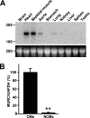
MURC expression in tissues. (A) Blots were made with total RNA (20 μg) isolated from tissues of adult mice. The MURC transcript was observed in heart, skeletal muscle, aorta, and lung. 28S RNA was used as a control for assessing RNA loading. (B) Real-time RT-PCR was performed with cDNAs from cultured rat neonatal cardiomyocytes (CMs) and noncardiomyocytes (NCMs). **, P < 0.01 compared with CMs.
FIG. 3.
FIG. 3.
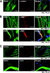
Localization of MURC in the heart. (A) Immunostaining was performed using adult mouse heart sections with anti-MURC and anti-α-actinin antibodies. (B and C) Immunostaining was performed using adult mouse heart sections with an anti-MURC antibody and an anti-α-smooth muscle actin (SMA) antibody (upper panels) or an anti-CD31 antibody (lower panels). Nuclei were stained by DAPI (blue). Higher-magnification images are shown in lower panels.
FIG. 4.
FIG. 4.
MURC expression in the heart. (A) RT-PCR was performed with cDNAs from hearts at E10.5 (left). Real-time RT-PCR was performed with cDNAs from embryonic (E10.5, E13.5, E15.5, and E17.5), neonatal, and adult hearts (right). *, P < 0.05 compared with E10.5; **, P < 0.01 compared with E10.5. (B) Real-time RT-PCR was performed with cDNAs from hearts of sham-operated mice (sham) and abdominal aortic-banded mice (band). Seven days after surgery, total RNA was isolated from hearts. **, P < 0.01 compared with sham.
FIG. 5.
FIG. 5.
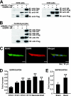
Identification of SDPR as a MURC-binding protein and SDPR expression in the heart. (A) Immunoprecipitation from COS cells expressing hMURC-Flag and/or hSDPR-HA. hMURC-Flag alone, hSDPR-HA alone, or both proteins were transiently expressed in COS cells. Each cell extract was subjected to immunoprecipitation with the anti-Flag or anti-HA monoclonal antibody. The immunoprecipitate was then subjected to SDS-PAGE, followed by Western blot analysis with the anti-Flag or anti-HA monoclonal antibody. IP, immunoprecipitation; IB, immunoblot assay. (B) Immunoprecipitation from cardiomyocytes infected with Ad-MURC and/or Ad-SDPR. LacZ, mMURC-Flag with LacZ, hSDPR-HA with LacZ, or mMURC-Flag with hSDPR-HA was transiently expressed in cardiomyocytes. Each cell extract was subjected to immunoprecipitation with the anti-Flag or anti-HA monoclonal antibody. The immunoprecipitate was then subjected to SDS-PAGE, followed by Western blot analysis with the anti-Flag or anti-HA monoclonal antibody. (C) Localization of SDPR in adult cardiomyocytes. Adult cardiomyocytes were infected with Ad-SDPR at an MOI of 10. Immunostaining was performed using isolated adult mouse cardiomyocytes with anti-MURC and anti-HA antibodies. Nuclei were stained by DAPI (blue). (D) Real-time RT-PCR was performed with cDNAs from embryonic (E10.5, E13.5, E15.5, and E17.5), neonatal, and adult hearts. **, P < 0.01 compared with E10.5. (E) Real-time RT-PCR was performed with cDNAs from hearts of sham-operated mice and abdominal aortic-banded mice. Seven days after surgery, total RNA was isolated from hearts. **, P < 0.01 compared with sham.
FIG. 6.
FIG. 6.
Induction of ANP expression by MURC through the Rho/ROCK signaling pathway. (A and C) The bar graph shows real-time RT-PCR analysis of ANP mRNA expression in rat neonatal cardiomyocytes. Cells were infected with Ad-LacZ or Ad-MURC at an MOI of 10 and then harvested 48 h after the infection. Y-27632 was added after infection. **, P < 0.01 compared with Ad-LacZ; ††, P < 0.01 compared with Y-27632-treated Ad-LacZ. (B) The bar graph shows RhoA activity in rat neonatal cardiomyocytes. Cells were infected with Ad-LacZ or Ad-MURC at an MOI of 10 and then harvested 48 h after infection. *, P < 0.05 compared with Ad-LacZ. (D to F) The bar graphs show a reporter assay using −638 ANP Luc or no-SRE1/SRE2 Luc. hMURC, mMURC, C3, and/or RhoA V14 were cotransfected with −638 ANP Luc or no-SRE1/SRE2 Luc in COS cells. Cells were pretreated with Y-27632 for 30 min before transfection. Cells were harvested 48 h after transfection. **, P < 0.01 compared with control; ††, P < 0.01 compared with hMURC. (G and H) The bar graphs show a reporter assay using −638 ANP Luc. mMURC, hSDPR, GFP-shRNA, and/or rSDPR-shRNA was cotransfected with −638 ANP Luc in rat neonatal cardiomyocytes. Cells were harvested 48 h after transfection. **, P < 0.01 compared with control; ††, P < 0.01 compared with mMURC. (I) The bar graph shows real-time RT-PCR analysis of endogenous MURC mRNA expression. Rat neonatal cardiomyocytes were treated with vehicle or PE (100 μM) for 48 h and then harvested. *, P < 0.05 compared with vehicle. (J and K) The endogenous MURC protein level in neonatal cardiomyocytes was assessed by Western blot analysis. Cells were infected with or without Ad-Luc shRNA or Ad-rMURC shRNA at an MOI of 20. Cardiomyocytes treated with vehicle or PE were incubated for 48 h and then harvested. (L) The bar graph shows real-time RT-PCR analysis of ANP mRNA expression in neonatal cardiomyocytes. Cells were infected with Ad-Luc shRNA or Ad-rMURC shRNA at an MOI of 20. After 72 h of incubation, cells were treated with PE for another 24 h. **, P < 0.01 compared with Ad-Luc shRNA; ††, P < 0.01 compared with PE-treated Ad-LacZ shRNA.
FIG. 7.
FIG. 7.
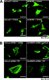
Induction of myofibrillar organization by MURC through the Rho/ROCK signaling pathway. (A) Cardiomyocytes were infected with Ad-LacZ or Ad-MURC at an MOI of 10. After infection, cells were treated with vehicle or 10 μM Y-27632. Ninety-six hours later, cells were stained with fluorescein isothiocyanate-conjugated phalloidin. (B) Cardiomyocytes were infected with Ad-Luc shRNA or Ad-rMURC shRNA at an MOI of 20. After 48 h of serum deprivation, cells were treated with vehicle or 100 μM PE. Forty-eight hours later, cells were stained with phalloidin.
FIG. 8.
FIG. 8.
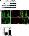
Generation of a transgenic mouse expressing MURC. (A) Expression level of MURC protein in the heart. Heart lysates from NTg and Tg-MURC mice (line 1 to 4) were immunoblotted with antibodies against Flag (top panel), MURC (middle panel), or GAPDH as an internal control (bottom panel). (B) Subcellular localization of Flag-tagged MURC in cardiomyocytes of Tg-MURC mice. Immunostaining was also performed using adult mouse heart sections with an anti-Flag antibody, an anti-MURC antibody (upper panels), or an anti-α-actinin antibody (lower panels). (C) The bar graph shows RhoA activity in the hearts of NTg and Tg-MURC mice. **, P < 0.01 compared with NTg mice.
FIG. 9.
FIG. 9.
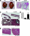
Characterization of cardiac phenotypes in a transgenic mouse expressing MURC. (A) Representative hearts from 13-week-old NTg (left) and Tg-MURC (right) mice. (B) Sagittal sections of hearts from 13-week-old NTg (left panel) and Tg-MURC (right panel) mice. Arrows indicate a thrombus in the left atrium. RA, right atrium; LA, left atrium; RV, right ventricle; LV, left ventricle. (C) Histological assessment of cardiac fibrosis by Masson's trichrome staining. Representative trichrome-stained sections of cardiac atria (upper panels) and ventricles (lower panels) in 13-week-old NTg (left) and Tg-MURC (right) mice. (D) Quantification of fibrosis area (blue) from trichrome-stained cardiac histological sections in 13-week-old NTg versus Tg-MURC mice. **, P < 0.01 compared with NTg mice. (E) Histological assessment of the size of cardiomyocytes. Representative Masson's trichrome-stained sections of cardiac ventricles in NTg (left) and Tg-MURC (right) mice at 5 weeks of age.
FIG. 10.
FIG. 10.
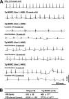
Conduction disturbance and increased vulnerability to atrial arrhythmias in transgenic mice expressing MURC. (A) Representative ECG recordings from anesthetized NTg and Tg-MURC mice. ECG samples of Tg-MURC mice are shown at 9 and 12 weeks of age. (B) Tg-MURC mice have a slow heart rate and prolongation of the PR interval. ECG data were collected at 12 weeks of age. **, P < 0.01 compared with NTg mice. AVB, atrioventricular block; HR, heart rate.
FIG. 11.
FIG. 11.
ANP mRNA expression in embryonic, neonatal, and adult hearts of NTg and Tg-MURC mice. Real-time RT-PCR was performed with cDNAs from embryonic (E10.5), neonatal, and adult hearts of NTg and Tg-MURC mice. **, P < 0.01 compared with Tg-MURC embryo at E10.5; ††, P < 0.01 compared with NTg adult; #, P < 0.05 compared with NTg neonate.

Similar articles

Cited by

References

    1. Adam, O., G. Frost, F. Custodis, M. A. Sussman, H. J. Schafers, M. Bohm, and U. Laufs. 2007. Role of Rac1 GTPase activation in atrial fibrillation. J. Am. Coll. Cardiol. 50359-367. - PubMed
    1. Amano, M., Y. Fukata, and K. Kaibuchi. 2000. Regulation and functions of Rho-associated kinase. Exp. Cell. Res. 26144-51. - PubMed
    1. Aoki, H., S. Izumo, and J. Sadoshima. 1998. Angiotensin II activates RhoA in cardiac myocytes: a critical role of RhoA in angiotensin II-induced premyofibril formation. Circ. Res. 82666-676. - PubMed
    1. Arai, A., J. A. Spencer, and E. N. Olson. 2002. STARS, a striated muscle activator of Rho signaling and serum response factor-dependent transcription. J. Biol. Chem. 27724453-24459. - PubMed
    1. Burgener, R., M. Wolf, T. Ganz, and M. Baggiolini. 1990. Purification and characterization of a major phosphatidylserine-binding phosphoprotein from human platelets. Biochem. J. 269729-734. - PMC - PubMed

Publication types

MeSH terms