. 2008 May;28(10):3114-26.
doi: 10.1128/MCB.02078-07. Epub 2008 Mar 10.

Transcription regulation of the rRNA gene by a multifunctional nucleolar protein, B23/nucleophosmin, through its histone chaperone activity

Affiliations

Transcription regulation of the rRNA gene by a multifunctional nucleolar protein, B23/nucleophosmin, through its histone chaperone activity

Kensaku Murano et al. Mol Cell Biol. 2008 May.

Abstract

It is well established that the transcription rate of the rRNA gene is closely associated with profound alterations in the cell growth rate. Regulation of rRNA gene transcription is likely to be dependent on the dynamic conversion of the chromatin structure. Previously, we identified B23/nucleophosmin, a multifunctional nucleolar phosphoprotein, as a component of template activating factor III that remodels the chromatin-like structure of the adenovirus genome complexed with viral basic proteins. It has also been shown that B23 has histone chaperone activity. Here, we examined the effect of B23 on rRNA gene transcription. B23 was found to be associated with the rRNA gene chromatin. Small-interfering-RNA-mediated down-regulation of the B23 expression level resulted in reduction of the transcription rate of the rRNA gene. We constructed a B23 mutant termed B23DeltaC, which lacks the domain essential for the histone chaperone activity and inhibited the histone binding activity of B23 in a dominant-negative manner. Expression of B23DeltaC decreased rRNA gene transcription and the rate of cell proliferation. These results suggest that B23 is involved in the transcription regulation of the rRNA gene as a nucleolar histone chaperone.


Figures

FIG. 1.
FIG. 1.
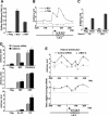
Relationship between cell growth and the level of rRNA gene transcription. (A) Expression level of B23. 293T cells were maintained in DMEM in the presence of FBS for 24 or 48 h and then pulse-labeled with [35S]methionine for 1 h. 35S-labeled B23 proteins were immunopurified using anti-B23 antibody and analyzed by 10% SDS-PAGE followed by autoradiography. As a loading control, 10% of input lysates was separated by 10% SDS-PAGE, and β-actin was detected by immunoblotting. (B) Expression level of nucleolus-related genes. 293T cells were maintained in DMEM in the presence of FBS for 24 or 48 h. Protein expression levels were analyzed by immunoblotting. (C) Transcription rate of the rRNA gene. 293T cells were maintained in DMEM in the presence of FBS for 72 h. The expression level of the rRNA gene was detected by run-on assays using [α-32P]GTP. Labeled rRNA was used as a probe for hybridization to membrane-blotted plasmid DNAs containing the rRNA gene (pUC-119-HurDNA) and pUC119 as a control. Results are means and standard deviations (SD) from three independent experiments. (D) Accumulation of pre-rRNA. 293T cells were maintained in DMEM in the presence of FBS for 72 h. The expression levels of pre-rRNA and β-actin mRNA as a loading control were determined by quantitative RT-PCR, and the amount of pre-rRNA normalized to that of β-actin mRNA is presented on the right. Results are means and SD from three independent experiments.
FIG. 2.
FIG. 2.
Repression of the rRNA gene transcription by siB23. (A) Expression level of B23.1. Immunoblotting was performed with lysates from HeLa cells treated with 40 pmol of control siRNA (lane 1) and siB23-A (lane 2), -B (lane 3), and -C (lane 4) (siRNA for B23.1). The mRNA level of B23.1 was also decreased by transfection of siB23-C compared with control siRNA (available on request). (B) Expression level of nucleolus-related proteins. Protein expression levels were determined by immunoblotting using HeLa cells treated with control siRNA and siB23-C. (C) Expression of B23 in nucleoli. Cellular B23 was stained by an indirect immunofluorescence method using anti-B23 antibody in HeLa cells treated with control siRNA or siB23-C. Nuclei were visualized with DAPI. Arrows indicate the positions of nucleoli. (D) Quantitative determination of pre-rRNA by RT-PCR. RNA isolated from HeLa cells treated with control siRNA or siB23-C was examined by RT-PCR using 5′ ETS-specific primers for pre-rRNA and primers for β-actin. The amount of pre-rRNA normalized to that of β-actin mRNA is shown on the right. Results are means and SD from three independent experiments.
FIG. 3.
FIG. 3.
Association of B23 with the chromatin around the rRNA gene. (A) Nucleolar ChIP analysis using quantitative PCR (Q-PCR). DNA was extracted from soluble nucleolar chromatin immunoprecipitated using anti-Flag, anti-B23, and anti-TBP antibodies. Q-PCR was performed using promoter-specific primers for the rRNA gene and chromatin-immunoprecipitated DNA as templates. Results are means and SD from three independent experiments. (B) ChIP analysis using Q-PCR through the whole rRNA gene unit. The structure of the human rRNA gene unit is shown under the graph. Boxes indicate rRNA-encoding sequences. The arrow indicates the transcription initiation site (+1) of the rRNA gene transcribed by Pol I. DNA was extracted from soluble chromatin immunoprecipitated with anti-Flag and anti-B23 antibodies. Q-PCR was performed using chromatin-immunoprecipitated DNA as the template and the primer sets in Table 1. Results are means ± SD from three independent experiments. (C) ChIP analysis using Q-PCR. 293T cells were maintained in DMEM in the presence of FBS for 72 h. DNA was extracted from chromatin that had been immunoprecipitated with anti-Flag and anti-B23 antibodies. Q-PCR was performed using promoter-specific primers for the rRNA gene and chromatin-immunoprecipitated DNA as templates. Results are means and SD from three independent experiments. (D) Histone density on the rRNA gene promoter. The soluble chromatin fraction from HeLa cells treated with control siRNA or siB23-C was examined by ChIP analyses with anti-Flag, anti-B23, anti-UBF, anti-TBP, anti-acetylated-histone H3 (Ac-H3), and anti-histone H3 antibodies. Q-PCR was performed with promoter-specific primers for the rRNA gene and chromatin-immunoprecipitated DNA as templates. Results are means and SD from three independent experiments. (E) Histone distribution over the rRNA gene. At the same time as the analysis for panel D, Q-PCR of the rRNA gene was carried out (top). The ratio of the histone density over the rRNA gene in cells treated with siB23-C relative to that in cells treated with control siRNA was plotted (middle). Results are means and SD from three independent experiments.
FIG. 4.
FIG. 4.
Inhibition of the histone binding activity of B23.1 by B23ΔC in vitro. (A) Schematic representation of the structure of B23.1, B23ΔA, and B23ΔC. Gray, black, and striped boxes indicate the oligomeric domain, acidic domain, and RNA binding domain, respectively. (B) Purification of recombinant proteins. Recombinant His-B23.1, His-B23ΔA, and His-B23ΔC were purified and separated (400 ng) by 12.5% SDS-PAGE followed by Coomassie brilliant blue staining. (C) Histone binding activity of B23 mutants. Recombinant B23 proteins (500 ng) were mixed with 300 ng core histones purified from HeLa cells. Immunoprecipitation was carried out with an anti-His antibody. Precipitated proteins were analyzed by 12.5% SDS-PAGE followed by immunoblotting with anti-His and anti-histone H3 antibodies. (D) Nucleosome assembly activity of B23 mutants. The nucleosome assembly activity of B23 mutants was examined by a supercoiling assay. Core histones (200 ng) preincubated without His-B23 (−) or with His-B23.1 (+, 167 ng; ++, 500 ng; +++, 1,500 ng), His-B23ΔA (142, 425, and 1,275 ng), and His-B23ΔC (66.8, 200, and 600 ng), were mixed with closed circular DNA relaxed by topoisomerase I and further incubated. The amounts of B23 mutant proteins were adjusted according to their sizes so as to be added at the same molecular numbers. The DNA was purified and separated by electrophoresis on a 1% agarose gel and visualized by staining with ethidium bromide. Positions of relaxed (R), supercoiled (S), and nicked (N) circular plasmid DNA are indicated. (E) Inhibition of the histone binding activity of B23.1 by B23ΔC. His-B23.1 (200 ng) was mixed without (lane 2) or with 200 ng (lane 3) or 400 ng (lane 4) of B23ΔC and subjected to the denature-renature protocol. Renatured B23 complexes were mixed with 300 ng core histones purified from HeLa cells. His-B23.1 was immunoprecipitated with an anti-B23 antibody that cannot recognize B23ΔC protein. Precipitated proteins were analyzed by 12.5% SDS-PAGE followed by silver staining and immunoblotting (IB) with an anti-His antibody. H and L indicate heavy and light chains of immunoglobulin, respectively.
FIG. 5.
FIG. 5.
Inhibition of the histone binding activity of B23.1 by B23ΔC in vivo. (A) Expression of B23 proteins in 293T cells. Immunoblotting was carried out using lysate prepared from 293T cells transfected with the indicated plasmids. (B) Histone binding activity of B23 mutants. Immunoprecipitation (IP) with anti-HA and anti-Flag antibodies was performed using lysates of 293T cells transfected with plasmids expressing B23 proteins. Immunoprecipitated proteins were analyzed by immunoblotting. (C) Lack of effect of B23ΔA on the histone binding activity of B23.1. Lysates of 293T cells (24 h posttransfection) transfected with pCHA-B23.1 (160 ng) and pCAGGS-Flag-B23ΔA (+, 160 ng; ++, 480 ng; +++, 1,440 ng) were subjected to immunoprecipitation with an anti-HA antibody. Immunoprecipitated proteins were analyzed by immunoblotting. (D) Inhibition of the histone binding activity of B23.1 by B23ΔC in vivo. Lysates of 293T cells (24 h posttransfection) transfected with pcDNA3-Flag-B23.1 (100 ng) and pCHA-B23ΔC (+, 300 ng; ++, 1,400 ng) were subjected to immunoprecipitation with anti-Flag antibody. Immunoprecipitated proteins were analyzed by immunoblotting.
FIG. 6.
FIG. 6.
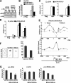
Inhibition of rRNA gene transcription by B23ΔC. (A) Interaction of B23ΔC with endogenous B23. 293T cells were transiently transfected with pCHA (control) and pCHA-B23ΔC and cultured for 72 h posttransfection. Lysates prepared from 293T cells were used for immunoprecipitation with anti-HA antibody. Immunoprecipitates (P) and flowthrough fractions (F) were resolved by 12.5% SDS-PAGE, transferred to a membrane, and probed with anti-B23 and anti-HA antibodies. (B) Inhibition of association of B23 with the rRNA gene by B23ΔC. 293T cells were cotransfected with pBabe-puro and the indicated plasmids, and 0.5 μg/ml puromycin was added 24 h posttransfection. After puromycin selection for 30 h, 293T cells were maintained in fresh medium for 18 h. Nucleolar ChIP was carried out with anti-B23 and anti-Flag antibodies. Quantitative PCR was performed with primers around the promoter region of the rRNA gene. Results are means and SD from three independent experiments. A paired Student's t test gave a P value of 0.02 between pCHA- and pCHA-B23ΔC-transfected cells. (C) Association of transcription factors on the rRNA gene promoter. ChIP was carried out with anti-UBF and anti-TBP antibodies, and lysates were prepared for mock-treated cells or cells expressing B23ΔC. Q-PCR was performed with primers around the promoter region of the rRNA gene. Results are means and SD from three independent experiments. (D) Histone density on the rRNA gene promoter. ChIP was carried out using anti-B23, anti-histone H3, and anti-acetylated-histone H3 (AcH3) antibodies in the presence or absence of B23ΔC. Q-PCR was performed with primers around the promoter region of the rRNA gene. Results are means and SD from three independent experiments. (E) Histone distribution over the rRNA gene. ChIP was performed with anti-histone H3 antibody using HA-B23ΔC-expressing or mock-treated cells. The amount of chromatin-immunoprecipitated DNA was determined by Q-PCR using primer sets for the rRNA gene (top). The ratio of the histone density over the rRNA gene in cells expressing B23ΔC relative to that in mock-treated cells was plotted (middle). Results are means and SD from three independent experiments. (F) Inhibition of the rRNA gene transcription by B23ΔC. 293T cells were transiently transfected with pCHA (control) and pCHA-B23ΔC and cultured for 72 h posttransfection. Nuclei isolated from these 293T cells were used for run-on transcription assays. Radiolabeled nuclear RNA was isolated and hybridized with 1 μg of pUC119 (control) and pUC119-HurDNA (rRNA) on a membrane. This assay was done in triplicate, and a typical pattern is shown. The right panel shows a densitometry quantitation of the hybridization signals. Results are means and SD from three independent experiments. (G) Inhibition of the rRNA gene transcription by B23ΔA. 293T cells were cotransfected with pBabe-puro and one of the indicated plasmids, and 0.5 μg/ml puromycin was added 24 h posttransfection. After puromycin selection for 30 h, 293T cells were maintained in fresh medium for 18 h. RNA isolated from 293T cells was examined by RT-PCR using 5′ ETS-specific primers for pre-rRNA and primers for β-actin mRNA. Results are means and SD from three independent experiments.
FIG. 7.
FIG. 7.
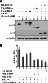
Involvement of acidic domains of B23 in rRNA gene transcription. (A) Expression of B23 proteins in HeLa cells transfected with siB23. HeLa cells were transfected with 40 pmol of control siRNA and 40 pmol of siB23-C. At 12 h after siRNAs transfection, HeLa cells were supertransfected with pCAGGS-Flag-B23.1 (+, 180 ng; ++, 360 ng), pCAGGS-Flag B23ΔA (180 and 360 ng), pCHA-B23ΔC (180 and 360 ng), and pEF321-T (40 ng) encoding SV40 T antigen for amplification of transfected plasmid. At 72 h after siRNA transfection, lysates of 2.5 × 10³ cells were analyzed by immunoblotting using anti-B23, anti-HA, and anti-β-actin antibodies. (B) Quantitative determination of pre-rRNA by Q-PCR. At the same time as the analysis for panel A, total RNA extracted from HeLa cells were examined by RT-PCR using 5′ ETS-specific primers for the rRNA gene. The amount of pre-rRNA was normalized to that of β-actin mRNA. Results are means and SD from three independent experiments.
FIG. 8.
FIG. 8.
Inhibition of cell proliferation by B23ΔC. (A) Proliferation inhibition of 293T cells by expression of B23ΔC. 293T cells were cotransfected with 1,400 ng of pCHA and 200 ng of pEGFP-N1 or with 1,400 ng of pCHA-B23ΔC and 200 ng of pEGFP-N1. The number of EGFP-positive cells was counted every 24 h as a function of culture time. Results are means and SD from three independent experiments. (B) B23 dose-dependent inhibition of the cell proliferation rate. The number of EGFP-positive cells was counted at 72 h after transfection of 200 ng of pEGFP-N1 with pCHA-B23ΔC (350 ng [lane 2], 700 ng [lane 3], and 1,400 ng [lane 4]), and pCHA (1,400 ng [lane 1], 700 ng [lane 2], and 350 ng [lane 3]). Results are means and SD from three independent experiments. (C) Cell cycle population of cells expressing B23ΔC. 293T cells were cotransfected with 1,400 ng of pCHA and 200 ng of pEGFP-N1 or with 1,400 ng of pCHA-B23ΔC and 200 ng of pEGFP-N1 and then incubated for 72 h. Cell population of 293T cells expressing EGFP was determined by fluorescence-activated cell sorting analysis. The sub-G1 fraction was quantitatively determined as a population of apoptotic cells (inset). (D) p53-independent effect of B23ΔC on cell proliferation. EGFP-positive p53−/− MEFs were counted at 72 h after transfection of pEGFP-N1 (125 ng) with pCHA-B23.1 (875 ng) or pCHA-B23ΔC (875 ng). Results are means and SD from three independent experiments.

Similar articles

Cited by

References

    1. Angelov, D., V. A. Bondarenko, S. Almagro, H. Menoni, F. Mongelard, F. Hans, F. Mietton, V. M. Studitsky, A. Hamiche, S. Dimitrov, and P. Bouvet. 2006. Nucleolin is a histone chaperone with FACT-like activity and assists remodeling of nucleosomes. EMBO J. 251669-1679. - PMC - PubMed
    1. Bertwistle, D., M. Sugimoto, and C. J. Sherr. 2004. Physical and functional interactions of the Arf tumor suppressor protein with nucleophosmin/B23. Mol. Cell. Biol. 24985-996. - PMC - PubMed
    1. Brady, S. N., Y. Yu, L. B. Maggi, Jr., and J. D. Weber. 2004. ARF impedes NPM/B23 shuttling in an Mdm2-sensitive tumor suppressor pathway. Mol. Cell. Biol. 249327-9338. - PMC - PubMed
    1. Chang, J. H., and M. O. Olson. 1990. Structure of the gene for rat nucleolar protein B23. J. Biol. Chem. 26518227-18233. - PubMed
    1. Colombo, E., J. C. Marine, D. Danovi, B. Falini, and P. G. Pelicci. 2002. Nucleophosmin regulates the stability and transcriptional activity of p53. Nat. Cell Biol. 4529-533. - PubMed

Publication types

MeSH terms

LinkOut - more resources