. 2008 May;28(10):3336-43.
doi: 10.1128/MCB.00567-07. Epub 2008 Mar 10.

Cdc1p is an endoplasmic reticulum-localized putative lipid phosphatase that affects Golgi inheritance and actin polarization by activating Ca2+ signaling

Affiliations

Cdc1p is an endoplasmic reticulum-localized putative lipid phosphatase that affects Golgi inheritance and actin polarization by activating Ca2+ signaling

Eugene Losev et al. Mol Cell Biol. 2008 May.

Abstract

In the budding yeast Saccharomyces cerevisiae, mutations in the essential gene CDC1 cause defects in Golgi inheritance and actin polarization. However, the biochemical function of Cdc1p is unknown. Previous work showed that cdc1 mutants accumulate intracellular Ca(2+) and display enhanced sensitivity to the extracellular Mn(2+) concentration, suggesting that Cdc1p might regulate divalent cation homeostasis. By contrast, our data indicate that Cdc1p is a Mn(2+)-dependent protein that can affect Ca(2+) levels. We identified a cdc1 allele that activates Ca(2+) signaling but does not show enhanced sensitivity to the Mn(2+) concentration. Furthermore, our studies show that Cdc1p is an endoplasmic reticulum-localized transmembrane protein with a putative phosphoesterase domain facing the lumen. cdc1 mutant cells accumulate an unidentified phospholipid, suggesting that Cdc1p may be a lipid phosphatase. Previous work showed that deletion of the plasma membrane Ca(2+) channel Cch1p partially suppressed the cdc1 growth phenotype, and we find that deletion of Cch1p also suppresses the Golgi inheritance and actin polarization phenotypes. The combined data fit a model in which the cdc1 mutant phenotypes result from accumulation of a phosphorylated lipid that activates Ca(2+) signaling.


Figures
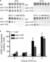
FIG. 1.
FIG. 1.
Two cdc1 alleles show different sensitivities to Mn2+ but still exhibit activated Ca2+ signaling at the restrictive temperature. (a) Wild-type (WT), cdc1-310, and cdc1-314 mutant strains were grown to stationary phase and serially diluted (1:10) five times. The strains were replica plated on YPD medium at 25°C and 37°C, at 37°C with 2 mM MnCl2, and at 25°C with 2 mM EGTA. (b) The activation of Ca2+ signaling was measured using a CDRE-lacZ reporter. Wild-type, cdc1-310, and cdc1-314 cells were grown in SD-URA medium to early log phase and shifted to 33.5°C for the indicated times. Reporter activity was measured every hour for 3 h.
FIG. 2.
FIG. 2.
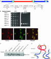
The putative phosphoesterase domain of Cdc1p is functionally important and faces the lumen of the ER. (a) Sequence analysis revealed a putative phosphoesterase domain (red box) and three hydrophobic putative transmembrane domains (green boxes). The conserved amino acid residues labeled in red are predicted to chelate the divalent metal ion. (b) Cdc1p mutants with alanine substitutions of the conserved residues indicated in panel a were expressed from a CEN plasmid in the cdc1-314 strain at 25°C and 37°C. A mutant of His287, which is not predicted to chelate the divalent metal ion, was used as a control. Lack of growth at 37°C shows that the mutated residues are important for Cdc1p function. (c) Cells expressing myc-tagged Cdc1p were immunolabeled with anti-myc antibody (red) and anti-Kar2p antibody (green). Scale bar, 5 μm. (d) The five potential N-linked glycosylation sites in Cdc1p were mutated separately or in combination in a myc-tagged Cdc1p protein, which was expressed as a second copy in the wild-type (WT) strain. The cell extracts were treated with EndoH and examined using SDS-PAGE. The lack of a mobility shift after EndoH treatment in the N215Q N233Q double mutant indicates that these residues are N glycosylated and are therefore in the lumen of the secretory pathway. (e) A diagram showing the inferred topology of Cdc1p.
FIG. 3.
FIG. 3.
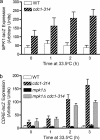
Mpk1p is activated in the cdc1-314 mutant but is not the major cause of Ca2+ signaling activation. (a) An MPK1-lacZ reporter was used to measure Mpk1p activity in cdc1-314 mutant cells. Wild-type (WT) and mutant cells were grown in SD-URA medium to log phase and then shifted to 33.5°C for the indicated times. Reporter activity was measured every hour for 3 h. (b) A CDRE-lacZ reporter was used to measure the activation of Ca2+ signaling. Wild-type, cdc1-314, mpk1Δ, and mpk1Δ cdc1-314 cells were grown in SD-URA medium to early log phase and shifted to 33.5°C for the indicated times. Reporter activity was measured every hour for 3 h.
FIG. 4.
FIG. 4.
Deletion of the Ca2+ channel Cch1p completely suppresses the Golgi inheritance and actin polarization phenotypes in the cdc1-314 mutant. (a) The inheritance of late Golgi cisternae was measured by assessing the presence of the late Golgi marker protein Sec7p-GFP in the buds of wild-type (WT), cdc1-314, cch1Δ cdc1-314, and cch1Δ cells after a shift to 33.5°C for 3 h. (b) Actin was visualized by staining with Alexa 594-phalloidin. Wild-type, cdc1-314, cch1Δ cdc1-314, cch1Δ, mpk1Δ, and mpk1Δ cdc1-314 cultures were grown at 25°C, shifted to 33.5°C for 3 h, fixed, and stained. Fluorescence images are shown. Most of the wild-type, cch1Δ cdc1-314, cch1Δ, and mpk1Δ cells contained polarized actin patches and cables, whereas most of the cdc1-314 and mpk1Δ cdc1-314 cells contained depolarized patches and few visible cables. Scale bar, 5 μm.
FIG. 5.
FIG. 5.
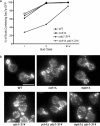
Treatment of cdc1-314 cells with the calcineurin inhibitor FK506 does not suppress the growth and actin polarization phenotypes. (a) Activation of Ca2+ signaling was measured using the CDRE-lacZ reporter. Wild-type (WT) and cdc1-314 cells were grown in 10 ml of SD-URA medium to early log phase and shifted to 33.5°C for the indicated times. Immediately before the shift, 10 μl of dimethyl sulfoxide or 1 mg/ml of FK506 in dimethyl sulfoxide was added. The final concentration of FK506 in cultures was 1 μg/ml. Reporter activity was measured every hour for 3 h. (b) Cell growth was measured by taking the absorbance at 600 nm. Cultures were treated as described in panel a. Readings were taken every hour for 3 h. (c) Actin was visualized by staining with Alexa 594-phalloidin. Cultures were treated as described in panel a. After 3 h, the cells were fixed and stained. Fluorescence images are shown. Most of the treated and untreated wild-type cells contained polarized actin patches and cables, whereas most of the treated and untreated cdc1-314 cells contained depolarized patches. Scale bar, 5 μm.
FIG. 6.
FIG. 6.
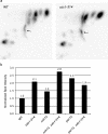
A phospholipid species accumulates in the cdc1-314 mutant at the restrictive temperature. (a) Wild-type (WT) and mutant strains were grown overnight in YPD medium supplemented with 32Pi. Upon reaching log phase, the cells were shifted to 33.5°C for 3 h. Lipids were extracted and run in two dimensions on TLC plates. These images from representative plates show the area where the major radioactive lipid species migrated. Arrows mark an unidentified phosphorylated lipid that was consistently present at higher levels in cdc1 cells than in wild-type cells. (b) The level of the unidentified radioactive lipid was measured in each of the indicated strains as a fraction of the total radioactive lipid, and the results were normalized by defining the signal from wild-type cells as 1.0.
FIG. 7.
FIG. 7.
A model for Cdc1p function and the link between Cdc1p and Golgi inheritance. (a) Cdc1p is presumed to be a lipid phosphatase, and we suggest that Per1p is needed to generate the substrate for Cdc1p. According to this model, inactivating Cdc1p causes its substrate to accumulate to toxic levels and also causes accumulation of a nontoxic precursor lipid. When Per1p is inactivated, only the nontoxic precursor accumulates, so a per1Δ mutation suppresses the lethality of cdc1 mutations. This nontoxic precursor may correspond to the unidentified lipid that was visualized in the experiment shown in Fig. 6. (b) In cdc1 cells, accumulation of the toxic phosphorylated lipid activates both Mpk1p- and Cch1p-dependent Ca2+ signaling. The Ca2+ signaling activates calcineurin. In a calcineurin-independent pathway, strong Ca2+ signaling depolarizes actin. We speculate that strong Ca2+ signaling also inhibits Golgi tethering. These two effects of strong Ca2+ signaling synergize to cause a pronounced defect in Golgi inheritance.

Similar articles

Cited by

References

    1. Abeijon, C., and C. B. Hirschberg. 1992. Topography of glycosylation reactions in the endoplasmic reticulum. Trends Biochem. Sci. 1732-36. - PubMed
    1. Ayscough, K. R., J. Stryker, N. Pokala, M. Sanders, P. Crews, and D. G. Drubin. 1997. High rates of actin filament turnover in budding yeast and roles for actin in establishment and maintenance of cell polarity revealed using the actin inhibitor latrunculin-A. J. Cell Biol. 137399-416. - PMC - PubMed
    1. Baudin, A., O. Ozier-Kalogeropoulos, A. Denouel, F. Lacroute, and C. Cullin. 1993. A simple and efficient method for direct gene deletion in Saccharomyces cerevisiae. Nucleic Acids Res. 213329-3330. - PMC - PubMed
    1. Bonilla, M., and K. Cunningham. 2003. Mitogen-activated protein kinase stimulation of Ca2+ signaling is required for survival of endoplasmic reticulum stress in yeast. Mol. Biol. Cell 144296-4305. - PMC - PubMed
    1. Bosson, R., M. Jaquenoud, and A. Conzelmann. 2006. GUP1 of Saccharomyces cerevisiae encodes an O-acyltransferase involved in remodeling of the GPI anchor. Mol. Biol. Cell 172636-2645. - PMC - PubMed

Publication types

MeSH terms