. 2008 May;28(10):3324-35.
doi: 10.1128/MCB.00144-08. Epub 2008 Mar 10.

Involvement of actinin-4 in the recruitment of JRAB/MICAL-L2 to cell-cell junctions and the formation of functional tight junctions

Affiliations

Involvement of actinin-4 in the recruitment of JRAB/MICAL-L2 to cell-cell junctions and the formation of functional tight junctions

Hiroyoshi Nakatsuji et al. Mol Cell Biol. 2008 May.

Abstract

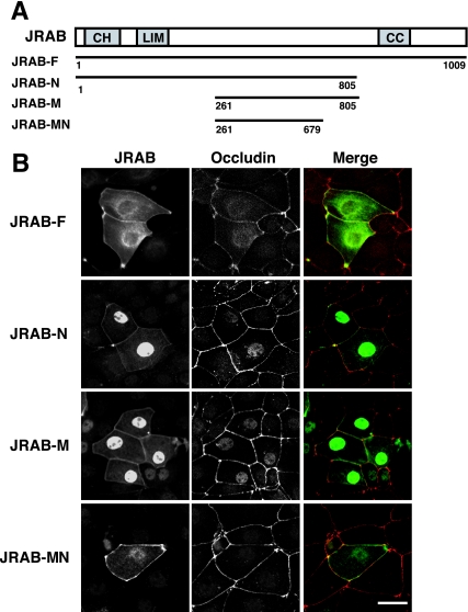
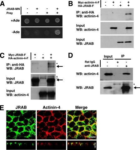
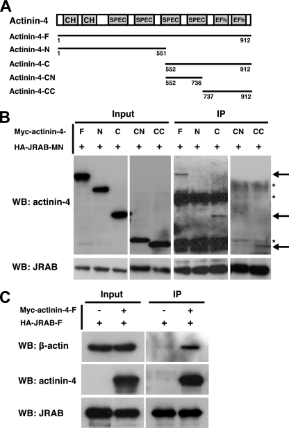
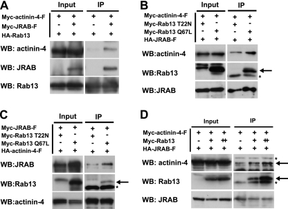
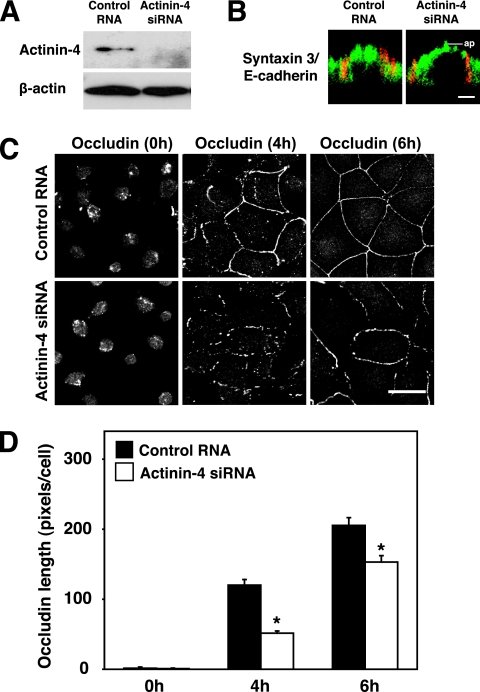
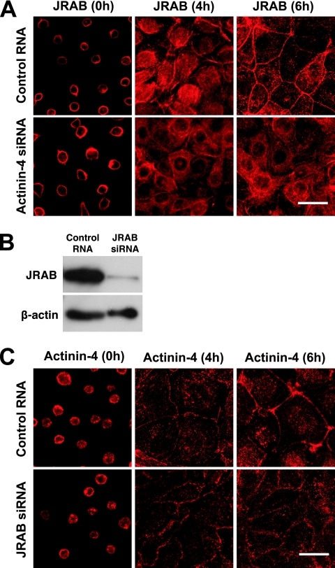
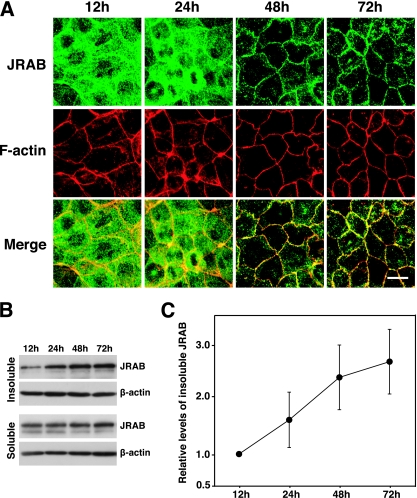
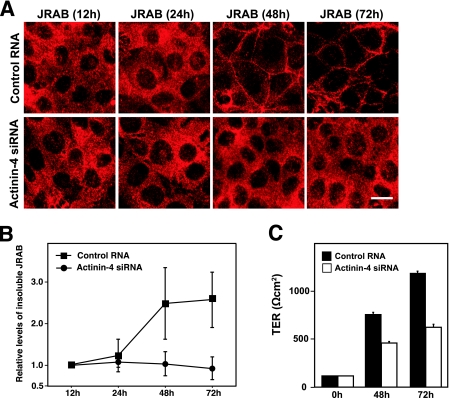
Tight junctions (TJs) are cell-cell adhesive structures that undergo continuous remodeling. We previously demonstrated that Rab13 and a junctional Rab13-binding protein (JRAB)/molecule interacting with CasL-like 2 (MICAL-L2) localized at TJs and mediated the endocytic recycling of the integral TJ protein occludin and the formation of functional TJs. Here, we investigated how JRAB/MICAL-L2 was targeted to TJs. Using a series of deletion mutants, we found the plasma membrane (PM)-targeting domain within JRAB/MICAL-L2. We then identified actinin-4, which was originally isolated as an actin-binding protein associated with cell motility and cancer invasion/metastasis, as a binding protein for the PM-targeting domain of JRAB/MICAL-L2, using a yeast two-hybrid system. Actinin-4 was colocalized with JRAB/MICAL-L2 at cell-cell junctions and linked JRAB/MICAL-L2 to F-actin. Although actinin-4 bound to JRAB/MICAL-L2 without Rab13, the actinin-4-JRAB/MICAL-L2 interaction was enhanced by Rab13 activation. Depletion of actinin-4 by using small interfering RNA inhibited the recruitment of occludin to TJs during the Ca(2+) switch. During the epithelial polarization after replating, JRAB/MICAL-L2 was recruited from the cytosol to cell-cell junctions. This JRAB/MICAL-L2 recruitment as well as the formation of functional TJs was delayed in actinin-4-depleted cells. These results indicate that actinin-4 is involved in recruiting JRAB/MICAL-L2 to cell-cell junctions and forming functional TJs.


Figures

FIG. 1.
FIG. 1.
Identification of the PM-targeting domain within JRAB/MICAL-L2. (A) Structures of the full-length and various fragments of JRAB/MICAL-L2. Numbers represent amino acid positions. CH, calponin homology domain; LIM, LIM domain; CC, coiled-coil domain. (B) MTD-1A cells were transfected with pCI-neo-Myc-JRAB/MICAL-L2-F, pCI-neo-Myc-JRAB/MICAL-L2-N, pCI-neo-Myc-JRAB/MICAL-L2-M, or pCI-neo-Myc-JRAB/MICAL-L2-MN and double immunolabeled with anti-Myc and anti-occludin antibodies. Bar, 20 μm. The results shown in panel B are representative of three independent experiments.
FIG. 2.
FIG. 2.
Identification of actinin-4 as a binding protein for the PM-targeting domain of JRAB/MICAL-L2. (A) Yeast transformants carrying the pGBDU vector encoding JRAB/MICAL-L2-MN and the pACT2 vector encoding a prey clone were spotted onto synthetic complete medium lacking adenine to score for the ADE2 reporter activity and incubated at 30°C for 3 days. (B and C) BHK cells cotransfected with pCI-neo-HA-JRAB/MICAL-L2 and pCI-neo-Myc-actinin-4 (B) or with pCI-neo-Myc-JRAB/MICAL-L2 and pCI-neo-HA-actinin-4 (C) were immunoprecipitated (IP) with anti-HA antibody and subjected to Western blotting (WB) analysis using anti-Myc and anti-HA antibodies. (D) MTD-1A cells were immunoprecipitated with anti-JRAB/MICAL-L2 antibody or rat immunoglobulin G and subjected to Western blotting analysis using anti-JRAB/MICAL-L2 and anti-actinin-4 antibodies. Arrows, Myc-JRAB and endogenous JRAB; asterisks, nonspecific bands. (E) MTD-1A cells grown on filters were double immunolabeled with anti-JRAB/MICAL-L2 and anti-actinin-4 antibodies and observed under a confocal microscope. Vertical sectional images are shown in the bottom panels. White arrow is equivalent in length to the thickness of the cellular sheet. Bar, 10 μm. The results shown in panels A to E are representative of three independent experiments.
FIG. 3.
FIG. 3.
Interaction of actinin-4 with JRAB/MICAL-L2 and F-actin. (A) Structures of the full-length and various fragments of actinin-4. Numbers represent amino acid positions. CH, calponin homology domain; SPEC, spectrin repeats; EFh, EF hands. (B) BHK cells cotransfected with pCI-neo-HA-JRAB/MICAL-L2-MN and pCI-neo-Myc-actinin-4-F, pCI-neo-Myc-actinin-4-N, pCI-neo-Myc-actinin-4-C, pCI-neo-Myc-actinin-4-CN, or pCI-neo-Myc-actinin-4-CC were immunoprecipitated (IP) with anti-HA antibody and subjected to Western blotting (WB) analysis using anti-Myc and anti-HA antibodies. Arrows indicate Myc-actinin-4-F, Myc-actinin-4-C, and Myc-actinin-4-CC, and asterisks indicate nonspecific bands. (C) BHK cells cotransfected with pCI-neo-HA-JRAB/MICAL-L2-F and pCI-neo-Myc-actinin-4-F were immunoprecipitated with anti-HA antibody and subjected to Western blotting analysis using anti-β-actin, anti-Myc, and anti-HA antibodies. The results shown in panels B and C are representative of three independent experiments.
FIG. 4.
FIG. 4.
Involvement of Rab13 activation in the regulation of the actinin-4-JRAB/MICAL-L2 interaction. BHK cells cotransfected with pCI-neo-HA-Rab13 and pCI-neo-Myc-actinin-4-F together with pCI-neo-Myc or pCI-neo-Myc-JRAB/MICAL-L2 (A), with pCI-neo-HA-JRAB/MICAL-L2 and pCI-neo-Myc-actinin-4-F together with pCI-neo-Myc-Rab13 T22N or pCI-neo-Myc-Rab13 Q67L (B), with pCI-neo-HA-actinin-4-F and pCI-neo-Myc-JRAB/MICAL-L2 together with pCI-neo-Myc-Rab13 T22N or pCI-neo-Myc-Rab13 Q67L (C) or with pCI-neo-HA-JRAB/MICAL-L2 (+, 1.5 μg) and pCI-neo-Myc-actinin-4-F (+, 3.0 μg) together with pCI-neo-Myc (−, 3.0 μg), pCI-neo-Myc plus pCI-neo-Myc-Rab13 (+, 1.5 plus 1.5 μg), or pCI-neo-Myc-Rab13 (++, 3.0 μg) (D) were immunoprecipitated (IP) with an anti-HA antibody and subjected to Western blotting (WB) analysis using anti-HA and anti-Myc antibodies. Arrows indicate Myc-Rab13 and Myc-actinin-4, and asterisks indicate nonspecific bands. The results shown in panels A to D are representative of three independent experiments.
FIG. 5.
FIG. 5.
Involvement of actinin-4 in the recruitment of occludin to TJs during the Ca2+ switch. (A) MTD-1A cells were transfected with control RNA or with actinin-4 siRNA and subjected to Western blotting analysis using anti-actinin-4 and anti-β-actin antibodies. (B) MTD-1A cells transfected with control RNA or actinin-4 siRNA were double immunolabeled for apical markers (syntaxin 3, green) and basolateral markers (E-cadherin, red). Bar, 5 μm. ap, level of the apical membranes. (C) MTD-1A cells transfected with control RNA or actinin-4 siRNA were subjected to a Ca2+-switch assay and then immunostained with antioccludin antibody at 0, 4, and 6 h after Ca2+ restoration. Bar, 20 μm. The results shown in panels A to C are representative of three independent experiments. (D) Occludin length per cell was quantitated and shown as the means and standard errors of the means of three independent experiments. Asterisks denote significant differences from control cells (P < 0.05).
FIG. 6.
FIG. 6.
Involvement of actinin-4 in the recruitment of JRAB/MICAL-L2 to the cell-cell junctions during the Ca2+ switch. (A) MTD-1A cells transfected with control RNA or actinin-4 siRNA were subjected to a Ca2+-switch assay and then immunostained with anti-JRAB/MICAL-L2 antibody at 0, 4, and 6 h after Ca2+ restoration. Bar, 20 μm. (B) MTD-1A cells were transfected with control RNA or JRAB/MICAL-L2 siRNA and subjected to Western blotting analysis using anti-JRAB/MICAL-L2 and anti-β-actin antibodies. (C) MTD-1A cells transfected with control RNA or JRAB/MICAL-L2 siRNA were subjected to a Ca2+-switch assay and then immunostained with anti-actinin-4 antibody at 0, 4, and 6 h after Ca2+ restoration. Bar, 20 μm. The results shown in panels A to C are representative of three independent experiments.
FIG. 7.
FIG. 7.
Recruitment of JRAB/MICAL-L2 from the cytosol to the cell-cell junctions during epithelial polarization after replating. (A) MTD-1A cells were dissociated into single cells, replated under confluent conditions, cultured for the indicated times, and then double labeled with rhodamine-phalloidin and anti-JRAB/MICAL-L2 antibody. Bar, 10 μm. The results shown are representative of three independent experiments. (B) Triton X-100-soluble and -insoluble fractions were prepared from MTD-1A cells cultured for the indicated times after replating. Equal amounts of proteins were subjected to Western blotting analysis using anti-JRAB/MICAL-L2 and anti-β-actin antibodies. (C) The levels of insoluble JRAB/MICAL-L2 protein were calculated as protein level at the indicated time/protein level at 12 h and shown as the means and standard errors of the means of three independent experiments.
FIG. 8.
FIG. 8.
Involvement of actinin-4 in the recruitment of JRAB/MICAL-L2 to the cell-cell junctions and the formation of functional TJs during epithelial polarization after replating. (A) MTD-1A cells transfected with control RNA or actinin-4 siRNA were dissociated into single cells, replated under confluent conditions, cultured for the indicated times, and then immunolabeled with anti-JRAB/MICAL-L2 antibody. Bar, 10 μm. The results shown are representative of three independent experiments. (B) Triton X-100-soluble and -insoluble fractions were prepared from MTD-1A cells transfected with control RNA or actinin-4 siRNA and cultured for the indicated times after replating. Equal amounts of proteins were subjected to Western blotting analysis using anti-JRAB/MICAL-L2 antibody. The levels of insoluble JRAB/MICAL-L2 protein were calculated as protein level at the indicated time/protein level at 12 h and are shown as the means and standard errors of the means of three independent experiments. (C) MTD-1A cells transfected with control RNA or actinin-4 siRNA were replated onto Transwell filters as an instant confluent monolayer and cultured. TER was measured at 0, 48, and 72 h after replating, and values are shown as the means and standard errors of the means of three independent experiments.

Similar articles

Cited by

References

    1. Anderson, J., C. Van Itallie, and A. Fanning. 2004. Setting up a selective barrier at the apical junction complex. Curr. Opin. Cell Biol. 16140-145. - PubMed
    1. Ando-Akatsuka, Y., S. Yonemura, M. Itoh, M. Furuse, and S. Tsukita. 1999. Differential behavior of E-cadherin and occludin in their colocalization with ZO-1 during the establishment of epithelial cell polarity. J. Cell Physiol. 179115-125. - PubMed
    1. Bertet, C., L. Sulak, and T. Lecuit. 2004. Myosin-dependent junction remodelling controls planar cell intercalation and axis elongation. Nature 429667-671. - PubMed
    1. Braga, V., and A. Yap. 2005. The challenges of abundance: epithelial junctions and small GTPase signalling. Curr. Opin. Cell Biol. 17466-474. - PubMed
    1. Broderick, M., and S. Winder. 2005. Spectrin, α-actinin, and dystrophin. Adv. Protein Chem. 70203-246. - PubMed

Publication types

MeSH terms