. 2008 May;28(10):3457-64.
doi: 10.1128/MCB.02019-07. Epub 2008 Mar 10.

SWI/SNF mediates polycomb eviction and epigenetic reprogramming of the INK4b-ARF-INK4a locus

Affiliations

SWI/SNF mediates polycomb eviction and epigenetic reprogramming of the INK4b-ARF-INK4a locus

Sima Kheradmand Kia et al. Mol Cell Biol. 2008 May.

Abstract

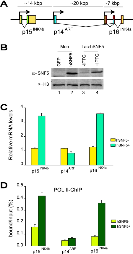
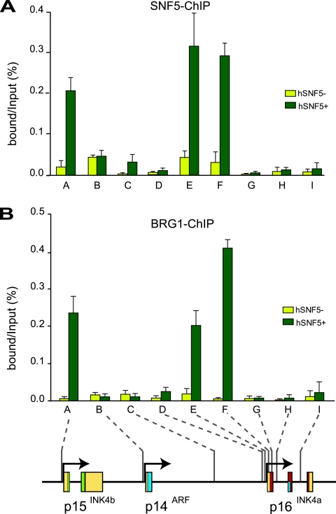
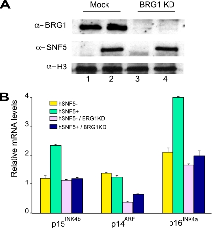
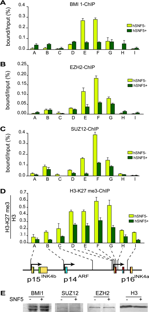
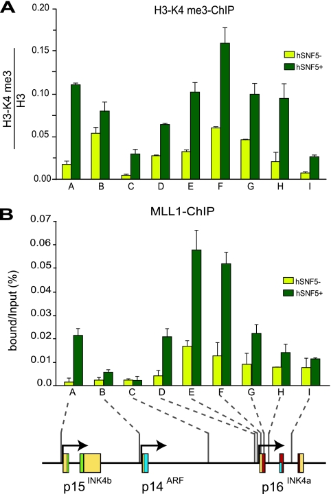
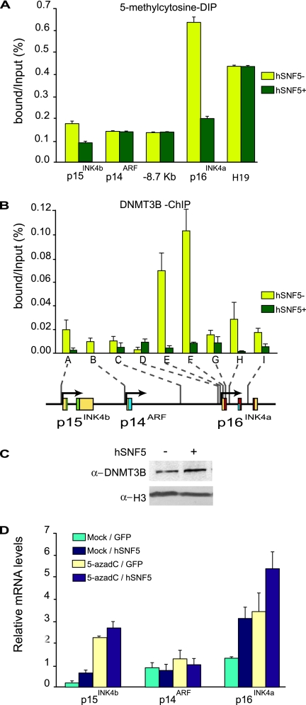
Stable silencing of the INK4b-ARF-INK4a tumor suppressor locus occurs in a variety of human cancers, including malignant rhabdoid tumors (MRTs). MRTs are extremely aggressive cancers caused by the loss of the hSNF5 subunit of the SWI/SNF chromatin-remodeling complex. We found previously that, in MRT cells, hSNF5 is required for p16(INK4a) induction, mitotic checkpoint activation, and cellular senescence. Here, we investigated how the balance between Polycomb group (PcG) silencing and SWI/SNF activation affects epigenetic control of the INK4b-ARF-INK4a locus in MRT cells. hSNF5 reexpression in MRT cells caused SWI/SNF recruitment and activation of p15(INK4b) and p16(INK4a), but not of p14(ARF). Gene activation by hSNF5 is strictly dependent on the SWI/SNF motor subunit BRG1. SWI/SNF mediates eviction of the PRC1 and PRC2 PcG silencers and extensive chromatin reprogramming. Concomitant with PcG complex removal, the mixed lineage leukemia 1 (MLL1) protein is recruited and active histone marks supplant repressive ones. Strikingly, loss of PcG complexes is accompanied by DNA methyltransferase DNMT3B dissociation and reduced DNA methylation. Thus, various chromatin states can be modulated by SWI/SNF action. Collectively, these findings emphasize the close interconnectivity and dynamics of diverse chromatin modifications in cancer and gene control.


Figures

FIG. 1.
FIG. 1.
Reexpression of hSNF5 in MRT cells induces p15INK4b and p16INK4a but not p14ARF. (A) Organization of the human INK4b-ARF-INK4a locus (not drawn to scale). The genomic locus spans approximately 40 kbp of human chromosome 9 and encodes three distinct proteins: p15INK4b, p14ARF, and p16INK4a. The 5′ and 3′ untranslated regions (yellow boxes), the coding sequences of p15INK4b (green), p14ARF (blue), and p16INK4a (red) are indicated. (B) Western immunoblotting analysis of hSNF5 expression in MON cells transduced with lentiviruses expressing either GFP (lane1) or hSNF5 (lane2) and either noninduced (lane 3) or induced (lane 4) G401-derived Lac-hSNF5 cells. Cell lysates were resolved by SDS-PAGE and analyzed by Western immunoblotting with antibodies directed against hSNF5. Histone H3 serves as a loading control. (C) RT-qPCR analysis of gene expression in MRT cells reveals hSNF5-dependent induction of p15INK4b and p16INK4a, whereas p14ARF transcription remains unaffected. Cells were collected 48 h after transduction with lentiviruses expressing either GFP (yellow bars) or hSNF5 (blue bars). RT-qPCR analysis of isolated mRNA was used to determine the relative expression levels of p15INK4b, p16INK4a, p14ARF, and USP14 (a control gene that is independent of hSNF5). mRNA levels were plotted as percentage of USP14 mRNA × 0.006. The bar graphs represent the mean of three independent biological replicates, each analyzed by three separate qPCR reactions. The standard deviations are indicated. (D) RNA Pol II promoter binding was analyzed by ChIP-qPCR. Cross-linked chromatin was prepared from MRT cells lacking hSNF5 but expressing GFP (light green bars) or from cells expressing hSNF5 (dark green bars). All ChIP data presented here are the result of at least three independent experiments. The abundance of specific DNA sequences in the immunoprecipitates was determined by qPCR and corrected for the independently determined amplification curves for each primer set. Background levels were determined by ChIP using species and isotype-matched immunoglobulins directed against an unrelated protein (GST). ChIPs with antibodies directed against RNA Pol II were analyzed by qPCR using primer sets corresponding to the p15INK4b, p14ARF, and p16INK4a promoters. ChIP signal levels for each region are presented as a percentage of input chromatin.
FIG. 2.
FIG. 2.
hSNF5 mediates BRG1 recruitment to the p15INK4b and p16INK4a promoters. (A) ChIP-qPCR analysis of hSNF5 binding to the INK4b-ARF-INK4a locus revealed that hSNF5 binds directly to the p15INK4b and p16INKa promoters, but not to p14ARF. Cross-linked chromatin was isolated from MRT cells that either lack (light green bars) or express (dark green bars) hSNF5. qPCR primer sets correspond to the p15INK4b promoter (A), the p14ARF promoter (B), an intergenic control region (C), and various regions of the p16INK4a locus (sets D to I). Primer sets E and F cover the p16INK4a promoter. The positions of the amplified regions on the INK4b-ARF-INK4a locus are indicated at the bottom. (B) BRG-1 binding to the p15INK4b and p16INK4a promoters is hSNF5 dependent, as revealed by ChIP-qPCR with antibodies directed against BRG-1. The procedures were as described in the legend to Fig. 1.
FIG. 3.
FIG. 3.
BRG1 is required for hSNF5-mediated induction of p15INK4b and p16INK4a. (A) Western blot analysis of the BRG1 protein levels in MON cell extracts prepared 4 days after BRG1 knockdown using lentiviral transduction with viruses expressing shRNA targeting BRG1 mRNA (clone 15549; Open Biosystems; top panel, lanes 3 and 4). As a control, cells were transduced with lentiviruses expressing GFP. One day after transduction with either GFP- or shRNA-expressing lentiviruses, cells were transduced again with viruses expressing either GFP (middle panel, lanes 1 and 3) or hSNF5 (lanes 2 and 3). Cell extracts were prepared 72 h after hSNF5 expression. Histone H3 serves as a loading control. (B) Loss of BRG1 abrogates transcriptional activation of p15INK4b and p16INK4a by hSNF5. Relative expression levels of p15INK4b, p14ARF, and p16INK4a in these cells were determined by RT-qPCR of isolated mRNA, 72 h after hSNF5 expression. The bar graphs represent the mean of three independent experiments, each analyzed in triplicate by RT-qPCR.
FIG. 4.
FIG. 4.
Restoration of SWI/SNF causes eviction of PcG silencers and loss of H3-K27 methylation. ChIPs using antibodies directed against BMI1 (A), EZH2 (B), SUZ12 (C), and H3-K27me3 (D) were performed. Cross-linked chromatin was isolated from MRT cells that either lack (yellow bars) or express (dark green bars) hSNF5. ChIPs were analyzed by qPCR using primer sets specific for the regions indicated by A to I along the INK4b-ARF-INK4a locus, revealing that PcG silencer binding peaks at the p16INK4a promoter. After hSNF5 induction both PRC1 (BMI1) and PRC2 (EZH2 and SUZ12) were removed, and H3-K27me3 was strongly reduced. H3-K27me3 ChIPs were normalized to H3 ChIP. Procedures were as described in the legend to Fig. 1. (E) hSNF5 expression does not affect BMI1, SUZ12, and EZH2 levels. Western immunoblotting analysis of BMI1, SUZ12, and EZH2 expression in MON cells transduced by either GFP- or SNF5-expressing lentiviruses. Cell lysates were resolved by SDS-PAGE and analyzed by Western immunoblotting with antibodies to BMI1, SUZ12, and EZH2, respectively. Histone H3 serves as a loading control.
FIG. 5.
FIG. 5.
hSNF5 induced recruitment of H3-K4 methylase MLL1. ChIPs with antibodies directed against H3-K4me3 (A) and MLL1 (B) reveal increased H3-K4me3 and MLL1 binding at p15INK4b and p16INK4a after hSNF5 expression. Cross-linked chromatin was isolated from MRT cells that either lack (yellow bars) or express (dark green bars) hSNF5. ChIPs were analyzed by qPCR using the primer sets specific for the regions indicated by A to I along the INK4b-ARF-INK4a locus. Histone H3-K4me3 ChIPs were normalized to histone H3 ChIP. The procedures were as described in the legend to Fig. 1.
FIG. 6.
FIG. 6.
Loss of DNA methylation at the p16INK4a promoter after SWI/SNF restoration. (A) MeDIP analysis of changes in CpG DNA methylation at the p16INK4a promoter, harboring a CpG island. A number of other regions (devoid of CpG islands) within the INK4b-ARF-INK4a locus were amplified as controls. The imprinted and hypermethylated H19 gene was used as a reference. Genomic DNA was isolated from MRT cells that either lack (yellow bars) or express (dark green bars) hSNF5. After MboI digestion and denaturation, MeDIP was performed with antibodies directed against 5-methylcytosine and quantified by using qPCR. Binding is expressed as a percentage of the input DNA. The bar graphs represent the mean of three independent MeDIP experiments, each analyzed in triplicate by qPCR. (B) Reactivation of SWI/SNF leads to loss of the DNA methyltransferase DNMT3B, as revealed by ChIPs on chromatin isolated from MRT cells that either lack (yellow bars) or express (dark green bars) hSNF5. The ChIPs were analyzed by qPCR using primer sets specific for the regions indicated by A-I along the INK4b-ARF-INK4a locus. The procedures were as described in the legend to Fig. 1. (C) hSNF5 expression does not affect DNMT3B expression levels. Western immunoblot analysis of DNMT3B in MON cells transduced by either GFP- or SNF5-expressing lentiviruses. Histone H3 serves as a loading control. (D) Treatment with a DNA methylation inhibitor and hSNF5 expression has additive effects on p15INK4b and p16INK4a induction. MON cells were either mock treated or incubated with 50 μmol of 5-azadC/liter. Approximately 48 h later the cells were transduced with lentiviruses expressing either GFP or hSNF5. Relative expression levels of p15INK4b, p14ARF, and p16INK4a were determined by the RT-qPCR of isolated mRNA. The bar graphs represent the mean of three independent experiments each analyzed in triplicate by RT-qPCR.

References

    1. Agger, K., P. A. C. Cloos, J. Christensen, D. Pasini, S. Rose, J. Rappsilber, I. Issaeva, E. Canaani, A. E. Salcini, and K. Helin. 2007. UTX and JMJD3 are histone H3K27 demethylases involved in HOX gene regulation and development. Nature 449731-734. - PubMed
    1. Bernard, D., J. F. Martinez-Leal, S. Rizzo, D. Martinez, D. Hudson, T. Visakorpi, G. Peters, A. Carnero, D. Beach, and J. Gil. 2005. CBX7 controls the growth of normal and tumor-derived prostate cells by repressing the Ink4a/Arf locus. Oncogene 245543-5551. - PubMed
    1. Betz, B. L., M. W. Strobeck, D. N. Reisman, E. S. Knudsen, and B. E. Weissman. 2002. Re-expression of hSNF5/INI1/BAF47 in pediatric tumor cells leads to G1 arrest associated with induction of p16ink4a and activation of RB. Oncogene 215193-5203. - PubMed
    1. Biegel, J. A., J. Y. Zhou, L. B. Rorke, C. Stenstrom, L. M. Wainwright, and B. Fogelgren. 1999. Germ-line and acquired mutations of INI1 in atypical teratoid and rhabdoid tumors. Cancer Res. 5974-79. - PubMed
    1. Bracken, A. P., D. Kleine-Kohlbrecher, N. Dietrich, D. Pasini, G. Gargiulo, C. Beekman, K. Theilgaard-Monch, S. Minucci, B. T. Porse, J.-C. Marine, K. H. Hansen, and K. Helin. 2007. The Polycomb group proteins bind throughout the INK4A-ARF locus and are disassociated in senescent cells. Genes Dev. 21525-530. - PMC - PubMed

Publication types

MeSH terms